



MKA031

Based on 1 Customer Validation
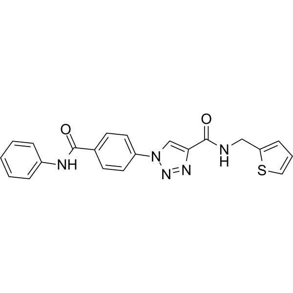
MKA031 (compound 6y) is a non-competitive MIF inhibitor with an IC50 value of 1.7 μM. MKA031 (compound 6y) interferes with MIF/AIF interaction, MIF nuclear translocation, and N-methyl-N'-nitro-N-nitrosoguanidine (MNNG)-induced dependent cell death. MKA031 can be used in the study of chronic hepatitis C virus infection.
For research use only. We do not sell to patients.
- Purity : 98.69%
- CAS No.: 2951012-00-1
- Formula: C21H17N5O2S
- Molecular Weight:403.46
-
Storage:Powder -20°C, 3 years ; In solvent -80°C, 6 months , -20°C, 1 month

To place orders, for customer services and technical support, please contact: MedChemExpress USA
Tel: 609-228-6898 E-mail: [email protected] [email protected]


-
 Biological Activity
-
Chemical Information
-
Solvent & Solubility
- Protocols
- Purity & Documentation
- References
-
 Help & FAQs




Biological Activity
Description
Cellular Effect
|
Cell Line

|
Type | Value | Description | References |
|---|---|---|---|---|
| HeLa | EC50 |
7.7 μM
Compound: 6y; MKA031
|
Protection against MNNG-induced parthanatos in human HeLa cells assessed as cell viability preincubated for 3 hrs prior to MNNG addition and measured for 15 mins followed by fresh media replacement for 24 hrs by Hoechst 33342 and propidium iodide staining
Protection against MNNG-induced parthanatos in human HeLa cells assessed as cell viability preincubated for 3 hrs prior to MNNG addition and measured for 15 mins followed by fresh media replacement for 24 hrs by Hoechst 33342 and propidium iodide staining
|
[PMID: 37352470] |
In Vitro
MKA031 (compound 6y) (1-100 μl; 3 h) prevents MNNG-Induced Cell Death[1]. MKA031 (compound 6y) (100 μl;3 h) protects Genomic DNA by Blocking the Recruitment of MIF to AIF[1].
MedChemExpress (MCE) has not independently confirmed the accuracy of these methods. They are for reference only.
-
Cell Line:Hela cells
-
Concentration:1-100 μM
-
Incubation Time:3 h
-
Result:Protected HeLa cells from MNNG-induced parthanatos in a dose dependent manner with an EC50 of 7.7 μM.
-
Cell Line:Hela cells
-
Concentration:100 μM
-
Incubation Time:3 h
-
Result:Interfered with the MIF/AIF interaction and blocks MIF nuclear translocation.


Chemical Information
-
CAS No. 2951012-00-1
-
Appearance Solid
-
Molecular Weight 403.46
-
Formula C21H17N5O2S
-
Color Off-white to light yellow
-
SMILES
O=C(NC1=CC=CC=C1)C2=CC=C(C=C2)N3N=NC(C(NCC4=CC=CS4)=O)=C3
-
Shipping
Room temperature in continental US; may vary elsewhere.
-
Storage
Powder -20°C 3 years In solvent -80°C 6 months -20°C 1 month


Solvent & Solubility
In Vitro:
DMSO : 25 mg/mL (61.96 mM; ultrasonic and warming and heat to 60°C; Hygroscopic DMSO has a significant impact on the solubility of product, please use newly opened DMSO)
Please refer to the solubility information to select the appropriate solvent. Once prepared, please aliquot and store the solution to prevent product inactivation from repeated freeze-thaw cycles.
Storage method and period of stock solution: -80°C, 6 months; -20°C, 1 month. When stored at -80°C, please use it within 6 months. When stored at -20°C, please use it within 1 month.
Please refer to the solubility information to select the appropriate solvent. Once prepared, please aliquot and store the solution to prevent product inactivation from repeated freeze-thaw cycles.
Storage method and period of stock solution: -80°C, 6 months; -20°C, 1 month. When stored at -80°C, please use it within 6 months. When stored at -20°C, please use it within 1 month.
Concentration (start) × Volume (start) = Concentration (final) × Volume (final)


Purity & Documentation
-
Data Sheet (271 KB)
-
SDS (251 KB)
- English - EN (251 KB)
- Français - FR (251 KB)
- Deutsch - DE (251 KB)
- Norwegian - NO (251 KB)
- Español - ES (251 KB)
- Swedish - SV (251 KB)
- Italian - IT (251 KB)
- Korean - KR (251 KB)
- Portuguese - PT (251 KB)
-
Handling Instructions (2659 KB)
References


Complete Stock Solution Preparation Table
Please refer to the solubility information to select the appropriate solvent. Once prepared, please aliquot and store the solution to prevent product inactivation from repeated freeze-thaw cycles.
Storage method and period of stock solution: -80°C, 6 months; -20°C, 1 month. When stored at -80°C, please use it within 6 months. When stored at -20°C, please use it within 1 month.
| Optional Solvent | Concentration Solvent Mass | 1 mg | 5 mg | 10 mg | 25 mg |
|---|---|---|---|---|---|
| DMSO | 1 mM | 2.4786 mL | 12.3928 mL | 24.7856 mL | 61.9640 mL |
| 5 mM | 0.4957 mL | 2.4786 mL | 4.9571 mL | 12.3928 mL | |
| 10 mM | 0.2479 mL | 1.2393 mL | 2.4786 mL | 6.1964 mL | |
| 15 mM | 0.1652 mL | 0.8262 mL | 1.6524 mL | 4.1309 mL | |
| 20 mM | 0.1239 mL | 0.6196 mL | 1.2393 mL | 3.0982 mL | |
| 25 mM | 0.0991 mL | 0.4957 mL | 0.9914 mL | 2.4786 mL | |
| 30 mM | 0.0826 mL | 0.4131 mL | 0.8262 mL | 2.0655 mL | |
| 40 mM | 0.0620 mL | 0.3098 mL | 0.6196 mL | 1.5491 mL | |
| 50 mM | 0.0496 mL | 0.2479 mL | 0.4957 mL | 1.2393 mL | |
| 60 mM | 0.0413 mL | 0.2065 mL | 0.4131 mL | 1.0327 mL |


MKA031 Related Classifications
HY-149308 Related Classifications
 Powered by Bioz