



MMT5-14
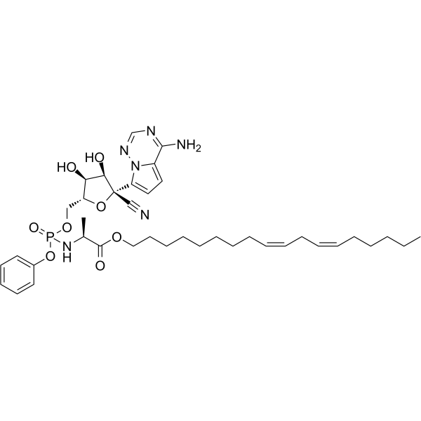
MMT5-14 is a remdesivir analogue with a higher antiviral activity in four variants of SARS-CoV-2 than Remdesivir (HY-104077). MMT5-14 inhibits SARS-CoV-2, α, β, γ and δ variants with EC50s of 0.4, 2.5, 15.9, 1.7 and 5.6 μM, respectively. MMT5-14 can be used for the research of COVID-19.
For research use only. We do not sell to patients.
- CAS No.: 2719679-31-7
- Formula: C39H55N6O8P
- Molecular Weight:766.86
-
Storage:
Please store the product under the recommended conditions in the Certificate of Analysis.

To place orders, for customer services and technical support, please contact: MedChemExpress USA
Tel: 609-228-6898 E-mail: [email protected] [email protected]


-
 Biological Activity
-
Chemical Information
- Protocols
- Purity & Documentation
- References
-
 Help & FAQs


-
HY-160554
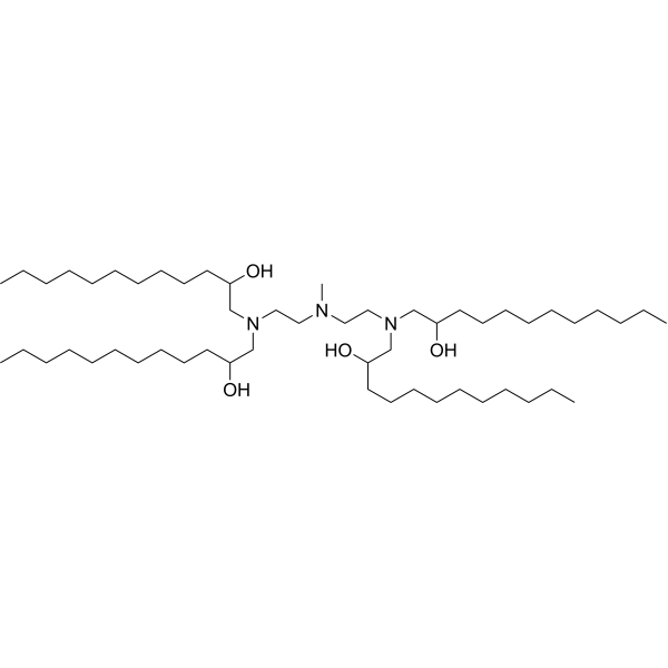
C12-113Lipidoid Delivery Agent -

HY-169497
MTDBMTDB-Alkyne Intermediate -
HY-136597
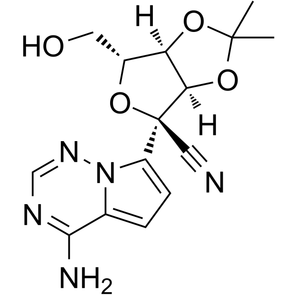
Remdesivir O-desphosphate acetonide impurity -

HY-W076298
Remdesivir intermediate-1 -

HY-136597R
Remdesivir O-desphosphate acetonide impurity (Standard)Remdisivir Impurity -

HY-139355
(S)-GS-621763GS-621763 S-enantiomer


Biological Activity
Description
In Vitro
MMT5-14 (10 μM; 0-12 h) increases epithelial cell uptake[1]. MMT5-14 (5 μM; 2 d) shows a better in vitro antiviral activity than remdesivir[1].
MedChemExpress (MCE) has not independently confirmed the accuracy of these methods. They are for reference only.
-
Cell Line:Vero-E6 cell line
-
Concentration:5 μM
-
Incubation Time:2 days
-
Result:Inhibited SARS-CoV-2, alpha, beta, gamma and delta variants with EC50s of 0.4, 2.5, 15.9, 1.7 and 5.6 μM, respectively.
In Vivo
MedChemExpress (MCE) has not independently confirmed the accuracy of these methods. They are for reference only.
-
Animal Model:Catheterized female hamsters[1]
-
Dosage:10 mg/kg
-
Administration:Intravenous injection; 10 mg/kg once
-
Result:Showed higher intact prodrugs concentration than remdesivir in lungs after 4 hours injection, increased tissue exposure in most of the tissues and showed 5- to 10-fold higher tissue selectivity in lungs compared to remdesivir.


Chemical Information
-
CAS No. 2719679-31-7
-
Molecular Weight 766.86
-
Formula C39H55N6O8P
-
SMILES
C[C@@H](C(OCCCCCCCC/C=C\C/C=C\CCCCC)=O)NP(OC1=CC=CC=C1)(OC[C@H]2O[C@@](C#N)(C3=CC=C4C(N)=NC=NN43)[C@H](O)[C@@H]2O)=O
-
Shipping
Room temperature in continental US; may vary elsewhere.
-
Storage
Please store the product under the recommended conditions in the Certificate of Analysis.


Purity & Documentation
References

Calculators
Concentration (start) × Volume (start) = Concentration (final) × Volume (final)


MMT5-14 Related Classifications
HY-151265 Related Classifications
 Powered by Bioz