



mPEG750-amine

Based on 1 Customer Validation
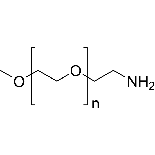
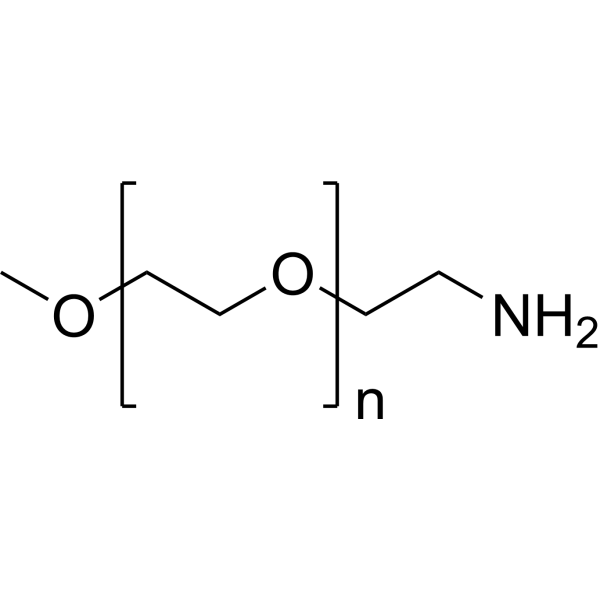
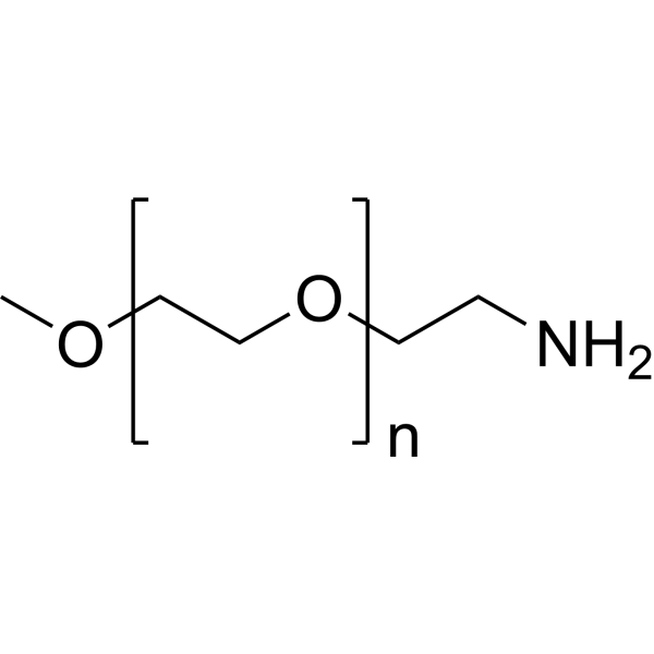
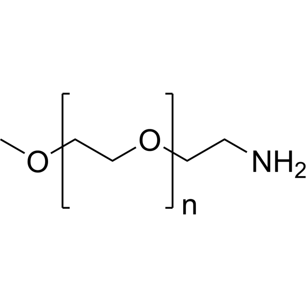
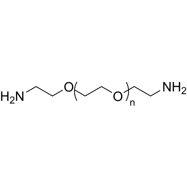
mPEG750-amine (mPEG750-NH2) is a chemical modification reagent for nanoparticles, capable of covalently binding to Ad-PVA to form Ad-PVA-PEG polymers. mPEG750-amine stabilizes gene delivery complexes by providing steric hindrance, reducing particle aggregation, while enhancing the water solubility and serum stability of the complex, reducing carrier cytotoxicity, and assisting in the efficient condensation of pDNA by cationic components to form nanoparticles that can be endocytosed by cells. mPEG750-amine can also be used to synthesize folate-conjugated polymer micelles for encapsulating the anticancer agent Camptothecin (HY-16560). Folate-conjugated polymer micelles are effective carriers for poorly soluble anticancer drugs, capable of avoiding macrophages and acting through folate receptor (FR)-mediated endocytosis to target tumor cells. mPEG750-amine can be applied to research in the field of non-viral gene delivery, as a component of gene delivery vectors, facilitating the safe and efficient delivery of nucleic acid drugs to target cells.
For research use only. We do not sell to patients.
- Purity : 90.0%
- CAS No.: 80506-64-5
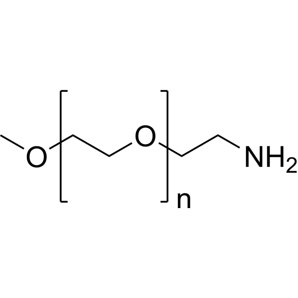
- Formula: (C2H4O)nC3H9NO
- Molecular Weight:750 (Average)
-
Storage:
4°C, stored under nitrogen, away from moisture
* In solvent : -80°C, 6 months; -20°C, 1 month (stored under nitrogen, away from moisture)

To place orders, for customer services and technical support, please contact: MedChemExpress USA
Tel: 609-228-6898 E-mail: [email protected] [email protected]


-
 Biological Activity
-
Chemical Information
-
Solvent & Solubility
- Protocols
- Purity & Documentation
- References
-
 Help & FAQs


-
HY-140676
mPEG2000-aminePEG Derivative -
HY-140677
mPEG5000-aminePEG Derivative -
HY-140675
mPEG1000-aminePEG Derivative -
HY-140679
mPEG20000-aminePEG Derivative -
HY-140678
mPEG10000-aminePEG Derivative -
HY-155881
mPEG550-aminePEG Derivative -
HY-155880
mPEG350-aminePEG Amine -
HY-155883
mPEG3400-amineMicellar Delivery -
HY-155884
mPEG4000-amineFolic Acid Micellar Delivery -
HY-W591632
PEG1000-bis-aminePEG Derivative -
HY-W115607
PEG4000-bis-amineLinker -
HY-140646
PEG2000-bis-aminePEG Derivative


Biological Activity
Description
In Vitro
Ad-PVA-PEG750, synthesized from mPEG750-amine via CDI activation reaction with Ad-PVA (stable for >5 days at pH=7, and 48 hours at pH=4), remains stable in a physiological environment at pH=7, but undergoes degradation via acetal bond cleavage under acidic conditions at pH=4, confirming its acid-responsive nature[2].
Ad-PVA-PEG750 synthesized from mPEG750-amine (forming complexes with cationic β-CD derivatives and pDNA at an N:P ratio ≥5) effectively concentrates pDNA. Compared to cationic β-CD alone, complete pDNA condensation and inhibition of its migration in agarose gel can be achieved at a lower N:P ratio[2].
The complex formed by Ad-PVA-PEG750 synthesized from mPEG750-amine and cationic β-CD exhibits 1000 times lower cytotoxicity to HeLa cells than 25 kDa bPEI, significantly reducing the cytotoxicity of the gene carrier[2].
MedChemExpress (MCE) has not independently confirmed the accuracy of these methods. They are for reference only. Further protocols information, click here.
In Vivo
It can also be used to evaluate the effects of mPEG-750 modified silicone nanoparticles on the mouse hematopoietic system (hemolysis, coagulation function)[1].
MedChemExpress (MCE) has not independently confirmed the accuracy of these methods. They are for reference only.


Chemical Information
-
CAS No. 80506-64-5
-
Appearance Oil
-
Molecular Weight 750 (Average)
-
Formula (C2H4O)nC3H9NO
-
Color Off-white to light yellow
-
SMILES
COCCOCCN.[n]
-
Synonyms
mPEG750-NH2
-
Shipping
Room temperature in continental US; may vary elsewhere.
-
Storage
4°C, stored under nitrogen, away from moisture
* In solvent : -80°C, 6 months; -20°C, 1 month (stored under nitrogen, away from moisture)


Solvent & Solubility
In Vitro:
DMSO : 100 mg/mL (Need ultrasonic; Hygroscopic DMSO has a significant impact on the solubility of product, please use newly opened DMSO)

Protocols
-
Cell Cytotoxicity Assay
Cytotoxicity assays are usually based on the assessment of cell membrane damage, which can also be indirectly detected by measuring cell viability. Detection methods include MTT assay, CKK-8 assay, LDH assay and ATP assay, etc.
-
Mammalian live/dead viability and cytotoxicity staining
Live/dead viability and cytotoxicity staining assays are based on the simultaneous detection of intracellular esterase activity in metabolically active (viable) cells and membrane integrity loss in non-viable cells. In commonly used dual-staining approaches, membrane-permeant fluorogenic substrates are converted by intracellular esterases into fluorescent products in live cells, while impermeant DNA-binding dyes selectively enter cells with compromised plasma membranes and label nucleic acids in dead or dying cells, enabling discrimination between viable and non-viable populations by fluorescence microscopy or flow cytometry.
-
Cell Viability Determination by MTT Colorimetric Assay
The following protocol uses the MTT colorimetric assay as a classic literature-established method for assessing cell viability/metabolic activity in cultured mammalian cells. MTT[3-(4,5-dimethylthiazol-2-yl)-2,5-diphenyltetrazolium bromide] is reduced by metabolically active cells to a colored formazan product; the amount of formazan is quantified spectrophotometrically and provides an indirect measure of metabolically active viable cells. Importantly, MTT reduction reflects cellular oxidoreductase/metabolic activity rather than an absolute direct count of living cells, so changes in cellular metabolism can alter the signal independently of cell number.


Purity & Documentation
-
Data Sheet (269 KB)
-
SDS (394 KB)
- English - EN (394 KB)
- Français - FR (394 KB)
- Deutsch - DE (394 KB)
- Norwegian - NO (394 KB)
- Español - ES (394 KB)
- Swedish - SV (394 KB)
- Italian - IT (394 KB)
- Korean - KR (394 KB)
- Portuguese - PT (394 KB)
-
Handling Instructions (2659 KB)
References

Calculators
Concentration (start) × Volume (start) = Concentration (final) × Volume (final)


mPEG750-amine Related Classifications
HY-155882 Related Classifications
 Powered by Bioz