



mPGES1-IN-3
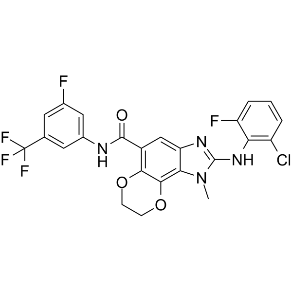
mPGES1-IN-3 is a selective, orally active mPGES-1 inhibitor with an IC50 of 8.0 nM for h-mPGES-1 and an IC50 of 10.79 nM for guinea pig mPGES1. mPGES1-IN-3 blocks the conversion of PGH2 (HY-136500) to PGE2 (HY-101952). mPGES1-IN-3 exhibits dose-dependent inhibition of LPS-induced thermal hyperalgesia. mPGES1-IN-3 can be used for research on rheumatoid arthritis.
For research use only. We do not sell to patients.
- CAS No.: 1469976-70-2
- Formula: C24H16ClF5N4O3
- Molecular Weight:538.85
-
Storage:
Please store the product under the recommended conditions in the Certificate of Analysis.

To place orders, for customer services and technical support, please contact: MedChemExpress USA
Tel: 609-228-6898 E-mail: [email protected] [email protected]


-
 Biological Activity
-
Chemical Information
- Protocols
- Purity & Documentation
- References
-
 Help & FAQs


-
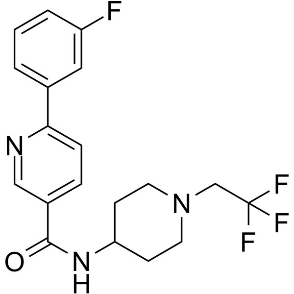
HY-10439
HPGDS inhibitor 1HPGDS Inhibitor -
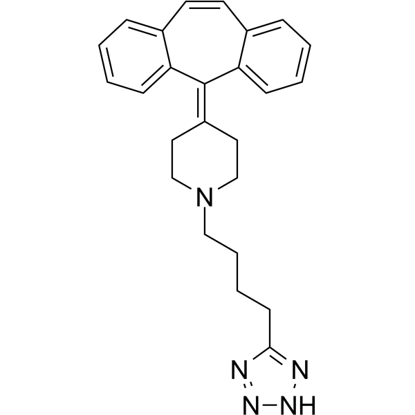
HY-13988
AT-56L-PGDS Inhibitor -
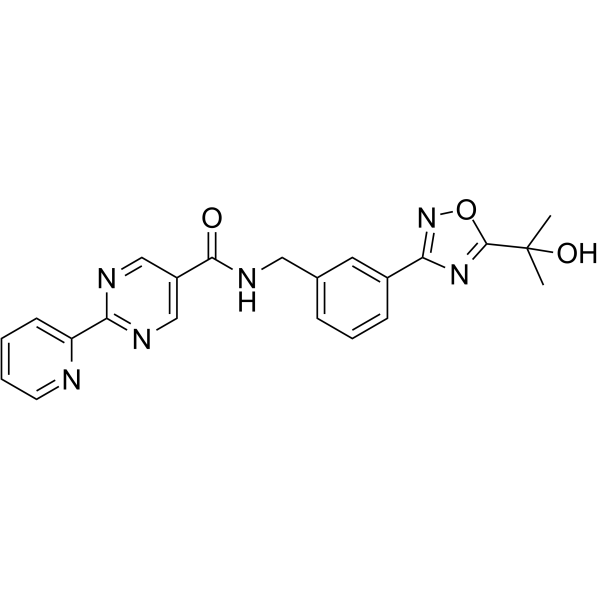
HY-12791
hPGDS-IN-1hPGDS Inhibitor -
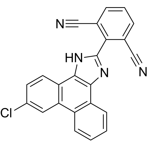
HY-13283
MF63MF63 Inhibitor -
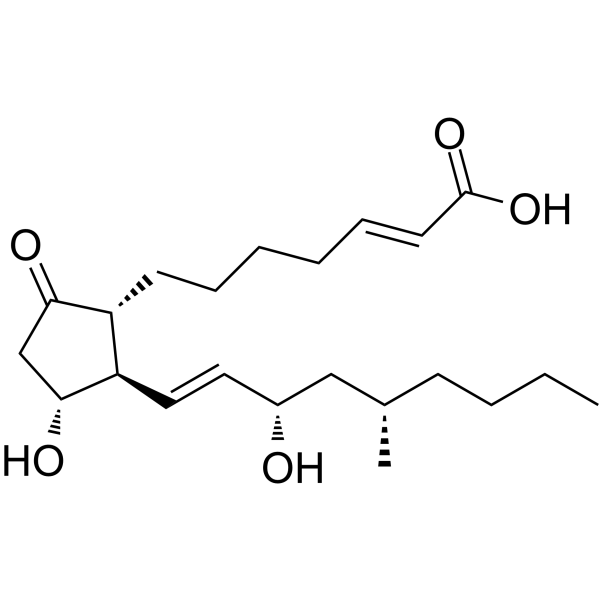
HY-B0683
LimaprostVasodilator Agent -

HY-10439R
HPGDS inhibitor 1 (Standard)Reference Standard -
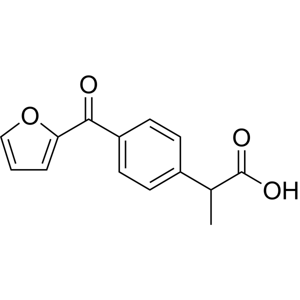
HY-106907
FurprofenAnti-Inflammatory Drug -

HY-13988R
AT-56 (Standard)L-PGDS Inhibitor -
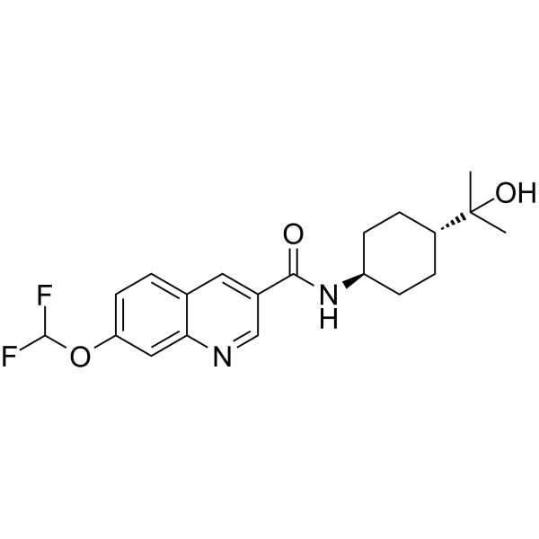
HY-126134
HPGDS inhibitor 2 -
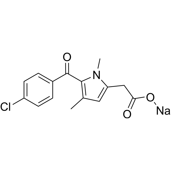
HY-B0890
Zomepirac sodium saltProstaglandin Synthetase Inhibitor -
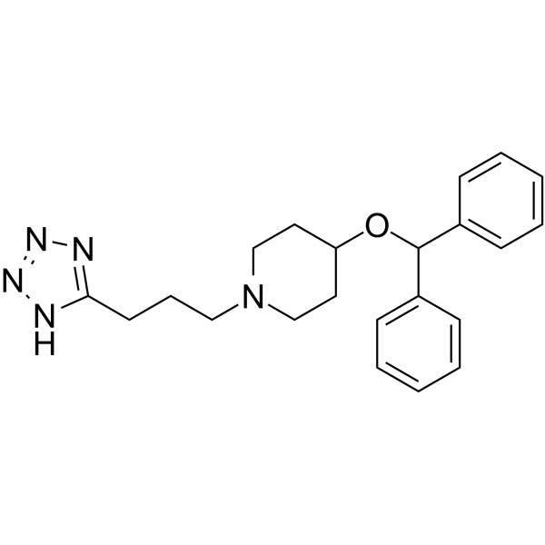
HY-108259
HQL-79Prostaglandin D Synthase Inhibitor -
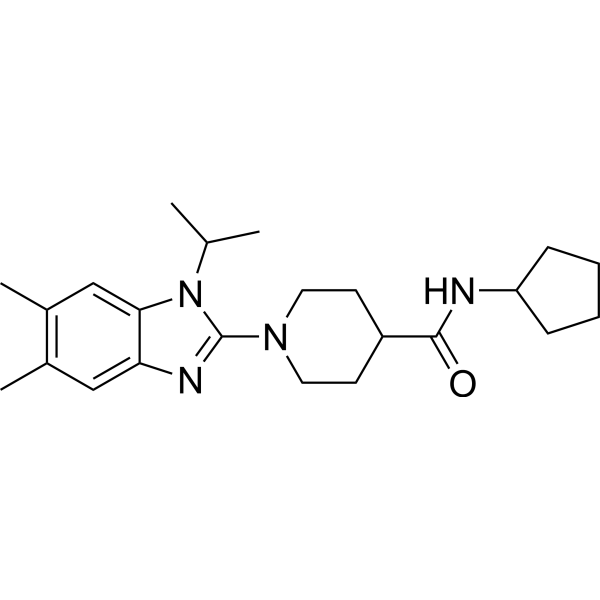
HY-118282
mPGES1-IN-7mPGES-1 Inhibitor


Biological Activity
Description
IC50 & Target
IC50: 8 nM (mPGES-1)[1]
Cellular Effect
|
Cell Line

|
Type | Value | Description | References |
|---|---|---|---|---|
| A549 | IC50 |
16.24 nM
|
Inhibition of PGE2 production in human A549 cells in the presence of 2% FBS.
Inhibition of PGE2 production in human A549 cells in the presence of 2% FBS.
|
27865703 |
In Vitro
mPGES1-IN-3 (Compound 17d) (10 μM; 60 min) exhibits favorable in vitro ADME properties, including good HWB potency, high metabolic stability across species, minimal CYP inhibition, and high plasma protein binding[1].
mPGES1-IN-3 (10 μM) exhibits much higher selectivity for mPGES-1 over other prostaglandin synthases and COX enzymes, and has a favorable hERG and off-target safety profile[1].
mPGES1-IN-3 is a potent inhibitor of guinea pig mPGES-1 and h-mPGES-1, but shows no inhibitory effect on rat or mouse mPGES-1[1].
MedChemExpress (MCE) has not independently confirmed the accuracy of these methods. They are for reference only. Further protocols information, click here.
Parmacokinetics
| Species | Dose | Route | T1/2 | CL | Vz | Cmax | AUC | Tmax | Bioavailability |
|---|---|---|---|---|---|---|---|---|---|
| Rat[1] | 1 mg/kg | i.v. | 6.2 h | 18.0 mL/min/kg | 10.24 L/kg | / | / | / | / |
| Rat[1] | 10 mg/kg | p.o. | / | / | / | 29.0 ng/mL | 149 ng·h/mL | 4 h | 1.5 % |
| Rat[1] | 10 mg/kg | p.o. | / | / | / | 163 ng/mL | 2259 ng·h/mL | 4 h | 23 % |
| Rat[1] | 30 mg/kg | p.o. | / | / | / | 598 ng/mL | 8403 ng·h/mL | 8 h | 29 % |
| Rat[1] | 100 mg/kg | p.o. | / | / | / | 2035 ng/mL | 23,114 ng·h/mL | 2 h | 24 % |
In Vivo
MedChemExpress (MCE) has not independently confirmed the accuracy of these methods. They are for reference only.
-
Animal Model:Guinea pig hyperalgesia pain model[1]
-
Dosage:10, 30, 60,100 mg/kg
-
Administration:p.o.
-
Result:Significantly inhibited
hyperalgesic response in a dose-dependent manner with maxi-
mum inhibition of 73% at 100 mg/kg.
Inhibited <50% of hyperalgesic response in comparison with Diclofenac (10 and 30 mg/kg)


Chemical Information
-
CAS No. 1469976-70-2
-
Molecular Weight 538.85
-
Formula C24H16ClF5N4O3
-
SMILES
O=C(NC1=CC(F)=CC(C(F)(F)F)=C1)C2=C(OCCO3)C3=C(N(C)C(NC4=C(Cl)C=CC=C4F)=N5)C5=C2
-
Shipping
Room temperature in continental US; may vary elsewhere.
-
Storage
Please store the product under the recommended conditions in the Certificate of Analysis.

Protocols
-
Collagen-Induced Arthritis
Collagen-induced arthritis (CIA) is an autoimmune murine model of rheumatoid arthritis in which immunization with type II collagen (CII) emulsified in an adjuvant induces a T cell- and autoantibody-driven inflammatory arthritis characterized by synovial hyperplasia, immune cell infiltration, and joint destruction. The model typically relies on genetically susceptible mouse strains (e. g. , DBA/1) and reproduces key features of human rheumatoid arthritis, including anti-collagen immune responses and progressive joint inflammation. Disease onset generally occurs within ~3-4 weeks after immunization, depending on antigen/adjuvant combinations and protocol variation. The immunopathology is driven by adaptive immune activation against CII, leading to systemic and local joint inflammation mediated by pro-inflammatory cytokines and effector immune cells, making CIA a standard preclinical platform for evaluating immunomodulatory and anti-arthritic interventions.
-
Cell Cytotoxicity Assay
Cytotoxicity assays are usually based on the assessment of cell membrane damage, which can also be indirectly detected by measuring cell viability. Detection methods include MTT assay, CKK-8 assay, LDH assay and ATP assay, etc.


Purity & Documentation
References

Calculators
Concentration (start) × Volume (start) = Concentration (final) × Volume (final)


mPGES1-IN-3 Related Classifications
HY-100864 Related Classifications
 Powered by Bioz