



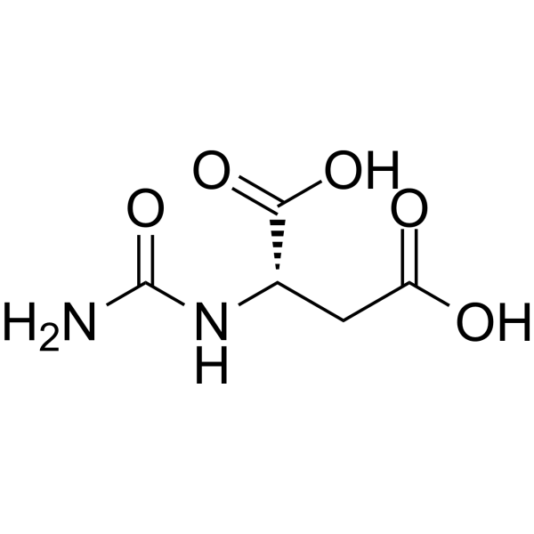
N-Carbamoyl-L-aspartic acid
Based on 1 publication(s) in Google Scholar
N-Carbamoyl-L-aspartic acid (L-Ureidosuccinic acid) is an important pyrimidine metabolic precursor and intermediate metabolite. N-Carbamoyl-L-aspartic acid reverses the growth inhibition of Ura+ strains induced by 2-thiouracil (with growth rate increasing linearly with its concentration), but fails to support the growth of uracil-requiring Ura- strains. N-Carbamoyl-L-aspartic acid inhibits the cell growth of *Saccharomyces cerevisiae* by suppressing the purine biosynthetic pathway at a pre-step of 5-aminoimidazole nucleotide synthesis. The growth inhibitory effect of N-Carbamoyl-L-aspartic acid on yeast can be alleviated by purines, and the sensitivity of strains is closely related to the activity level of dihydroorotase.
For research use only. We do not sell to patients.
- Purity : 99.83%
- CAS No.: 13184-27-5
- Formula: C5H8N2O5
- Molecular Weight:176.13
-
Storage:Powder -20°C, 3 years , 4°C, 2 years ; In solvent -80°C, 6 months , -20°C, 1 month

To place orders, for customer services and technical support, please contact: MedChemExpress USA
Tel: 609-228-6898 E-mail: [email protected] [email protected]


-
 Biological Activity
-
Chemical Information
-
Solvent & Solubility
- Protocols
- Purity & Documentation
- References
-
 Help & FAQs


-
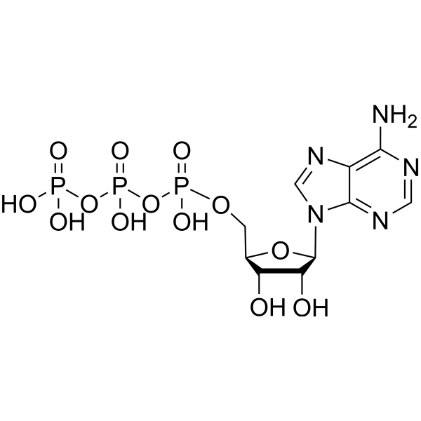
HY-B2176
ATPEndogenous Metabolite -
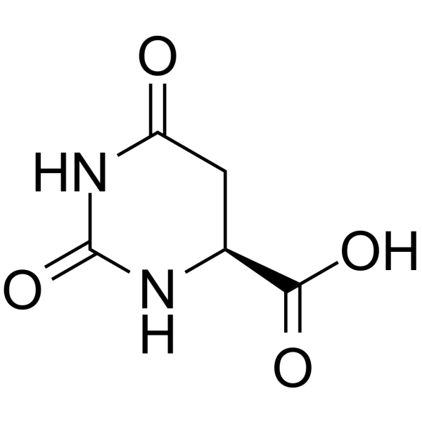
HY-W015495
L-Dihydroorotic acidMetabolic Product -
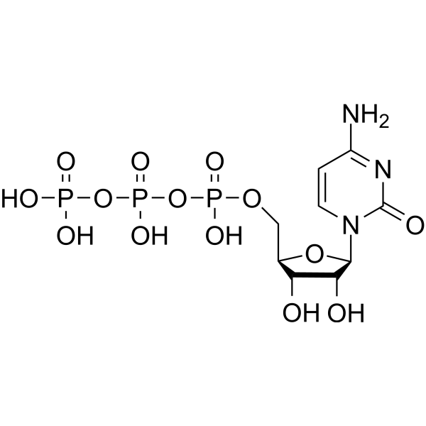
HY-125818
Cytidine-5'-triphosphateNucleoside Triphosphate -
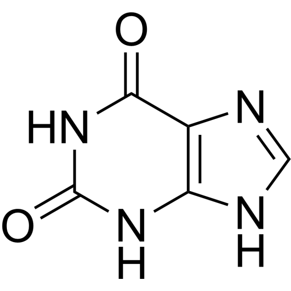
HY-W017389
Xanthine -
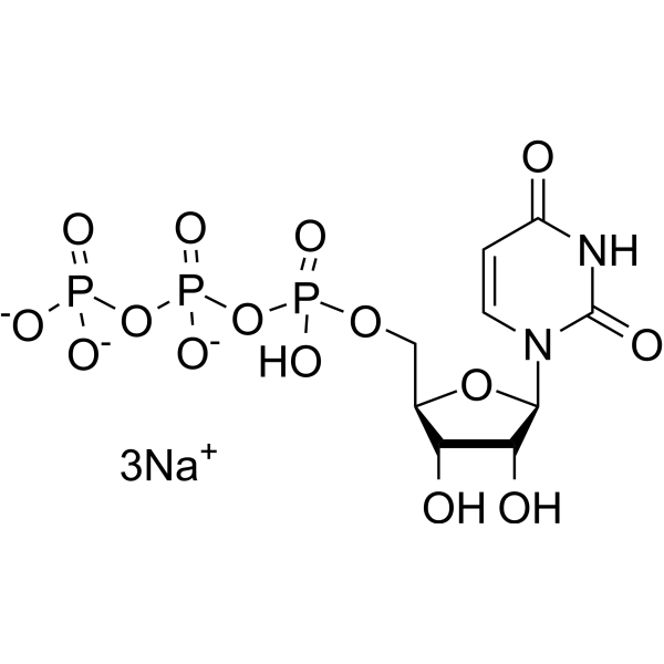
HY-W013093
Uridine triphosphate trisodium saltUTP -
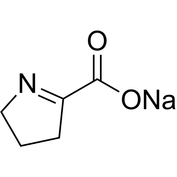
HY-138354A
Pyrroline-5-carboxylate sodiumIntermediate -
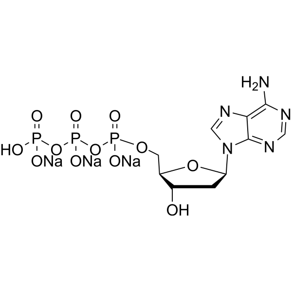
HY-136648A
2'-Deoxyadenosine-5'-triphosphate trisodiumDNA Polymerase Substrate -
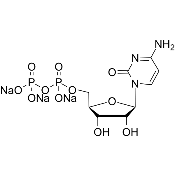
HY-W008915
Cytidine 5'-diphosphate trisodium salt -
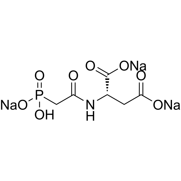
HY-112732B
Sparfosic acid trisodiumAspartate Transcarbamoyl Transferase Inhibitor -

HY-160610A
Carbamoyl phosphate dilithium hydrate, ≥85% (enzymatic)Antisickling Agent
Publications Citing Use of MedChemExpress (MCE) N-Carbamoyl-L-aspartic acid
More

Biological Activity
Description
In Vitro
N-Carbamoyl-L-aspartic acid (25-100 μg/mL, 10 mg/mL; up to 70 hrs) potently inhibits growth of Saccharomyces cerevisiae wild type FL 100, ura2-60, ura4-20, and ure1 strains, while ura1-8, ura3-6, and ura5-5 strains are highly resistant to this inhibition[1].
MedChemExpress (MCE) has not independently confirmed the accuracy of these methods. They are for reference only. Further protocols information, click here.
-
Cell Line:Saccharomyces cerevisiae strains (wild type FL 100, ura2-60, ura4-20, ure1, ura1-8, ura3-6, ura5-5)
-
Concentration:25 μg/mL, 50 μg/mL, 100 μg/mL, 10 mg/mL; 100 μg/mL
-
Incubation Time:up to 70 hrs; 375 min
-
Result:Dramatically inhibited growth of wild type FL 100, ura2-60, ura4-20, and ure1 strains, with initial reduced growth rates that returned to normal after 12-20 hrs (depending on concentration).
Matched untreated cell density by 48 hrs with 100 μg/mL treatment.
Showed no concentration-dependent recovery of normal growth rate in ura4-20 strain.
Only slightly or not inhibited ura1-8, ura3-6, and ura5-5 strains.
Created a halo of no growth surrounding ura4-20 streak near ura2-60 lawns, confirming it is the inhibitory enantiomorph.
Showed unimpeded growth of USA-resistant ura2-60 derivative adjacent to ura4-20.


Chemical Information
-
CAS No. 13184-27-5
-
Appearance Solid
-
Molecular Weight 176.13
-
Formula C5H8N2O5
-
Color White to off-white
-
SMILES
O=C(O)C[C@@H](C(O)=O)NC(N)=O
-
Synonyms
L-Ureidosuccinic acid
-
Structure Classification
-
Initial Source
-
Shipping
Room temperature in continental US; may vary elsewhere.
-
Storage
Powder -20°C 3 years 4°C 2 years In solvent -80°C 6 months -20°C 1 month


Publications (1)
-
Journal Impact Factor
-
Most Recent
-
Mol Cell
2025 Dec 4;85(23):4347-4364.e12. PMID: 41270758


Solvent & Solubility
In Vitro:
H2O : 31.25 mg/mL (177.43 mM; Need ultrasonic)
Please refer to the solubility information to select the appropriate solvent. Once prepared, please aliquot and store the solution to prevent product inactivation from repeated freeze-thaw cycles.
Storage method and period of stock solution: -80°C, 6 months; -20°C, 1 month. When stored at -80°C, please use it within 6 months. When stored at -20°C, please use it within 1 month.
* Note: If you choose water as the stock solution, please dilute it to the working solution, then filter and sterilize it with a 0.22 μm filter before use.
Please refer to the solubility information to select the appropriate solvent. Once prepared, please aliquot and store the solution to prevent product inactivation from repeated freeze-thaw cycles.
Storage method and period of stock solution: -80°C, 6 months; -20°C, 1 month. When stored at -80°C, please use it within 6 months. When stored at -20°C, please use it within 1 month.
* Note: If you choose water as the stock solution, please dilute it to the working solution, then filter and sterilize it with a 0.22 μm filter before use.
Concentration (start) × Volume (start) = Concentration (final) × Volume (final)

Protocols
-
Protocol for Cell Counting and Cell Density Analysis
Cell counting and cell-density analysis estimate the number of cells in a known volume or field area. Manual hemocytometer counting uses a chamber of defined geometry to convert counted cells into cells/mL, while automated counters and image-analysis workflows detect cell objects from optical, brightfield, fluorescence, impedance, or digital-image features. Trypan blue viability counting is based on dye exclusion: viable cells with intact membranes exclude dye, while non-viable cells with compromised membranes stain blue. The readout is total cell density, viable-cell density, dead-cell density, and percent viability. Cell density can also be estimated from microscopy images by counting objects per image area, from flow cytometry using calibrated volume or reference particles, or from in situ microscopy in bioreactors after calibration against reference methods such as hemocytometer or flow cytometry.
-
Somatic Cell Culture
A method of simulating the in vivo environment in vitro to maintain the cell growth, differentation and main functions.
-
CCK-8/WST-8 Cell Proliferation Assay
The CCK-8/WST-8 assay is based on the reduction of the water-soluble tetrazolium salt WST-8 to a water-soluble formazan product by cellular dehydrogenases in metabolically active cells, where the generated formazan amount is proportional to the number of living cells and is quantified by measuring absorbance in the visible range, providing a colorimetric readout for cell viability and proliferation assessment. This class of tetrazolium-based assays improves upon earlier MTT-based systems by producing a water-soluble formazan, eliminating the need for organic solubilization steps and enabling direct spectrophotometric measurement in culture medium.
-
Cell Counting-Based Growth Curve Assay
Cell counting-based growth curve assays quantify cell proliferation by directly measuring changes in viable cell number over time using manual or automated counting methods such as hemocytometer-based counting or instrument-assisted cell enumeration, enabling construction of growth curves that reflect population expansion dynamics in response to culture conditions. A widely used approach is trypan blue exclusion with hemocytometer counting, where membrane-compromised (non-viable) cells take up the dye, allowing discrimination between viable and non-viable cells while simultaneously enabling total cell number quantification. Repeated sampling across time points allows estimation of proliferation rate, growth phases, and comparative growth kinetics between experimental conditions.
-
MTT Cell Proliferation Assay
The MTT assay is a colorimetric endpoint assay for estimating viable cell number, cell growth, cytotoxicity, or cell activation in cultured mammalian cells. Living cells reduce the yellow tetrazolium salt MTT into purple/blue formazan, while dead cells do not generate the same signal; the resulting color can be quantified with a multiwell spectrophotometer. MTT reduction is commonly interpreted as a readout of metabolic activity that often correlates with viable cell number, but it should not be treated as a direct cell-counting method unless the assay is optimized for the cell type and experimental condition. Studies show that MTT reduction can involve mitochondrial and non-mitochondrial reducing systems, and formazan may accumulate in intracellular lipid droplets rather than simply marking mitochondria.
-
Cell Viability Determination by MTT Colorimetric Assay
The following protocol uses the MTT colorimetric assay as a classic literature-established method for assessing cell viability/metabolic activity in cultured mammalian cells. MTT[3-(4,5-dimethylthiazol-2-yl)-2,5-diphenyltetrazolium bromide] is reduced by metabolically active cells to a colored formazan product; the amount of formazan is quantified spectrophotometrically and provides an indirect measure of metabolically active viable cells. Importantly, MTT reduction reflects cellular oxidoreductase/metabolic activity rather than an absolute direct count of living cells, so changes in cellular metabolism can alter the signal independently of cell number.


Purity & Documentation
-
Data Sheet (273 KB)
-
SDS (393 KB)
- English - EN (393 KB)
- Français - FR (393 KB)
- Deutsch - DE (393 KB)
- Norwegian - NO (393 KB)
- Español - ES (393 KB)
- Swedish - SV (393 KB)
- Italian - IT (393 KB)
- Korean - KR (393 KB)
- Portuguese - PT (393 KB)
-
Handling Instructions (2659 KB)
References


Complete Stock Solution Preparation Table
Please refer to the solubility information to select the appropriate solvent. Once prepared, please aliquot and store the solution to prevent product inactivation from repeated freeze-thaw cycles.
Storage method and period of stock solution: -80°C, 6 months; -20°C, 1 month. When stored at -80°C, please use it within 6 months. When stored at -20°C, please use it within 1 month.
| Optional Solvent | Concentration Solvent Mass | 1 mg | 5 mg | 10 mg | 25 mg |
|---|---|---|---|---|---|
| H2O | 1 mM | 5.6776 mL | 28.3881 mL | 56.7762 mL | 141.9406 mL |
| 5 mM | 1.1355 mL | 5.6776 mL | 11.3552 mL | 28.3881 mL | |
| 10 mM | 0.5678 mL | 2.8388 mL | 5.6776 mL | 14.1941 mL | |
| 15 mM | 0.3785 mL | 1.8925 mL | 3.7851 mL | 9.4627 mL | |
| 20 mM | 0.2839 mL | 1.4194 mL | 2.8388 mL | 7.0970 mL | |
| 25 mM | 0.2271 mL | 1.1355 mL | 2.2710 mL | 5.6776 mL | |
| 30 mM | 0.1893 mL | 0.9463 mL | 1.8925 mL | 4.7314 mL | |
| 40 mM | 0.1419 mL | 0.7097 mL | 1.4194 mL | 3.5485 mL | |
| 50 mM | 0.1136 mL | 0.5678 mL | 1.1355 mL | 2.8388 mL | |
| 60 mM | 0.0946 mL | 0.4731 mL | 0.9463 mL | 2.3657 mL | |
| 80 mM | 0.0710 mL | 0.3549 mL | 0.7097 mL | 1.7743 mL | |
| 100 mM | 0.0568 mL | 0.2839 mL | 0.5678 mL | 1.4194 mL |
* Note: If you choose water as the stock solution, please dilute it to the working solution, then filter and sterilize it with a 0.22 μm filter before use.


N-Carbamoyl-L-aspartic acid Related Classifications
HY-128425A Related Classifications
 Powered by Bioz