



Naphthofluorescein

Based on 1 Customer Validation
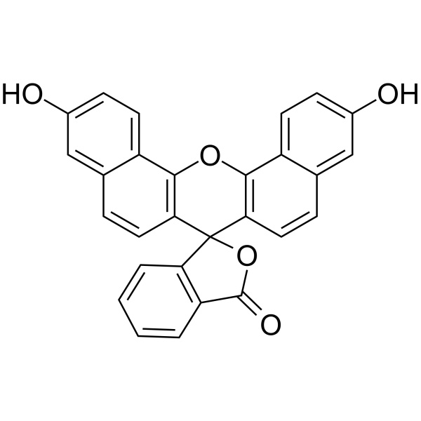
Naphthofluorescein inhibits the interaction between HIF-1 and Mint3. Naphthofluorescein suppresses Mint3-dependent HIF-1 activity and glycolysis in cancer cells and macrophages without cytotoxicity in vitro and adverse effect in vivo. Naphthofluorescein is also a fluorescent pH-sensitive probe that can be used for functional Cerenkov imaging.
For research use only. We do not sell to patients.
- Purity : 99.02%
- CAS No.: 61419-02-1
- Formula: C28H16O5
- Molecular Weight:432.42
-
Storage:Powder -20°C, 3 years , 4°C, 2 years ; In solvent -80°C, 6 months , -20°C, 1 month

To place orders, for customer services and technical support, please contact: MedChemExpress USA
Tel: 609-228-6898 E-mail: [email protected] [email protected]


-
 Biological Activity
-
Chemical Information
-
Solvent & Solubility
- Protocols
- Purity & Documentation
- References
-
 Help & FAQs


-
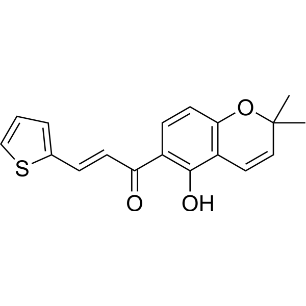
HY-100693
SYP-5HIF-1 Inhibitor -
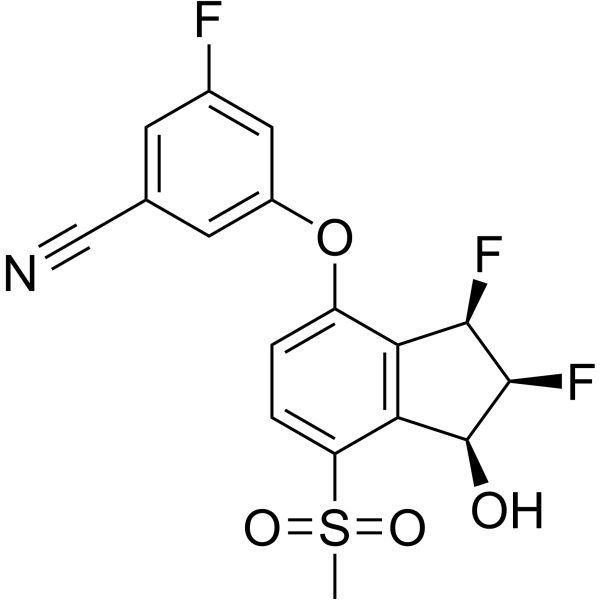
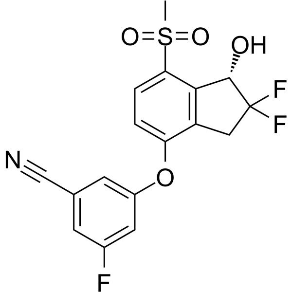
HY-125840
BelzutifanHIF-2α Inhibitor -
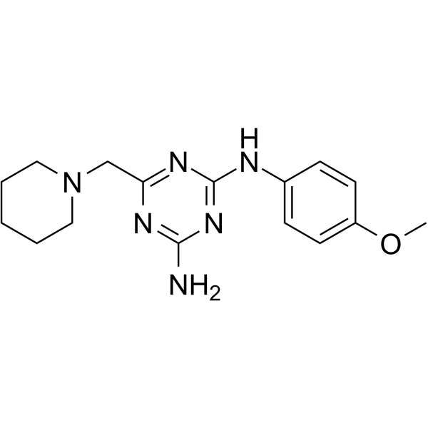
HY-116785
BRD32048ETV1 Direct Binder -

HY-100693R
SYP-5 (Standard)HIF-1 Inhibitor -
HY-12867
PT-2385HIF-2α Inhibitor -
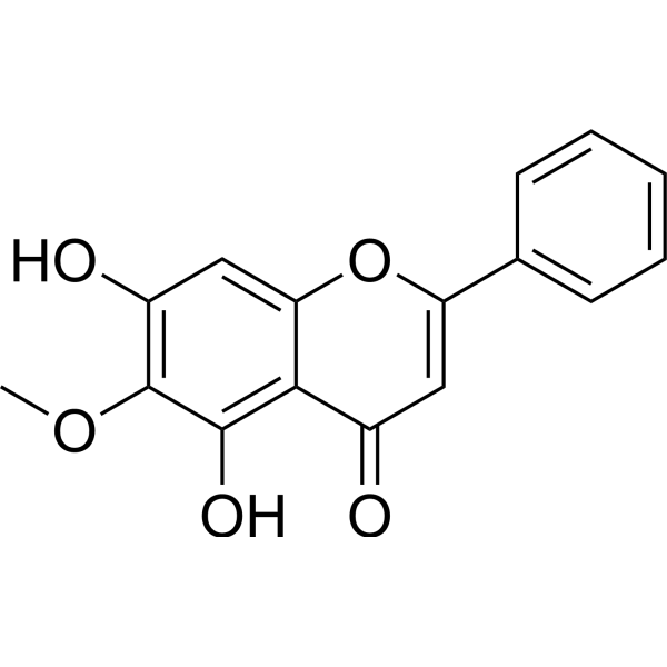
HY-N0560
Oroxylin AAutophagy Inducer -
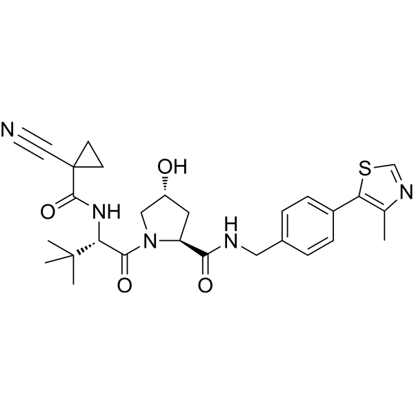
HY-100947
VH-298VHL:HIF-α Interaction Inhibitor -
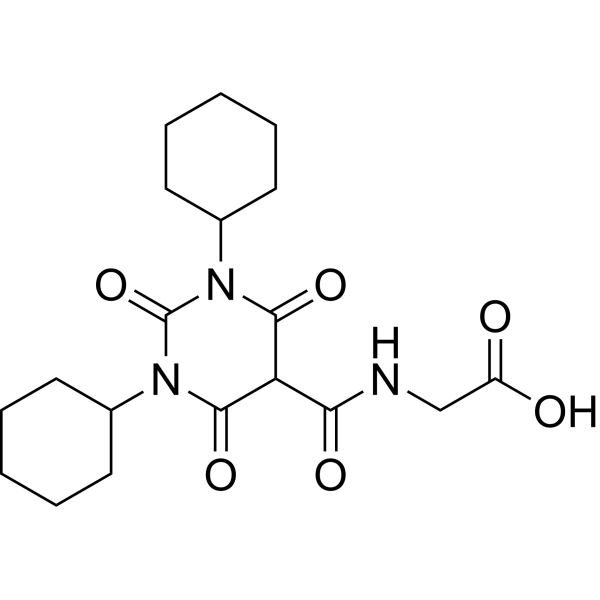
HY-17608
DaprodustatHIF-PH Inhibitor -
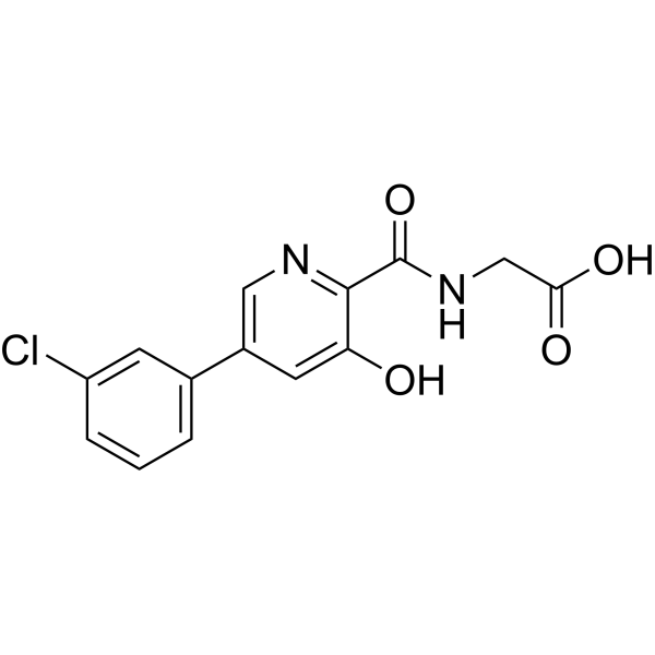
HY-101277
VadadustatHIF PH Inhibitor -
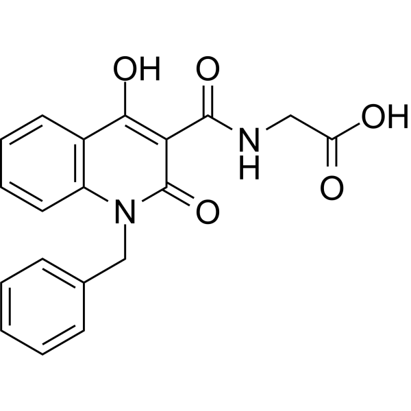
HY-15468
IOX2HIF/HIF Prolyl-Hydroxylase Inhibitor -
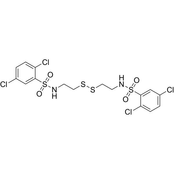
HY-18777
KC7F2HIF/HIF Prolyl-Hydroxylase Inhibitor -
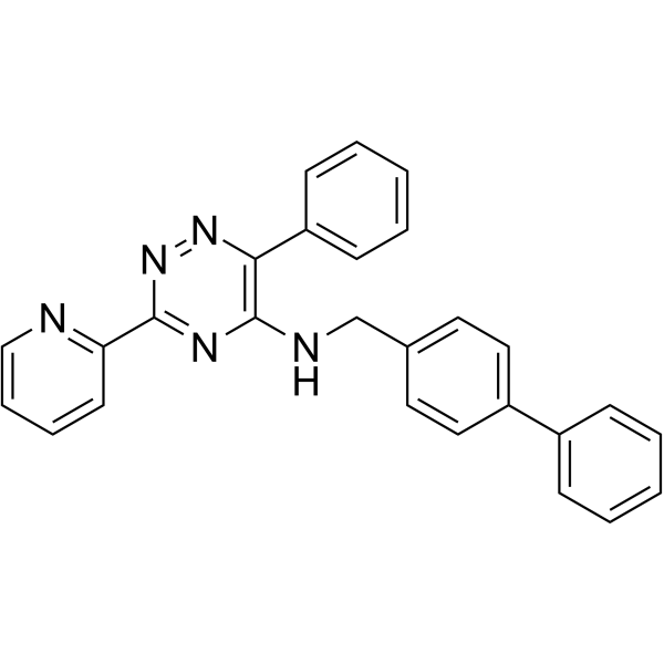
HY-12754
ML228HIF Activator


Biological Activity
Description
IC50 & Target
HIF-1[1]
In Vitro
Naphthofluorescein (compound 19) (0-10 μM; 24 hours) suppresses the HIF-1 reporter activity in a concentration-dependent manner[1].
MedChemExpress (MCE) has not independently confirmed the accuracy of these methods. They are for reference only. Further protocols information, click here.
-
Cell Line:HT1080 cells
-
Concentration:0-10 μM
-
Incubation Time:24 hours
-
Result:Significantly suppressed the HIF-1 reporter activity in a concentration-dependent manner.
In Vivo
Naphthofluorescein (compound 19) (100 mg/kg; i.p.; once daily for 5 consecutive days followed by 2 days off for 2 weeks) strikingly suppresses the tumour growth of subcutaneously injected E0771 cells and significantly attenuates tumour growth of MDA-MB-231 and AsPC-1 cells in immunodeficient mice. In turn, naphthofluorescein does not attenuate tumour growth of FIH-1-depleted MDA-MB-231 cells[1].
Naphthofluorescein (compound 19) (100 mg/kg; i.p.; once daily for 5 consecutive days followed by 2 days off for 2 weeks) suppresses tumour growth in human cancer cells in an FIH-1-dependent manner[1].
MedChemExpress (MCE) has not independently confirmed the accuracy of these methods. They are for reference only.
-
Animal Model:Male C57BL/6J mice (8 weeks)[1]
-
Dosage:100 mg/kg
-
Administration:i.p.; once daily for 5 consecutive days followed by 2 days off for 2 weeks
-
Result:Neither showed weight loss or apparent histological abnormalities in the lung, liver, and kidney, or caused cause severe adverse effects for at least 2 weeks in mice.
-
Animal Model:Female C57BL/6J mice (E0771) and BALB/c nude mice (HT1080, MDA-MB-231, and AsPC-1) (6 weeks)[1]
-
Dosage:100 mg/kg
-
Administration:i.p.; once daily for 5 consecutive days followed by 2 days off for 2 weeks
-
Result:Strikingly suppressed the tumour growth of subcutaneously injected E0771 cells and significantly attenuated tumour growth of MDA-MB-231 and AsPC-1 cells in immunodeficient mice. In turn, naphthofluorescein did not attenuate tumour growth of FIH-1-depleted MDA-MB-231 cells.


Chemical Information
-
CAS No. 61419-02-1
-
Appearance Solid
-
Molecular Weight 432.42
-
Formula C28H16O5
-
Color Purple to purplish red
-
SMILES
O=C(C1=C2C=CC=C1)OC32C4=C(OC5=C3C=CC6=CC(O)=CC=C56)C7=CC=C(O)C=C7C=C4
-
Synonyms
Naphthafluorescein
-
Shipping
Room temperature in continental US; may vary elsewhere.
-
Storage
Powder -20°C 3 years 4°C 2 years In solvent -80°C 6 months -20°C 1 month


Solvent & Solubility
In Vitro:
DMSO : 125 mg/mL (289.07 mM; Need ultrasonic; Hygroscopic DMSO has a significant impact on the solubility of product, please use newly opened DMSO)
Please refer to the solubility information to select the appropriate solvent. Once prepared, please aliquot and store the solution to prevent product inactivation from repeated freeze-thaw cycles.
Storage method and period of stock solution: -80°C, 6 months; -20°C, 1 month. When stored at -80°C, please use it within 6 months. When stored at -20°C, please use it within 1 month.
Please refer to the solubility information to select the appropriate solvent. Once prepared, please aliquot and store the solution to prevent product inactivation from repeated freeze-thaw cycles.
Storage method and period of stock solution: -80°C, 6 months; -20°C, 1 month. When stored at -80°C, please use it within 6 months. When stored at -20°C, please use it within 1 month.
Concentration (start) × Volume (start) = Concentration (final) × Volume (final)

Protocols
-
Cell Cytotoxicity Assay
Cytotoxicity assays are usually based on the assessment of cell membrane damage, which can also be indirectly detected by measuring cell viability. Detection methods include MTT assay, CKK-8 assay, LDH assay and ATP assay, etc.
-
Mammalian live/dead viability and cytotoxicity staining
Live/dead viability and cytotoxicity staining assays are based on the simultaneous detection of intracellular esterase activity in metabolically active (viable) cells and membrane integrity loss in non-viable cells. In commonly used dual-staining approaches, membrane-permeant fluorogenic substrates are converted by intracellular esterases into fluorescent products in live cells, while impermeant DNA-binding dyes selectively enter cells with compromised plasma membranes and label nucleic acids in dead or dying cells, enabling discrimination between viable and non-viable populations by fluorescence microscopy or flow cytometry.
-
Cell Viability Determination by MTT Colorimetric Assay
The following protocol uses the MTT colorimetric assay as a classic literature-established method for assessing cell viability/metabolic activity in cultured mammalian cells. MTT[3-(4,5-dimethylthiazol-2-yl)-2,5-diphenyltetrazolium bromide] is reduced by metabolically active cells to a colored formazan product; the amount of formazan is quantified spectrophotometrically and provides an indirect measure of metabolically active viable cells. Importantly, MTT reduction reflects cellular oxidoreductase/metabolic activity rather than an absolute direct count of living cells, so changes in cellular metabolism can alter the signal independently of cell number.


Purity & Documentation
-
Data Sheet (278 KB)
-
SDS (393 KB)
- English - EN (393 KB)
- Français - FR (393 KB)
- Deutsch - DE (393 KB)
- Norwegian - NO (393 KB)
- Español - ES (393 KB)
- Swedish - SV (393 KB)
- Italian - IT (393 KB)
- Korean - KR (393 KB)
- Portuguese - PT (393 KB)
-
Handling Instructions (2659 KB)
References
[1]. Sakamoto T, et al. Pharmacological inhibition of Mint3 attenuates tumour growth, metastasis, and endotoxic shock. Commun Biol. 2021;4(1):1165. Published 2021 Oct 7. [Content Brief]
[2]. Arroyo AD, et al. Development of fluorinated naphthofluoresceins for Cerenkov imaging. J Fluor Chem. 2019;225:27-34. [Content Brief]


Complete Stock Solution Preparation Table
Please refer to the solubility information to select the appropriate solvent. Once prepared, please aliquot and store the solution to prevent product inactivation from repeated freeze-thaw cycles.
Storage method and period of stock solution: -80°C, 6 months; -20°C, 1 month. When stored at -80°C, please use it within 6 months. When stored at -20°C, please use it within 1 month.
| Optional Solvent | Concentration Solvent Mass | 1 mg | 5 mg | 10 mg | 25 mg |
|---|---|---|---|---|---|
| DMSO | 1 mM | 2.3126 mL | 11.5628 mL | 23.1257 mL | 57.8142 mL |
| 5 mM | 0.4625 mL | 2.3126 mL | 4.6251 mL | 11.5628 mL | |
| 10 mM | 0.2313 mL | 1.1563 mL | 2.3126 mL | 5.7814 mL | |
| 15 mM | 0.1542 mL | 0.7709 mL | 1.5417 mL | 3.8543 mL | |
| 20 mM | 0.1156 mL | 0.5781 mL | 1.1563 mL | 2.8907 mL | |
| 25 mM | 0.0925 mL | 0.4625 mL | 0.9250 mL | 2.3126 mL | |
| 30 mM | 0.0771 mL | 0.3854 mL | 0.7709 mL | 1.9271 mL | |
| 40 mM | 0.0578 mL | 0.2891 mL | 0.5781 mL | 1.4454 mL | |
| 50 mM | 0.0463 mL | 0.2313 mL | 0.4625 mL | 1.1563 mL | |
| 60 mM | 0.0385 mL | 0.1927 mL | 0.3854 mL | 0.9636 mL | |
| 80 mM | 0.0289 mL | 0.1445 mL | 0.2891 mL | 0.7227 mL | |
| 100 mM | 0.0231 mL | 0.1156 mL | 0.2313 mL | 0.5781 mL |


Naphthofluorescein Related Classifications
HY-124473 Related Classifications
 Powered by Bioz