



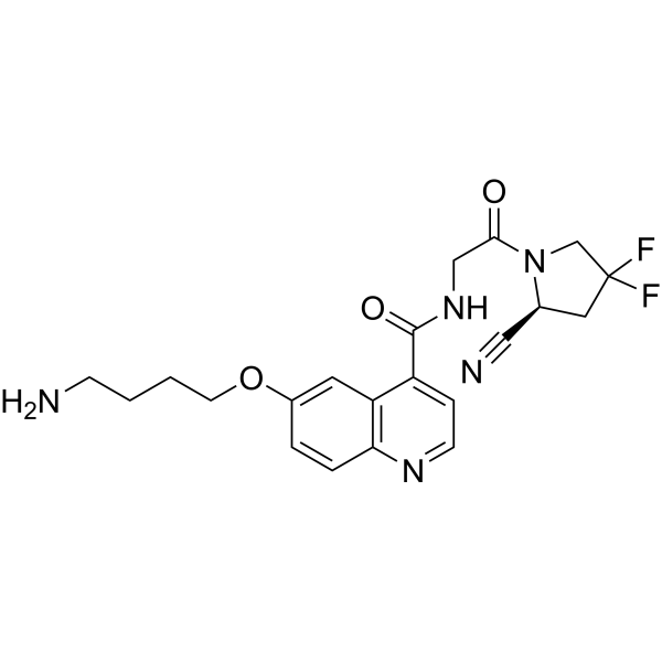
NH2-UAMC1110
NH2-UAMC1110 is an aminobutoxy derivative of the fibroblast activation protein (FAP) inhibitor UAMC1110 (HY-100684), and is a precursor compound for the synthesis of FAP inhibitor probes, not directly used in bioactivity experiments. For example, NH2-UAMC1110 is involved in the synthesis of the radiotracer FAPI-QS, which exhibits high tumor selectivity and high dose-response, and has been used for tumor diagnosis. NH2-UAMC1110 introduces an active amino group into its structure, enabling it to form covalent bonds with various molecules (such as DOTA, DATA5m, radionuclide chelators, etc.), thereby synthesizing molecular imaging probes or targeted compounds with the ability to target FAP. NH2-UAMC1110 specifically binds to the FAP active site, inhibiting its proline-selective serine protease activity (including dipeptidyl peptidase and endopeptidase activity), blocking FAP-mediated tissue remodeling processes. Its key activity is high targeting and high affinity, and its core function is to be coupled with bifunctional chelators (such as DOTA, DATA5m) as a targeting module. NH2-UAMC1110 can be applied to diagnostic imaging studies of tumors expressing FAP (such as colorectal cancer, pancreatic cancer, etc.), and also provides molecular tools for targeted research of FAP-related diseases with high FAP expression, such as fibrosis and arthritis.
Nos produits utilisent uniquement pour la recherche. Nous ne vendons pas aux patients.
- Pureté : 99.86%
- CAS No.: 2758337-19-6
- Formule: C21H23F2N5O3
- Masse moléculaire:431.44
-
Stockage:
-20°C, sealed storage, away from moisture
* In solvent : -80°C, 6 months; -20°C, 1 month (sealed storage, away from moisture)

To place orders, for customer services and technical support, please contact: MedChemExpress USA
Tel: 609-228-6898 E-mail: [email protected] [email protected]


-
 Activité biologique
-
Chemical Information
- Protocole
- Pureté et documentation
- Références
-
 Help & FAQs




Activité biologique
Description
In Vitro
NH2-UAMC1110 was coupled with DOTA.SA in phosphate buffer at pH 9 to successfully synthesize the FAP-targeting probe DOTA.SA.FAPi. The product was purified by HPLC to achieve the required purity[1].
MedChemExpress (MCE) has not independently confirmed the accuracy of these methods. They are for reference only. Further protocols information, click here.


Chemical Information
-
CAS No. 2758337-19-6
-
Appearance Solid
-
Masse moléculaire 431.44
-
Formule C21H23F2N5O3
-
Color White to off-white
-
SMILES
O=C(N1[C@@H](CC(F)(C1)F)C#N)CNC(C2=CC=NC3=C2C=C(OCCCCN)C=C3)=O
-
Livraison
Room temperature in continental US; may vary elsewhere.
-
Stockage
-20°C, sealed storage, away from moisture
* In solvent : -80°C, 6 months; -20°C, 1 month (sealed storage, away from moisture)

Protocole
-
Research Protocol for Drug Screening technologies
Drug screening technologies are experimental and computational strategies used to identify small molecules or chemical probes that modulate a defined molecular target, signaling pathway, cellular phenotype, disease model, or patient-derived response profile. High-throughput screening tests many compounds in miniaturized assay formats, while quantitative high-throughput screening tests compounds across concentration ranges so that potency and efficacy can be inferred from concentration-response behavior rather than from a single-point signal. The core biological function of a drug-screening strategy is to connect compound exposure with measurable pathway activity, target modulation, cell-state change, viability, cytotoxicity, morphology, or disease-relevant phenotype. Assay performance must be evaluated before screening because hit identification depends on the separation between positive and negative controls, control variability, plate effects, outliers, and the statistical framework
-
Fibrosis/Collagen Morphometry
Fibrosis and collagen morphometry is based on the quantitative visualization of fibrillar collagen deposition in tissue sections using histochemical stains such as Sirius Red (Picrosirius Red) or Masson's trichrome, followed by image-based or polarization-enhanced analysis to estimate collagen proportional area as a surrogate of extracellular matrix accumulation during fibrotic remodeling. Sirius Red combined with polarized light microscopy enhances detection of collagen fibers due to birefringence properties, enabling more specific visualization of collagen type I and III fibrils compared to conventional bright-field histology, while whole-section or region-restricted digital morphometry reduces field-selection bias in fibrosis assessment. Alternative quantitative approaches include second harmonic generation (SHG) and two-photon excited fluorescence microscopy, which enable label-free detection of fibrillar collagen and have been validated against histological staining and biochemica
-
RNA extraction experimental
By lysing cells, releasing RNA, and removing impurities such as proteins and DNA, high-purity RNA products are finally obtained. The commonly used traditional method is the guanidine isothiocyanate/phenol/chloroform method (Trizol), which is suitable for a variety of animal materials including animal tissues, microorganisms, cultured cells, etc., and most plant materials.
-
Collagen: Sirius Red Staining
Sirius Red or picrosirius red staining is a histochemical method for visualizing collagen-rich extracellular matrix in tissue sections, and collagen fibers are detected as red-stained structures under bright-field microscopy with enhanced birefringence under polarized light. Picrosirius red is useful for assessing total collagen organization, distribution, and fibrosis burden, but polarized color should not be interpreted as a definitive collagen type I versus type III readout because color is affected by fiber orientation, thickness, and packing.
-
Collagen-Induced Arthritis
Collagen-induced arthritis (CIA) is an autoimmune murine model of rheumatoid arthritis in which immunization with type II collagen (CII) emulsified in an adjuvant induces a T cell- and autoantibody-driven inflammatory arthritis characterized by synovial hyperplasia, immune cell infiltration, and joint destruction. The model typically relies on genetically susceptible mouse strains (e. g. , DBA/1) and reproduces key features of human rheumatoid arthritis, including anti-collagen immune responses and progressive joint inflammation. Disease onset generally occurs within ~3-4 weeks after immunization, depending on antigen/adjuvant combinations and protocol variation. The immunopathology is driven by adaptive immune activation against CII, leading to systemic and local joint inflammation mediated by pro-inflammatory cytokines and effector immune cells, making CIA a standard preclinical platform for evaluating immunomodulatory and anti-arthritic interventions.
-
Connective Tissue: Masson's Trichrome/Collagen Trichrome Staining
Masson’s Trichrome (collagen/trichrome staining) is a histological technique that differentially stains tissue compartments using sequential acidic dyes to distinguish collagen from muscle and cytoplasmic components based on dye affinity and tissue permeability differences, enabling visualization of fibrosis and connective tissue architecture in histological sections. The classical formulation typically uses Weigert's iron hematoxylin for nuclear staining, Biebrich scarlet-acid fuchsin for cytoplasm and muscle, and aniline blue (or light green variants) for collagen, producing a characteristic blue/green collagen signal contrasted against red cytoplasm and dark nuclei. The staining principle relies on selective displacement of smaller dye molecules by larger anionic dyes in collagen-rich regions under controlled acidified conditions, which enhances collagen-specific dye retention. This property makes the method widely used for fibrosis assessment in organs such as heart, liver, lung, a


Pureté et documentation
-
Fiche technique (267 KB)
-
SDS (252 KB)
- English - EN (252 KB)
- Français - FR (252 KB)
- Deutsch - DE (252 KB)
- Norwegian - NO (252 KB)
- Español - ES (252 KB)
- Swedish - SV (252 KB)
- Italian - IT (252 KB)
- Korean - KR (252 KB)
- Portuguese - PT (252 KB)
-
Instruction de manipulation (2659 KB)
Références

Calculators
Concentration (start) × Volume (start) = Concentration (final) × Volume (final)


NH2-UAMC1110 Related Classifications
HY-153552 Related Classifications
 Powered by Bioz