



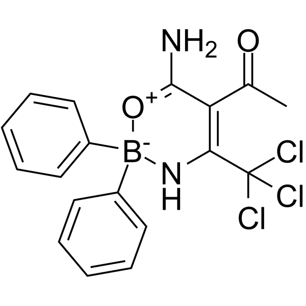
NLRP3-IN-NBC6
NLRP3-IN-NBC6 is a potent, selective NLRP3 inflammasome inhibitor (IC50= 574 nM) that acts independently of Ca2+. NLRP3-IN-NBC6 inhibits Nigericin (HY-127019)-induced inflammasome activation in THP-1 cells and Imiquimod (HY-B0180)-induced IL-1β release from LPS-primed bone marrow-derived macrophages (BMDMs).
For research use only. We do not sell to patients.
- CAS No.: 2068818-02-8
- Formula: C18H16BCl3N2O2
- Molecular Weight:409.50
-
Storage:Powder -20°C, 3 years , 4°C, 2 years ; In solvent -80°C, 6 months , -20°C, 1 month

To place orders, for customer services and technical support, please contact: MedChemExpress USA
Tel: 609-228-6898 E-mail: [email protected] [email protected]


-
 Biological Activity
-
Chemical Information
- Protocols
- Purity & Documentation
- References
-
 Help & FAQs


-
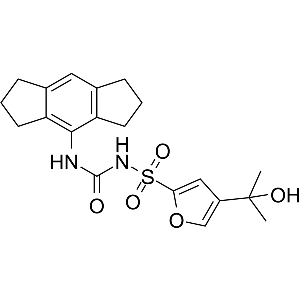
HY-12815
MCC950NLRP3 Inhibitor -
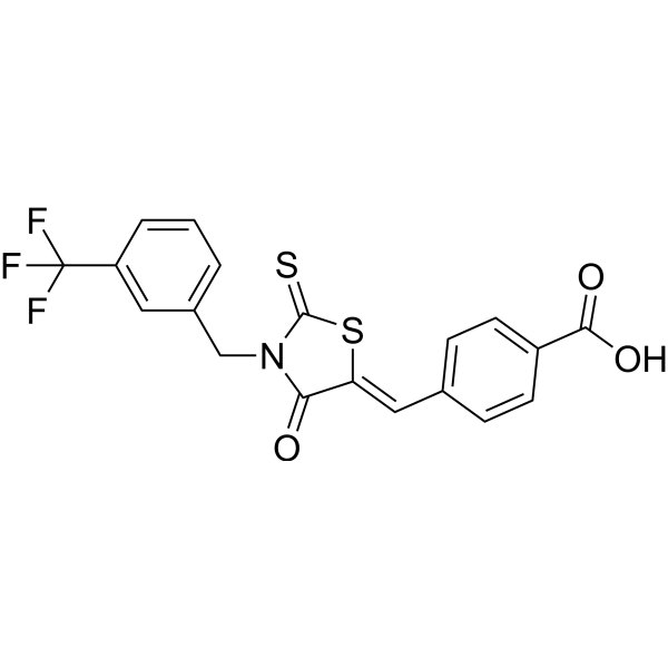
HY-103666
CY-09NLRP3 Inhibitor -
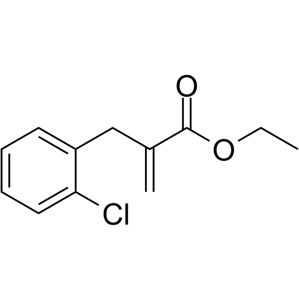
HY-101868
INF39NLRP3 Inhibitor -
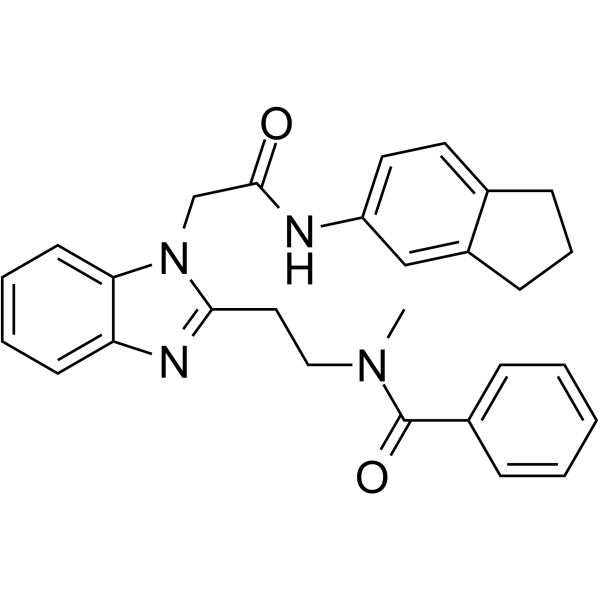
HY-136555
GSK717NOD2 Inhibitor -
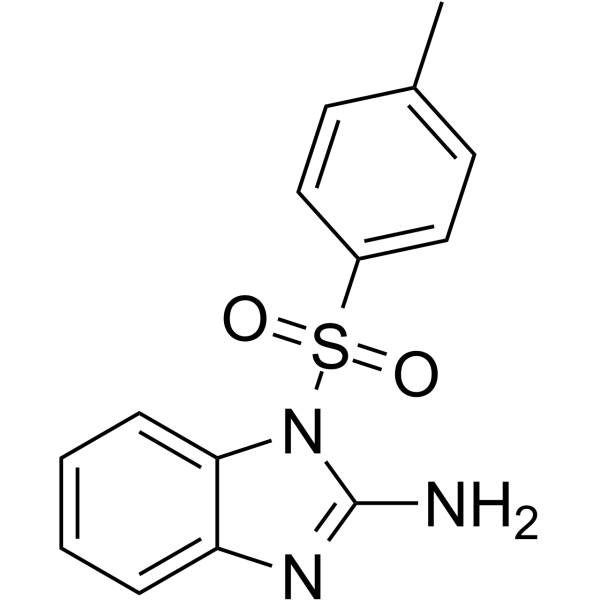
HY-18639
Nodinitib-1NOD-like Receptor (NLR) Inhibitor -
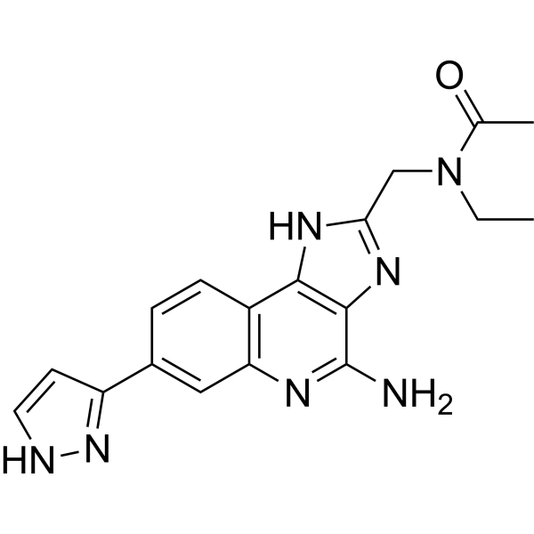
HY-139396
BMS-986299NLRP3 Agonist -
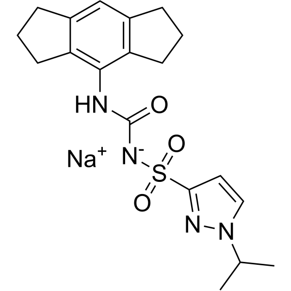
HY-137245A
Emlenoflast sodiumNLRP3 Inhibitor -
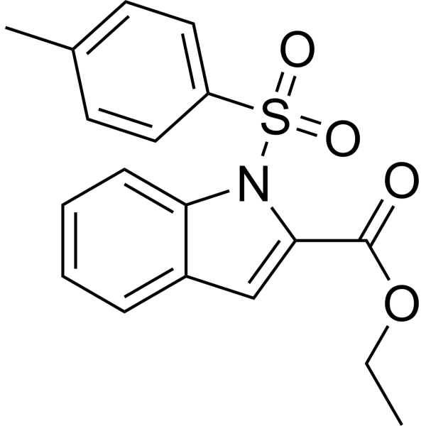
HY-100691
NOD-IN-1NLR Inhibitor -
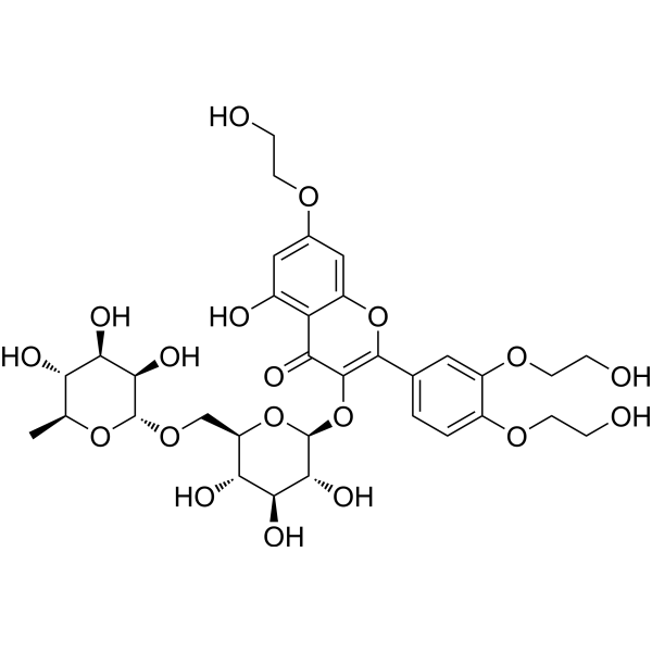
HY-N0139
TroxerutinNOD-like Receptor (NLR) Inhibitor -
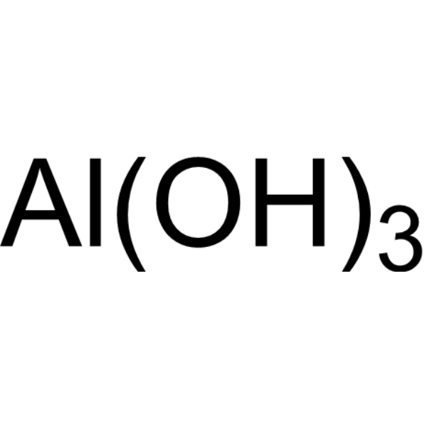
HY-B1521
Aluminum HydroxideAntacid Aegnt -
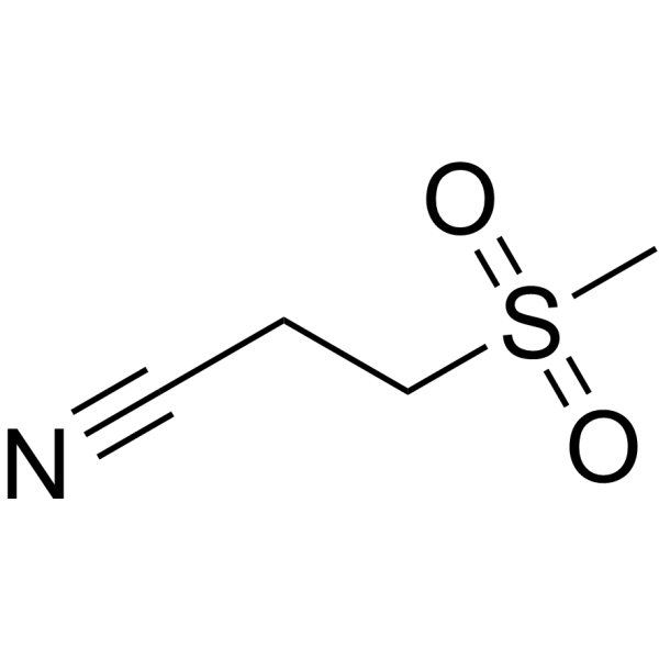
HY-17629
DapansutrileNLRP3 Inhibitor -
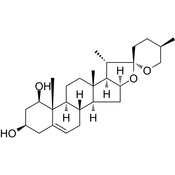
HY-N0496
RuscogeninNOD-like Receptor (NLR) Inhibitor


Biological Activity
Description
IC50 & Target
[1]|
NLRP3 574 nM (IC50) |
In Vitro
In LPS-primed primary BMDMs, NLRP3-IN-NBC6 (10 μM; 15 minutes) inhibits Imiquimod-induced IL-1β release. NLRP3-IN-NBC6 completely inhibits NLRP3-dependent IL-1β release in neutrophils (neutrophils were isolated from WT and NLRP3 KO murine bone marrow and primed with LPS followed by Nigericin treatment in the presence of 10 μM NLRP3-IN-NBC6)[1].
MedChemExpress (MCE) has not independently confirmed the accuracy of these methods. They are for reference only. Further protocols information, click here.


Chemical Information
-
CAS No. 2068818-02-8
-
Appearance Solid
-
Molecular Weight 409.50
-
Formula C18H16BCl3N2O2
-
Color White to off-white
-
SMILES
CC(C1=C(N[B-](C2=CC=CC=C2)([O+]=C1N)C3=CC=CC=C3)C(Cl)(Cl)Cl)=O
-
Shipping
Room temperature in continental US; may vary elsewhere.
-
Storage
Powder -20°C 3 years 4°C 2 years In solvent -80°C 6 months -20°C 1 month

Protocols
-
Cell Cytotoxicity Assay
Cytotoxicity assays are usually based on the assessment of cell membrane damage, which can also be indirectly detected by measuring cell viability. Detection methods include MTT assay, CKK-8 assay, LDH assay and ATP assay, etc.
-
Ca2+ Staining Technique
Ca2+ staining is an experimental technique that utilizes specific fluorescent probes (such as Fluo-4 AM, Fura-2, etc.) to qualitatively or quantitatively detect dynamic changes in intracellular Ca2+ concentrations; this is achieved by monitoring the changes in fluorescent signals generated when these probes bind to free intracellular calcium ions. The underlying principle relies primarily on the presence of chelating groups within the probe's molecular structure that possess high affinity for calcium ions.
-
Pyroptosis Solutions
Pyroptosis is a lytic inflammatory cell-death pathway executed by gasdermin pores, most classically through inflammasome-mediated activation of caspase-1, cleavage of gasdermin D, membrane pore formation, LDH release, and secretion of IL-1β and IL-18. The canonical pathway is commonly modeled by priming cells with an inflammatory signal such as LPS to induce pro-IL-1β and inflammasome components, followed by an activation signal such as ATP or nigericin to activate NLRP3, ASC speck formation, caspase-1 cleavage, GSDMD cleavage, cytokine release, and pyroptotic membrane rupture. The non-canonical pathway is triggered when cytosolic LPS activates mouse caspase-11 or human caspase-4/5, leading to GSDMD cleavage and pyroptosis, and this can secondarily activate NLRP3-dependent IL-1β release. Pyroptosis is linked to inflammatory injury, infection, cancer, liver disease, ocular disease, placental inflammation, and other disease phenotypes, but unresolved questions include which gasdermin fam


Purity & Documentation
-
Data Sheet (277 KB)
-
SDS (251 KB)
- English - EN (251 KB)
- Français - FR (251 KB)
- Deutsch - DE (251 KB)
- Norwegian - NO (251 KB)
- Español - ES (251 KB)
- Swedish - SV (251 KB)
- Italian - IT (251 KB)
- Korean - KR (251 KB)
- Portuguese - PT (251 KB)
-
Handling Instructions (2659 KB)
References

Calculators
Concentration (start) × Volume (start) = Concentration (final) × Volume (final)


NLRP3-IN-NBC6 Related Classifications
HY-131040 Related Classifications
 Powered by Bioz