



NOC-5

Based on 1 Customer Validation
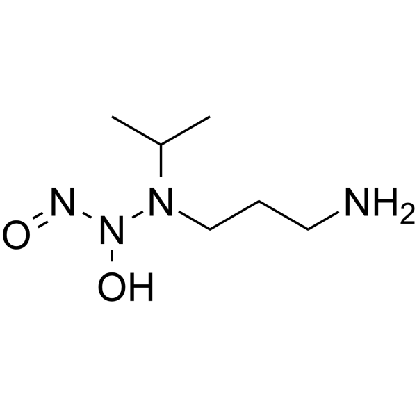
NOC-5 is a diazeniumdiolate compound that belongs to the same family of NO donors. NOC-5 is able to increase intracellular XIAP and Aven levels, potentially leading to the inhibition of caspase-9 activity following increased mitochondrial permeability.
For research use only. We do not sell to patients.
- Purity : 98.50%
- CAS No.: 146724-82-5
- Formula: C6H16N4O2
- Molecular Weight:176.22
-
Storage:
-80°C

To place orders, for customer services and technical support, please contact: MedChemExpress USA
Tel: 609-228-6898 E-mail: [email protected] [email protected]


-
 Biological Activity
-
Chemical Information
- Protocols
- Purity & Documentation
- References
-
 Help & FAQs






Biological Activity
Description
In Vitro
NOC-5 (5-20 μM) in PBS led to a rapid increase in NO release on day one[1].
NOC-5 (100 μM, 24 h) activates only caspase-3; an increase in caspase-3 activity occurred after NO donor treatment: 1.62 (1.57-1.66). NOC-5 has pro-apoptotic action in Jurkat cells, but at the same time increased the level of anti-apoptotic proteins[2].
MedChemExpress (MCE) has not independently confirmed the accuracy of these methods. They are for reference only. Further protocols information, click here.


Chemical Information
-
CAS No. 146724-82-5
-
Appearance Solid
-
Molecular Weight 176.22
-
Formula C6H16N4O2
-
Color Light yellow to yellow
-
SMILES
CC(N(CCCN)N(N=O)O)C
-
Shipping
Shipping with dry ice.
-
Storage
-80°C

Protocols
-
Apoptosis Solutions
Apoptosis is a regulated, generally non-lytic cell-death pathway that removes unwanted, damaged, infected, or abnormal cells through coordinated morphological changes, caspase activation, DNA fragmentation, and membrane remodeling. The intrinsic apoptosis pathway is controlled mainly by mitochondrial outer membrane permeabilization, BCL-2 family proteins, cytochrome c release, apoptosome formation, caspase-9 activation, and downstream executioner caspase-3/7 activation. The extrinsic apoptosis pathway is initiated by death receptors such as Fas, TNFR, and TRAIL receptors, which recruit adaptor proteins and activate caspase-8 before engaging executioner caspases or mitochondrial amplification through BID cleavage. Apoptosis is linked to many phenotypes, including cancer cell killing, tissue homeostasis, immune regulation, neurodegeneration, infection response, and treatment-induced cytotoxicity; unresolved questions include how apoptosis interacts with necroptosis, pyroptosis, ferroptos
-
Transepithelial/transendothelial electrical resistance assay
TEER measures electrical resistance across epithelial or endothelial monolayers cultured on permeable supports, and the readout reflects ionic conductance through the cell barrier, especially the paracellular pathway regulated by junctional integrity. TEER can be measured without destroying the monolayer and is commonly used before or during transport, permeability, barrier-disruption, and barrier-maturation experiments. TEER values are influenced by biological maturation and technical conditions; reported factors include temperature, medium formulation, passage number, electrode geometry, membrane properties, and junctional length during early monolayer maturation. Therefore, TEER should be interpreted with blank-insert subtraction, area normalization, repeated readings, and, when possible, orthogonal barrier readouts such as FITC-dextran flux or tight-junction staining.


Purity & Documentation
-
Data Sheet (265 KB)
-
SDS (466 KB)
- English - EN (466 KB)
- Français - FR (466 KB)
- Deutsch - DE (466 KB)
- Norwegian - NO (466 KB)
- Español - ES (466 KB)
- Swedish - SV (466 KB)
- Italian - IT (466 KB)
- Korean - KR (466 KB)
- Portuguese - PT (466 KB)
-
Handling Instructions (2659 KB)
References
[1]. Starikova EG, et al. Nitric oxide donor NOC-5 increases XIAP and Aven level in Jurkat cells. Cell Biol Int. 2014 Jul;38(7):799-802. [Content Brief]
[2]. Sophie A Bradley, et al. Characterisation and comparison of temporal release profiles of nitric oxide generating donors. J Neurosci Methods. 2015 Apr 30;245:116-24. [Content Brief]
[3]. Elena G Starikova, et al. Nitric oxide donor NOC-5 increases XIAP and Aven level in Jurkat cells. Cell Biol Int. 2014 Jul;38(7):799-802. [Content Brief]

Calculators
Concentration (start) × Volume (start) = Concentration (final) × Volume (final)


NOC-5 Related Classifications
HY-126166 Related Classifications
 Powered by Bioz