



Olive oil
Based on 7 publication(s) in Google Scholar
Olive oil (Cropure OL) is an oleaginous compound found in the fruit of the Olea europaea tree. Olive oil contains many phenolic components and exerts antioxidant activity. Olive oil exhibits hydroxyl radical scavenging, platelet aggregation inhibition and xanthine oxidase inhibitory activity. Olive oil can promote wound healing and relieve inflammation. Olive oil can be used for the research of inflammation, cancer, metabolic and cardiovascular disease, such as diabetic foot ulcers and inflammatory bowel disease.
For research use only. We do not sell to patients.
- CAS No.: 8001-25-0
-
Storage:
Please store the product under the recommended conditions in the Certificate of Analysis.

To place orders, for customer services and technical support, please contact: MedChemExpress USA
Tel: 609-228-6898 E-mail: [email protected] [email protected]


-
 Biological Activity
-
Chemical Information
-
Solvent & Solubility
- Protocols
- Purity & Documentation
- References
-
 Help & FAQs


-

HY-Y0320
Dimethyl sulfoxide, meets analytical specification of Ch.P.Aprotic Solvent -

HY-Y1891
Tween 80 -
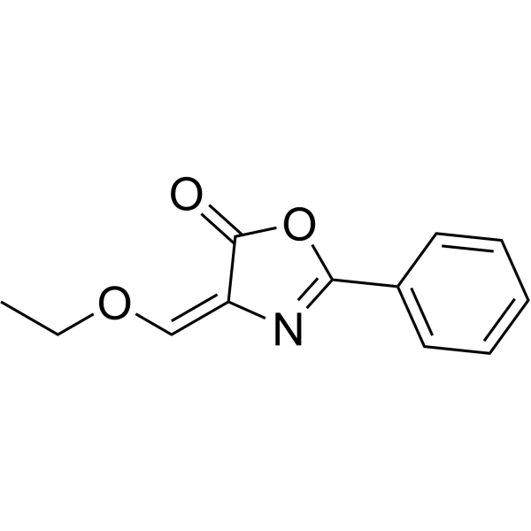
HY-126360
OxazoloneColitis Inducer -

HY-154629
Sesame OilNatural Product -
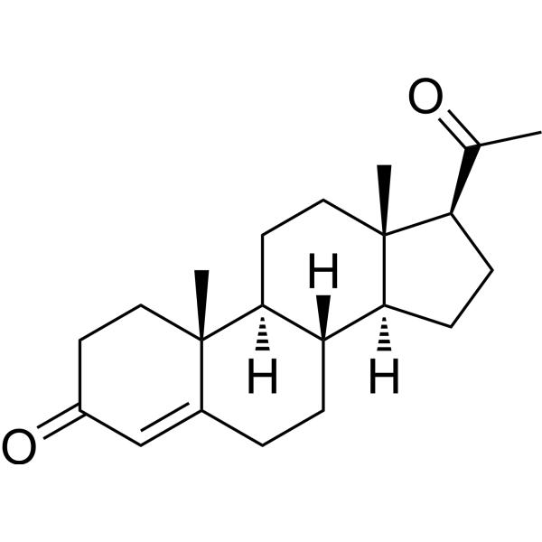
HY-N0437
ProgesteroneEndogenous Metabolite -
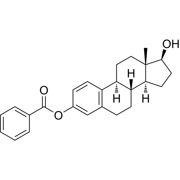
HY-B1192
Estradiol benzoateHBx inhibitor -

HY-Y1888
Corn oil -
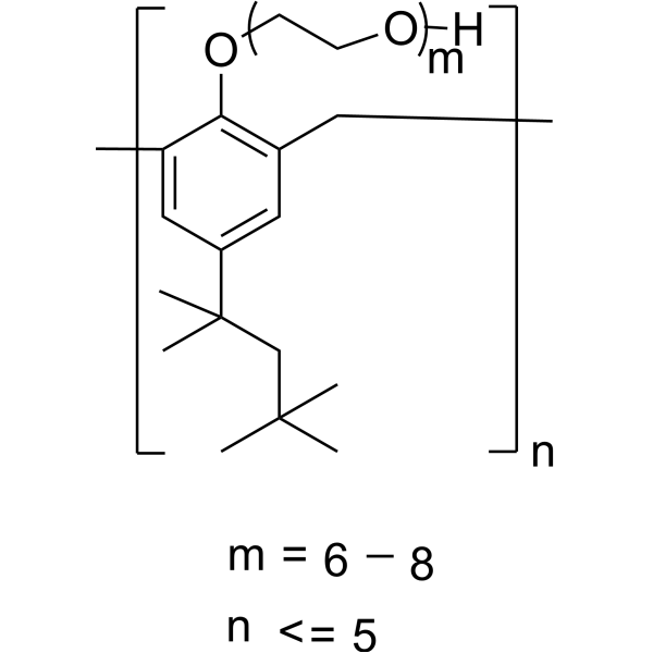
HY-B1068
TyloxapolHyperlipidemia Inducer -
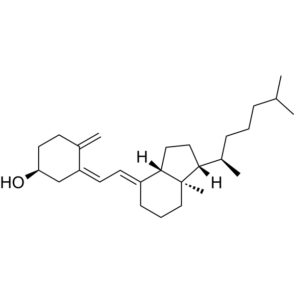
HY-15398
Vitamin D3Vitamin -
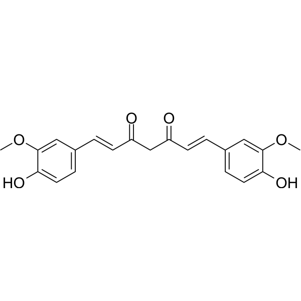
HY-N0005
Curcuminp300 Histone Acetylatransferase Inhibitor -
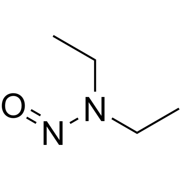
HY-N7434
N-NitrosodiethylamineDNA Repair/Replicase Modulator
Publications Citing Use of MedChemExpress (MCE) Olive oil
More- Engineering. 2026 Mar 4.
- Phytother Res. 2024 Nov;38(11):5184-5202. [Abstract]
- Environ Sci Eur. 2026 Feb 21;38(1):71.
- Food Funct. 2026 Apr 14;17(7):3229-3247. [Abstract]
- J Inflamm Res. 2026 Feb 6:19:573709. [Abstract]
- Immunobiology. 2026 May 7;231(3):153189. [Abstract]
- Chin J Integr Med. 2025 Dec 9. [Abstract]


Biological Activity
Description
In Vitro
Olive oil exhibits hydroxyl radical scavenging and xanthine oxidase inhibitory activity, with 73% inhibition of xanthine oxidase activity[2].
Olive oil contains phenolic components (hydroxytyrosol and oleuropein) potently inhibit copper sulfate-induced oxidation of isolated LDL in vitro[3].
Olive oil contains phenolic components (hydroxytyrosol and oleuropein) exhibit non-antioxidant biological activities in vitro, including inhibition of human platelet aggregation and leukocyte eicosanoid production (hydroxytyrosol, with an EC50 in the 10−5 M range) and increased nitric oxide production by lipopolysaccharide-treated murine macrophages (oleuropein)[3].
MedChemExpress (MCE) has not independently confirmed the accuracy of these methods. They are for reference only. Further protocols information, click here.
In Vivo
MedChemExpress (MCE) has not independently confirmed the accuracy of these methods. They are for reference only.
-
Animal Model:Wistar[4]
-
Dosage:Aqueous solution containing olive oil and HT
-
Administration:intrarectal
-
Result:Reduced the severity of inflammatory damage in the TNBS-induced colitis model, with reduced inflammatory infiltrate.
Clinical Trial
| NCT Number | Sponsor | Condition | Start Date |
Phase

|
|---|---|---|---|---|
| NCT01329991 | Plexxikon| | 2011-05 | PHASE1 |


Chemical Information
-
CAS No. 8001-25-0
-
Appearance Liquid (Density: 0.9135 g/cm3)
-
Color Colorless to light yellow
-
SMILES
[Olive oil]
-
Synonyms
Cropure OL
-
Shipping
Room temperature in continental US; may vary elsewhere.
-
Storage
Please store the product under the recommended conditions in the Certificate of Analysis.


Publications (7)
-
Journal Impact Factor
-
Most Recent
-
-
Phytother Res
Geniposide modulates GSK3β to inhibit Th17 differentiation and mitigate endothelial damage in intracranial aneurysm. [Abstract]2024 Nov;38(11):5184-5202. PMID: 39180344 -
-
Food Funct
Dual improvement effects of ω-3 on bone loss and liver injury in rats with liver-bone axis damage. [Abstract]2026 Apr 14;17(7):3229-3247. PMID: 41834729 -
J Inflamm Res
Gut Microbiota Mitigates Chronic Itch and Cutaneous Inflammation in DNFB-Induced Atopic Dermatitis Mice. [Abstract]2026 Feb 6:19:573709. PMID: 41890815 -
Immunobiology
Mannose receptor modulates hepatic stellate cell activation and alleviates liver fibrosis through immune-fibrotic crosstalk. [Abstract]2026 May 7;231(3):153189. PMID: 42107443 -
Chin J Integr Med
Total Flavonoids of Litchi chinensis Sonn. Seed Improves Hepatic Fibrosis by Inhibiting PANoptosis. [Abstract]2025 Dec 9. PMID: 41361707


Solvent & Solubility
In Vitro:
DMSO : 100 mg/mL (ultrasonic and warming and heat to 60°C; Hygroscopic DMSO has a significant impact on the solubility of product, please use newly opened DMSO)
In Vivo:
Select the appropriate dissolution method based on your experimental animal and administration route.
- For the following dissolution methods, please ensure to first prepare a clear stock solution using an In Vitro approach and then sequentially add co-solvents:
- To ensure reliable experimental results, the clarified stock solution can be appropriately stored based on storage conditions. As for the working solution for In Vivo experiments, it is recommended to prepare freshly and use it on the same day.
- The percentages shown for the solvents indicate their volumetric ratio in the final prepared solution. If precipitation or phase separation occurs during preparation, heat and/or sonication can be used to aid dissolution.
Add each solvent one by one: 10% DMSO 40% PEG300 5% Tween-80 45% Saline
Solubility: ≥ 2.5 mg/mL; Clear solution
This protocol yields a clear solution of ≥ 2.5 mg/mL (saturation unknown).
Taking 1 mL working solution as an example, add 100 μL DMSO stock solution (25.0 mg/mL) to 400 μL PEG300, and mix evenly; then add 50 μL Tween-80 and mix evenly; then add 450 μL Saline to adjust the volume to 1 mL.
Preparation of Saline: Dissolve 0.9 g sodium chloride in ddH₂O and dilute to 100 mL to obtain a clear Saline solution.
In Vivo Dissolution Calculator
Please enter the basic information of animal experiments:
-
-
-
-
Recommended: Prepare an additional quantity of animals to account for potential losses during experiments.
Please enter your animal formula composition:
-
%DMSO +
Recommended: Keep the proportion of DMSO in working solution below 2% if your animal is weak.
-
%+
-
+%Tween-80 + +
-
%Saline +
The co-solvents required include: DMSO, . All of co-solvents are available by MedChemExpress (MCE). , Tween 80. All of co-solvents are available by MedChemExpress (MCE).
Working solution concentration: 0.22 mg/mL
Method for preparing stock solution: mg drug dissolved in μL DMSO. Stock solution concentration: mg/mL.
1. Take μL DMSO stock solution;
2. Add μL .
μL , mix evenly;
3. Then add μL Tween 80, mix evenly;
4. Then add μL
Please ensure that the stock solution in the first step is dissolved to a clear state, and add co-solvents in sequence. You can use ultrasonic heating (ultrasonic cleaner, recommended frequency 20-40 kHz), vortexing, etc. to assist dissolution.

Protocols
-
Cell migration
Cell migration is a method that plays an important role in wound healing, cell differentiation, embryonic development, etc.
-
DSS-Induced Colitis
Dextran sulfate sodium (DSS)-induced colitis is generated by administering DSS in mouse drinking water, producing epithelial injury, barrier disruption, weight loss, diarrhea, fecal blood, colon shortening, histologic mucosal damage, and inflammatory mediator changes; the model is mainly used to study acute or chronic intestinal inflammation resembling selected features of ulcerative colitis. DSS injury is interpreted through clinical and tissue readouts rather than a single molecular endpoint: daily body weight, stool consistency, and bleeding are combined into a disease activity index, while colon length, histology, cytokines, myeloperoxidase activity, intestinal permeability, and tight-junction markers provide complementary measures of inflammation and barrier damage.
-
TNBS-Induced Colitis
TNBS-induced colitis is produced by intrarectal delivery of 2,4,6-trinitrobenzene sulfonic acid in ethanol, where ethanol disrupts the mucosal barrier and TNBS haptenates colonic proteins, generating immune-mediated colonic inflammation with weight loss, diarrhea, ulceration, transmural injury, inflammatory-cell infiltration, and cytokine responses. The model is used as an experimental intestinal inflammation model with Crohn’s disease–like features, especially when Th1-type responses, IL-12–dependent inflammation, chronic relapsing inflammation, or fibrosis-related endpoints are studied.
-
Research Protocol for Cardiovascular Diseases
Cardiovascular disease can be modeled as maladaptive cardiac remodeling, where ischemic injury or pressure overload activates inflammatory signaling, fibroblast activation, extracellular-matrix deposition, cardiomyocyte hypertrophy, vascular remodeling, and progressive ventricular dysfunction. The TGF-β/SMAD axis is a central profibrotic pathway after myocardial injury and pressure overload, while innate immune and cytokine pathways regulate leukocyte recruitment, scar formation, and adverse remodeling. Key unresolved questions include which inflammatory signals are reparative versus harmful, when fibrosis is protective versus maladaptive, and whether pathway inhibition improves function without weakening necessary infarct healing or compensatory remodeling.
-
Research Protocol for Inflammation-related Diseases
The NLRP3 inflammasome is a cytosolic innate immune signaling platform that integrates priming signals and danger-signal activation to promote caspase-1 activation, maturation of IL-1β and IL-18, and gasdermin D-mediated pyroptotic cell death. The core experimental logic is to determine whether inflammatory disease phenotypes are driven by increased NLRP3 expression, ASC-containing inflammasome assembly, caspase-1 cleavage, GSDMD cleavage, and extracellular release of IL-1β/IL-18 rather than by nonspecific cell injury alone. The pathway is strongly linked to inflammation-related disease phenotypes because monosodium urate crystals activate NALP3/NLRP3 inflammasome signaling in gout-like crystal inflammation, cholesterol crystals activate NLRP3 inflammasomes in atherogenesis models, and DSS-induced intestinal inflammation has been reported to involve NLRP3 inflammasome activity. However, experimental colitis studies also show context-dependent protective effects of NLRP3 inflammasome co
-
Cell-Exclusion Zone Migration Assay
The Cell-Exclusion Zone (CEZ) migration assay is an in vitro 2D cell migration method in which a defined cell-free area is created using removable physical barriers such as silicone stoppers, allowing cells to be seeded around the barrier and subsequently migrate into the cleared zone after barrier removal. This approach enables quantification of collective cell migration by monitoring repopulation of the initially cell-free region over time using microscopy-based imaging. Compared with scratch-based wound healing assays, barrier-based exclusion methods are designed to avoid mechanical damage to the extracellular matrix and reduce injury-induced effects on boundary cells, thereby improving interpretability of migration behavior in vitro. The assay readout is typically the progressive reduction in the cell-free area or the number of cells invading the exclusion zone, reflecting coordinated cell motility relevant to physiological processes such as wound healing, epithelial repair, and ca


Purity & Documentation
-
Data Sheet (272 KB)
-
SDS (393 KB)
- English - EN (393 KB)
- Français - FR (393 KB)
- Deutsch - DE (393 KB)
- Norwegian - NO (393 KB)
- Español - ES (393 KB)
- Swedish - SV (393 KB)
- Italian - IT (393 KB)
- Korean - KR (393 KB)
- Portuguese - PT (393 KB)
-
Handling Instructions (2659 KB)
References
[1]. Malik S, et al. Olive Oil Shows Promise for Wound Healing of Ulcers. Cutis. 2024;113(6):260-263. [Content Brief]
[2]. Owen RW, et al. Olives and olive oil in cancer prevention. Eur J Cancer Prev. 2004 Aug;13(4):319-26. [Content Brief]
[3]. Visioli F, et al. Antiatherogenic components of olive oil. Curr Atheroscler Rep. 2001;3(1):64-67. [Content Brief]
[4]. Vrdoljak J, et al. Effects of Olive Oil and Its Components on Intestinal Inflammation and Inflammatory Bowel Disease. Nutrients. 2022;14(4):757. Published 2022 Feb 11. [Content Brief]

Calculators
Concentration (start) × Volume (start) = Concentration (final) × Volume (final)


Olive oil Related Classifications
HY-108749 Related Classifications
 Powered by Bioz