



OX2R-IN-1
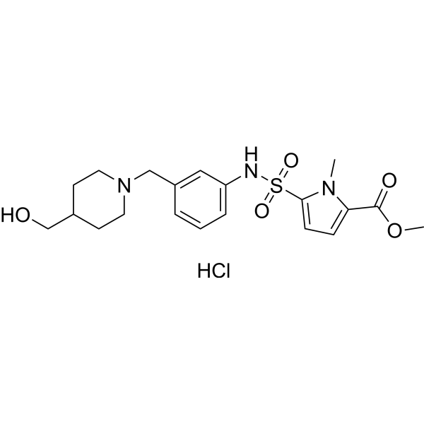
OX2R-IN-1 (compound 15) is a low cytotoxicity profile OX2R-IN-1 antagonist (a potential OX2R binder) with an IC50 value of 484 μM. OX2R-IN-1 (compound 15) can cross the BBB into the brain with a short half-life.
For research use only. We do not sell to patients.
- CAS No.: 2639148-08-4
- Formula: C20H28ClN3O5S
- Molecular Weight:457.97
-
Storage:
Please store the product under the recommended conditions in the Certificate of Analysis.

To place orders, for customer services and technical support, please contact: MedChemExpress USA
Tel: 609-228-6898 E-mail: [email protected] [email protected]


-
 Biological Activity
-
Chemical Information
- Protocols
- Purity & Documentation
- References
-
 Help & FAQs


All Orexin Receptor (OX Receptor) Isoforms
More

Biological Activity
Description
IC50 & Target
|
OX2 Receptor |
In Vitro
OX2R-IN-1 (compound 15) has low cytotoxic withIC50 values of 484 μM. OX2R-IN-1 (compound 15) is significant and dose-dependently reduce the signal of orexin A-evoked response (0.2 μM) in CHO-K1 cell line. OX2R-IN-1 (compound 15) has uncertain permeation through the BBB, since the PAMPA assay is limited by several drawbacks[1].
MedChemExpress (MCE) has not independently confirmed the accuracy of these methods. They are for reference only. Further protocols information, click here.
-
Cell Line:CHO-K1 cell line
-
Concentration:100-1000 µM
-
Incubation Time:24h
-
Result:Exhibited low cytotoxic with IC50values of 484 μM
In Vivo
MedChemExpress (MCE) has not independently confirmed the accuracy of these methods. They are for reference only.
-
Animal Model:Albino male Wistar rats[1]
-
Dosage:75 mg/kg
-
Administration:OX2R-IN-1 (75 mg/kg; intraperitoneal injection) blood samples are collected at 0, 5, 10, 20, 30, 40, 60, 90, 120 and 240 min with a short half-life and poor bioavailability
-
Result:
Parameter Units Plasma Brain tissuer t1/2,β (h) .......t1/2 min 9.63 14.85 tmax min 5 40 Cmax μmol/L 29.40 0.199 AUC μmol/L*min 1362.68 18.51 MRT min 39.94 68.93


Chemical Information
-
CAS No. 2639148-08-4
-
Molecular Weight 457.97
-
Formula C20H28ClN3O5S
-
SMILES
O=C(C1=CC=C(S(=O)(NC2=CC=CC(CN3CCC(CC3)CO)=C2)=O)N1C)OC.Cl
-
Shipping
Room temperature in continental US; may vary elsewhere.
-
Storage
Please store the product under the recommended conditions in the Certificate of Analysis.

Protocols
-
Cell Cytotoxicity Assay
Cytotoxicity assays are usually based on the assessment of cell membrane damage, which can also be indirectly detected by measuring cell viability. Detection methods include MTT assay, CKK-8 assay, LDH assay and ATP assay, etc.
-
Mammalian live/dead viability and cytotoxicity staining
Live/dead viability and cytotoxicity staining assays are based on the simultaneous detection of intracellular esterase activity in metabolically active (viable) cells and membrane integrity loss in non-viable cells. In commonly used dual-staining approaches, membrane-permeant fluorogenic substrates are converted by intracellular esterases into fluorescent products in live cells, while impermeant DNA-binding dyes selectively enter cells with compromised plasma membranes and label nucleic acids in dead or dying cells, enabling discrimination between viable and non-viable populations by fluorescence microscopy or flow cytometry.
-
Protocol for Pharmacokinetic Study
Pharmacokinetic studies quantify how an organism handles a drug over time through absorption, distribution, metabolism, and excretion, and the core experimental readout is the concentration-time profile of parent drug and, when relevant, metabolites in biological matrices such as plasma, whole blood, urine, bile, or tissue. Pharmacokinetic analysis links dose, route, exposure, clearance, half-life, distribution, bioavailability, and systemic exposure to drug efficacy and toxicity hypotheses rather than measuring a signaling pathway directly. The literature links pharmacokinetics to drug-development phenotypes by showing that drug metabolism and pharmacokinetics influence compound progression, exposure-response interpretation, safety margins, dosing strategy, and failure risk during discovery and development. DMPK science contributes to compound optimization by integrating physicochemical properties, in vitro metabolism, transporter behavior, in vivo exposure, and pharmacodynamic contex
-
Cell Viability Determination by MTT Colorimetric Assay
The following protocol uses the MTT colorimetric assay as a classic literature-established method for assessing cell viability/metabolic activity in cultured mammalian cells. MTT[3-(4,5-dimethylthiazol-2-yl)-2,5-diphenyltetrazolium bromide] is reduced by metabolically active cells to a colored formazan product; the amount of formazan is quantified spectrophotometrically and provides an indirect measure of metabolically active viable cells. Importantly, MTT reduction reflects cellular oxidoreductase/metabolic activity rather than an absolute direct count of living cells, so changes in cellular metabolism can alter the signal independently of cell number.


Purity & Documentation
References

Calculators
Concentration (start) × Volume (start) = Concentration (final) × Volume (final)


OX2R-IN-1 Related Classifications
HY-149014 Related Classifications
 Powered by Bioz