



PCSK9 degrader 1

Based on 1 Customer Validation
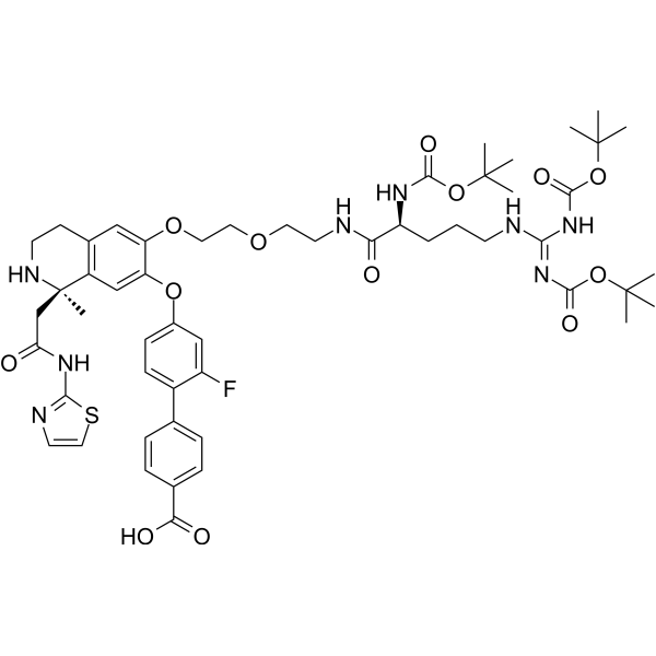
PCSK9 degrader 1 is a HyT-like PCSK9 degrader with a Ki of 107 nM. PCSK9 degrader 1 binds to the allosteric pocket between the catalytic domain and C-terminal domain of PCSK9. While maintaining key interactions with this protein, it extends the proteasome-recruiting moiety, thereby driving degradation via the proteasomal pathway. PCSK9 degrader 1 induces the degradation of both the precursor and mature forms of PCSK9, with DC50 values of 4.8 μM and 3.4 μM respectively, and does not significantly affect cell viability or the expression of housekeeping proteins. PCSK9 degrader 1 can be used in the research of coronary heart disease.
For research use only. We do not sell to patients.
- Purity : 98.33%
- Formula: C53H69FN8O13S
- Molecular Weight:1077.22
-
Storage:Powder -20°C, 3 years ; In solvent -80°C, 6 months , -20°C, 1 month

To place orders, for customer services and technical support, please contact: MedChemExpress USA
Tel: 609-228-6898 E-mail: [email protected] [email protected]


-
 Biological Activity
-
Chemical Information
-
Solvent & Solubility
- Protocols
- Purity & Documentation
- References
-
 Help & FAQs




Biological Activity
Description
IC50 & Target
Ki: 107 nM (PCSK9)[1]
Cellular Effect
|
Cell Line

|
Type | Value | Description | References |
|---|---|---|---|---|
| HEK293 | DC50 |
4.8 μM
|
Half-maximal degradation concentration of pro-PCSK9 in PCSK9-overexpressing HEK293 cells assessed via simple western blotting after 24 h incubation in serum-free DMEM.
Half-maximal degradation concentration of pro-PCSK9 in PCSK9-overexpressing HEK293 cells assessed via simple western blotting after 24 h incubation in serum-free DMEM.
|
31653597 |
| HEK293 | DC50 |
3.4 μM
|
Half-maximal degradation concentration of mature PCSK9 in PCSK9-overexpressing HEK293 cells assessed via simple western blotting after 24 h incubation in serum-free DMEM.
Half-maximal degradation concentration of mature PCSK9 in PCSK9-overexpressing HEK293 cells assessed via simple western blotting after 24 h incubation in serum-free DMEM.
|
31653597 |
In Vitro
PCSK9 degrader 1 (compound 16) (overnight) binds to recombinant human PCSK9 with high affinity, displaying a Ki of 107 nM, while preserving key interactions with the protein's allosteric pocket[1].
PCSK9 degrader 1 (1.25-20 μM; 24 h) induces concentration-dependent degradation of pro and mature PCSK9 in PCSK9-overexpressing HEK293 cells, with DC50 values of 4.8 μM and 3.4 μM, respectively, and up to 61% maximum degradation at 20 μM[1].
MedChemExpress (MCE) has not independently confirmed the accuracy of these methods. They are for reference only.
-
Cell Line:HEK293 cells overexpressing PCSK9
-
Concentration:1.25-20 μM
-
Incubation Time:24 h
-
Result:Induced concentration-dependent degradation of both pro and mature forms of PCSK9.
Achieved a DC50 of 4.8 μM for pro-PCSK9.
Achieved a DC50 of 3.4 μM for mature PCSK9.
Achieved 58% degradation of pro-PCSK9 at 20 μM.
Achieved 61% degradation of mature PCSK9 at 20 μM.
Did not significantly reduce cell viability at 20 μM.
Did not alter expression of the housekeeping protein GAPDH at 20 μM.


Chemical Information
-
Appearance Solid
-
Molecular Weight 1077.22
-
Formula C53H69FN8O13S
-
Color White to off-white
-
SMILES
OC(C(C=C1)=CC=C1C(C(F)=C2)=CC=C2OC(C(OCCOCCNC([C@@H](NC(OC(C)(C)C)=O)CCCN/C(NC(OC(C)(C)C)=O)=N/C(OC(C)(C)C)=O)=O)=C3)=CC4=C3CCN[C@]4(C)CC(NC5=NC=CS5)=O)=O
-
Shipping
Room temperature in continental US; may vary elsewhere.
-
Storage
Powder -20°C 3 years In solvent -80°C 6 months -20°C 1 month


Solvent & Solubility
In Vitro:
DMSO : 100 mg/mL (92.83 mM; ultrasonic and warming and heat to 60°C; Hygroscopic DMSO has a significant impact on the solubility of product, please use newly opened DMSO)
Please refer to the solubility information to select the appropriate solvent. Once prepared, please aliquot and store the solution to prevent product inactivation from repeated freeze-thaw cycles.
Storage method and period of stock solution: -80°C, 6 months; -20°C, 1 month. When stored at -80°C, please use it within 6 months. When stored at -20°C, please use it within 1 month.
Please refer to the solubility information to select the appropriate solvent. Once prepared, please aliquot and store the solution to prevent product inactivation from repeated freeze-thaw cycles.
Storage method and period of stock solution: -80°C, 6 months; -20°C, 1 month. When stored at -80°C, please use it within 6 months. When stored at -20°C, please use it within 1 month.
Concentration (start) × Volume (start) = Concentration (final) × Volume (final)


Purity & Documentation
-
Data Sheet (287 KB)
-
SDS (251 KB)
- English - EN (251 KB)
- Français - FR (251 KB)
- Deutsch - DE (251 KB)
- Norwegian - NO (251 KB)
- Español - ES (251 KB)
- Swedish - SV (251 KB)
- Italian - IT (251 KB)
- Korean - KR (251 KB)
- Portuguese - PT (251 KB)
-
Handling Instructions (2659 KB)
References


Complete Stock Solution Preparation Table
Please refer to the solubility information to select the appropriate solvent. Once prepared, please aliquot and store the solution to prevent product inactivation from repeated freeze-thaw cycles.
Storage method and period of stock solution: -80°C, 6 months; -20°C, 1 month. When stored at -80°C, please use it within 6 months. When stored at -20°C, please use it within 1 month.
| Optional Solvent | Concentration Solvent Mass | 1 mg | 5 mg | 10 mg | 25 mg |
|---|---|---|---|---|---|
| DMSO | 1 mM | 0.9283 mL | 4.6416 mL | 9.2832 mL | 23.2079 mL |
| 5 mM | 0.1857 mL | 0.9283 mL | 1.8566 mL | 4.6416 mL | |
| 10 mM | 0.0928 mL | 0.4642 mL | 0.9283 mL | 2.3208 mL | |
| 15 mM | 0.0619 mL | 0.3094 mL | 0.6189 mL | 1.5472 mL | |
| 20 mM | 0.0464 mL | 0.2321 mL | 0.4642 mL | 1.1604 mL | |
| 25 mM | 0.0371 mL | 0.1857 mL | 0.3713 mL | 0.9283 mL | |
| 30 mM | 0.0309 mL | 0.1547 mL | 0.3094 mL | 0.7736 mL | |
| 40 mM | 0.0232 mL | 0.1160 mL | 0.2321 mL | 0.5802 mL | |
| 50 mM | 0.0186 mL | 0.0928 mL | 0.1857 mL | 0.4642 mL | |
| 60 mM | 0.0155 mL | 0.0774 mL | 0.1547 mL | 0.3868 mL | |
| 80 mM | 0.0116 mL | 0.0580 mL | 0.1160 mL | 0.2901 mL |


PCSK9 degrader 1 Related Classifications
HY-130245 Related Classifications
 Powered by Bioz