



Polygalacin D
Based on 3 publication(s) in Google Scholar
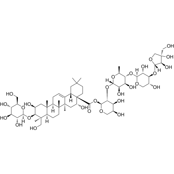
Polygalacin D (PGD) is a bioactive compound isolated from Platycodon grandiflorum with anticancer and anti-proliferative properties. PGD suppresses the expression of the IAP family of proteins including survivin, cIAP-1 and cIAP-2 and blocks the PI3K/Akt pathway by inhibiting the phosphorylation of GSK3β, Akt and the expression of PI3K. Polygalacin D induces apoptosis
Nos produits utilisent uniquement pour la recherche. Nous ne vendons pas aux patients.
- Pureté : 99.68%
- CAS No.: 66663-91-0
- Formule: C57H92O27
- Masse moléculaire:1209.32
-
Stockage:
4°C, protect from light
* In solvent : -80°C, 6 months; -20°C, 1 month (protect from light)

To place orders, for customer services and technical support, please contact: MedChemExpress USA
Tel: 609-228-6898 E-mail: [email protected] [email protected]


-
 Activité biologique
-
Chemical Information
-
Solvant et solubilité
- Protocole
- Pureté et documentation
- Références
-
 Help & FAQs


-
HY-N1067
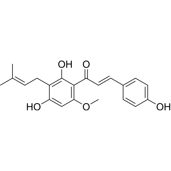
XanthohumolCOX Inhibitor -
HY-N0121
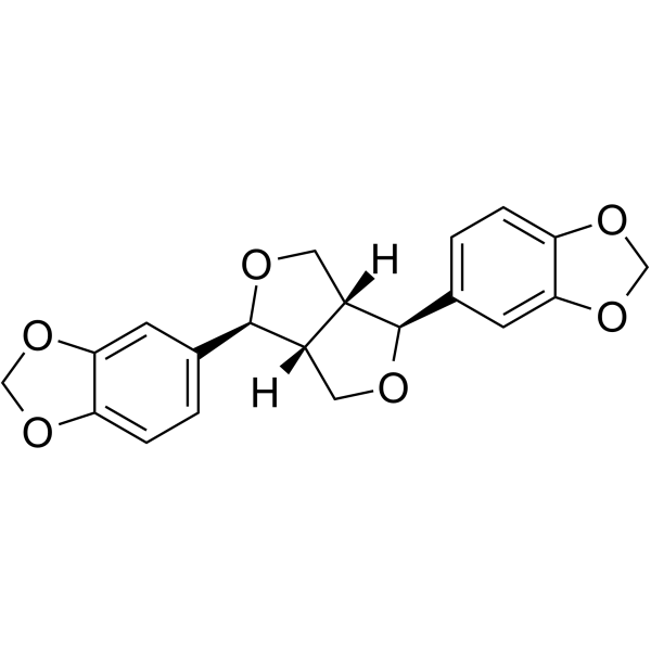
SesaminDelta 5 Desaturase Inhibitor -
HY-N6624
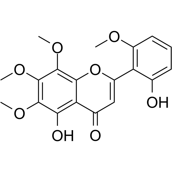
Skullcapflavone IIBacterial Inhibitor -
HY-N0735
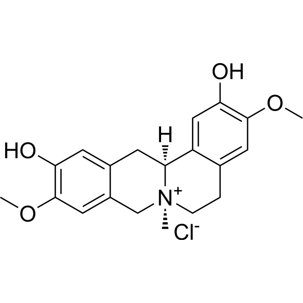
Phellodendrine chlorideAnti-inflammatory/Anti-tumor/Anti-allergic Agent -
HY-N0747
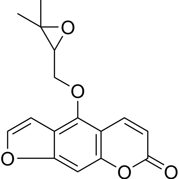
OxypeucedaninHKv1.5 Current Blocker -
HY-N8213
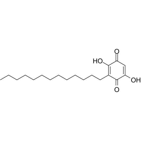
RapanoneAntioxidant Agent -
HY-N7004
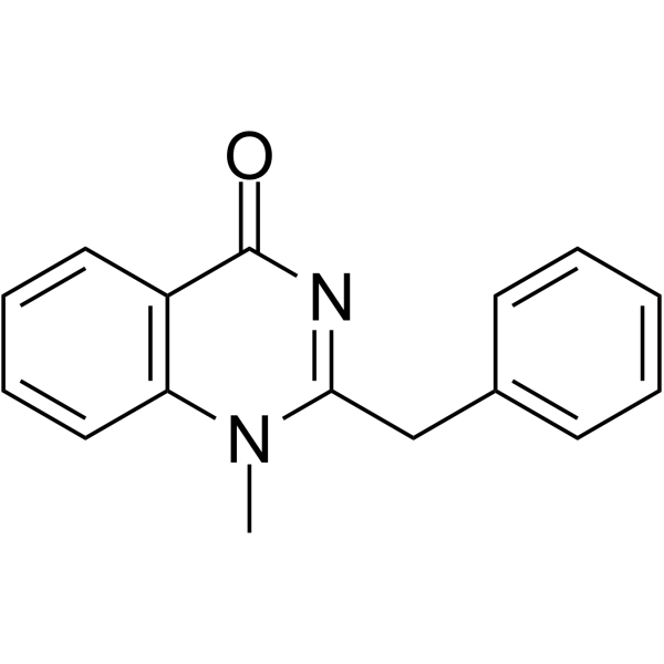
ArborinemAChR Inhibitor -
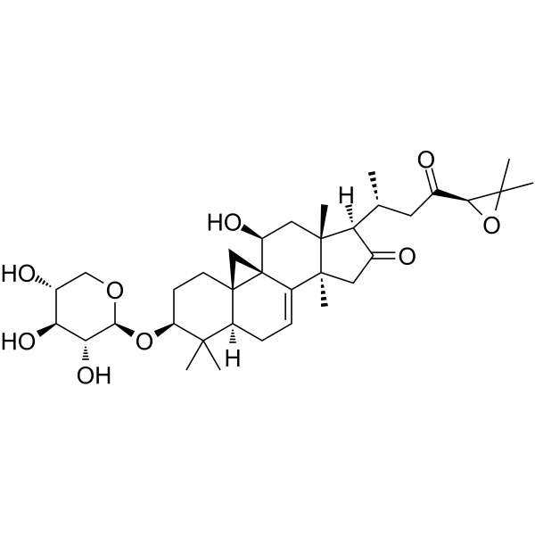
HY-N9331
Cimicifugoside H-1 -
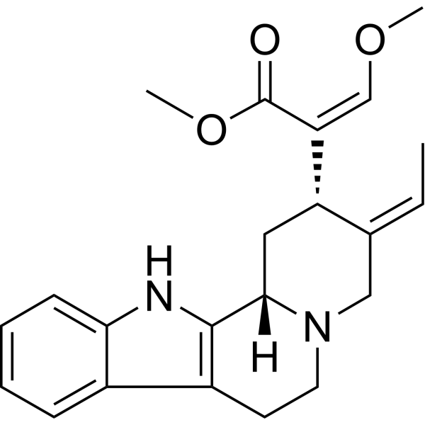
HY-N2411
Geissoschizine methyl ether5-HT1A Receptor Agonist -
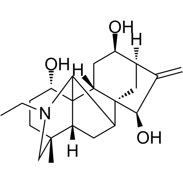
HY-N2162
12-EpinapellineAnti-Inflammatory Agent -
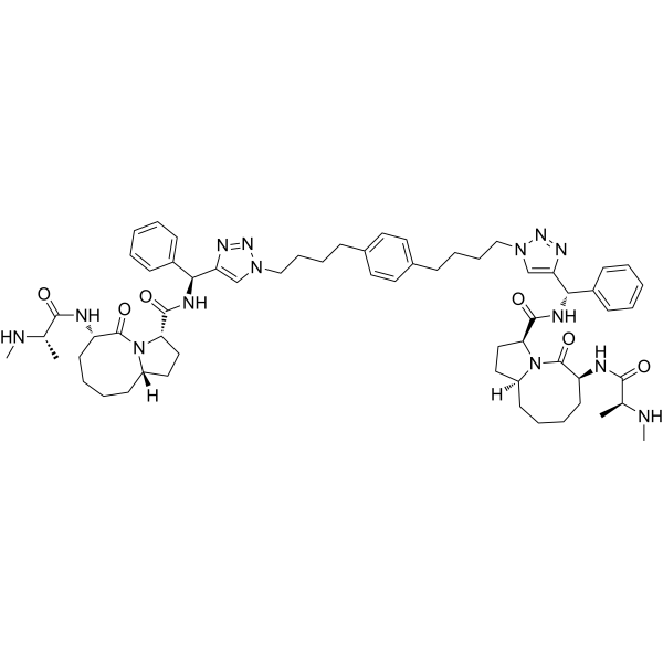
HY-15989
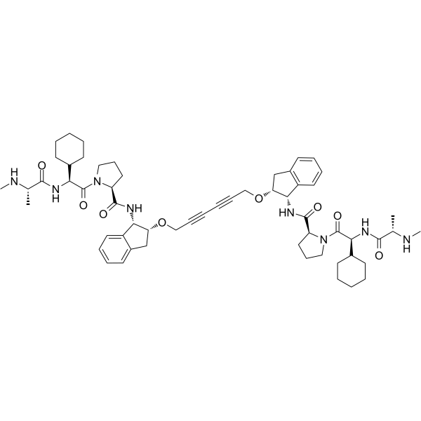
SM-164IAP Antagonist -
HY-12600
AZD5582
Publications Citing Use of MedChemExpress (MCE) Polygalacin D
More

Activité biologique
Description
IC50 & Target
IC50: IAP[1]
Cellular Effect
|
Cell Line

|
Type | Value | Description | References |
|---|---|---|---|---|
| HCT-15 | IC50 |
>10 μM
Compound: 9
|
Antiproliferative activity against human HCT15/CL02 cells after 48 hrs by sulforhodamine B assay
Antiproliferative activity against human HCT15/CL02 cells after 48 hrs by sulforhodamine B assay
|
[PMID: 20939516] |
| HCT-15 | IC50 |
8.5 μM
Compound: 9
|
Antiproliferative activity against human HCT15 cells after 48 hrs by sulforhodamine B assay
Antiproliferative activity against human HCT15 cells after 48 hrs by sulforhodamine B assay
|
[PMID: 20939516] |
| HSC-T6 | IC50 |
1.04 μM
Compound: 151
|
Antiproliferative activity against rat HSC-T6 cells assessed as reduction in cell viability incubated for 48 hrs by MTT assay
Antiproliferative activity against rat HSC-T6 cells assessed as reduction in cell viability incubated for 48 hrs by MTT assay
|
[PMID: 29353722] |
| MES-SA | IC50 |
5 μM
Compound: 9
|
Antiproliferative activity against human MESSA cells after 48 hrs by sulforhodamine B assay
Antiproliferative activity against human MESSA cells after 48 hrs by sulforhodamine B assay
|
[PMID: 20939516] |
| MES-SA/Dx5 | IC50 |
7.7 μM
Compound: 9
|
Antiproliferative activity against human MESSA/DX5 cells after 48 hrs by sulforhodamine B assay
Antiproliferative activity against human MESSA/DX5 cells after 48 hrs by sulforhodamine B assay
|
[PMID: 20939516] |
In Vitro
Polygalacin D (0-40 µM; 48 hours) inhibits cell proliferation with IC50s of 26.49 µM in the A549 cells and 20.52 µM in the H460 cells[1]. Polygalacin D (0-20 µM; 48 hours) increases the proportion of early and late apoptotic cells, and nuclear condensation is observed in A549 and H460 cells[1]. Polygalacin D (0-20 µM; 48 hours) may exerts its apoptotic effects by regulating the apoptotic proteins and the IAP family of proteins in A549 and H460 cells[1].
MedChemExpress (MCE) has not independently confirmed the accuracy of these methods. They are for reference only.
-
Cell Line:A549 and H460 cancer cell lines
-
Concentration:10 µM, 20 µM, 40µM
-
Incubation Time:48 hours
-
Result:Inhibited the proliferation of NSCLC cell lines.
-
Cell Line:A549 and H460 cancer cell lines
-
Concentration:10 µM, 20 µM
-
Incubation Time:48 hours
-
Result:Induced apoptosis of NSCLC cell lines.
-
Cell Line:A549 and H460 cancer cell lines
-
Concentration:10 µM, 20 µM
-
Incubation Time:48 hours
-
Result:Decreased survivin, c-IAP-1 and c-IAP-2 expression.
- Kinase activity and phosphorylation assays
- RNA extraction experimental
- Western Blot
- Detection of Apoptosis
- TUNEL staining for apoptotic DNA fragmentation
- Annexin V plus membrane-impermeant dye apoptosis staining
- Apoptosis Solutions
- Protocol for Kinase activity and phosphorylation assays
- Cell Viability Determination by MTT Colorimetric Assay


Chemical Information
-
CAS No. 66663-91-0
-
Appearance Solid
-
Masse moléculaire 1209.32
-
Formule C57H92O27
-
Color White to off-white
-
SMILES
O[C@H]([C@@H]([C@@H](O[C@@]1([H])[C@@H]([C@H]([C@H](O)CO1)O[C@@]2([H])[C@@H]([C@](CO)(O)CO2)O)O)[C@H](C)O3)O)[C@]3([H])O[C@H]([C@H]([C@@H](O)CO4)O)[C@@H]4OC([C@]56[C@](CC(C)(C)CC6)([H])C7=CC[C@@]([C@@]8([C@@]([C@](C)([C@@H](O[C@]9([H])O[C@@H]([C@@H](O)[C@H](O)[C@H]9O)CO)[C@@H](O)C8)CO)([H])CC%10)C)([H])[C@]%10(C)[C@]7(C)C[C@H]5O)=O
-
Structure Classification
-
Livraison
Room temperature in continental US; may vary elsewhere.
-
Stockage
4°C, protect from light
* In solvent : -80°C, 6 months; -20°C, 1 month (protect from light)


Publications (3)
-
Journal Impact Factor
-
Most Recent
-
Free Radic Biol Med
Polygalacin D suppresses esophageal squamous cell carcinoma growth and metastasis through regulating miR-142-5p/Nrf2 axis. [Abstract]2021 Feb 20:164:58-75. PMID: 33307164 -
Cancer Gene Ther
2025 Oct 2. PMID: 41039015 -
Biol Pharm Bull
Polygalacin D Prevents Breast Cancer Progression through Proteasome-Mediated Degradation of HDAC1 and HDAC2. [Abstract]2025;48(12):1897-1905. PMID: 41371635


Solvant et solubilité
In Vitro:
DMSO : 100 mg/mL (82.69 mM; Need ultrasonic; Hygroscopic DMSO has a significant impact on the solubility of product, please use newly opened DMSO)
Please refer to the solubility information to select the appropriate solvent. Once prepared, please aliquot and store the solution to prevent product inactivation from repeated freeze-thaw cycles.
Storage method and period of stock solution: -80°C, 6 months; -20°C, 1 month (protect from light). When stored at -80°C, please use it within 6 months. When stored at -20°C, please use it within 1 month.
Please refer to the solubility information to select the appropriate solvent. Once prepared, please aliquot and store the solution to prevent product inactivation from repeated freeze-thaw cycles.
Storage method and period of stock solution: -80°C, 6 months; -20°C, 1 month (protect from light). When stored at -80°C, please use it within 6 months. When stored at -20°C, please use it within 1 month.
Concentration (start) × Volume (start) = Concentration (final) × Volume (final)
In Vivo:
Select the appropriate dissolution method based on your experimental animal and administration route.
- For the following dissolution methods, please ensure to first prepare a clear stock solution using an In Vitro approach and then sequentially add co-solvents:
- To ensure reliable experimental results, the clarified stock solution can be appropriately stored based on storage conditions. As for the working solution for In Vivo experiments, it is recommended to prepare freshly and use it on the same day.
- The percentages shown for the solvents indicate their volumetric ratio in the final prepared solution. If precipitation or phase separation occurs during preparation, heat and/or sonication can be used to aid dissolution.
Add each solvent one by one: 10% DMSO 40% PEG300 5% Tween-80 45% Saline
Solubility: ≥ 2.5 mg/mL (2.07 mM); Clear solution
This protocol yields a clear solution of ≥ 2.5 mg/mL (saturation unknown).
Taking 1 mL working solution as an example, add 100 μL DMSO stock solution (25.0 mg/mL) to 400 μL PEG300, and mix evenly; then add 50 μL Tween-80 and mix evenly; then add 450 μL Saline to adjust the volume to 1 mL.
Preparation of Saline: Dissolve 0.9 g sodium chloride in ddH₂O and dilute to 100 mL to obtain a clear Saline solution.
Add each solvent one by one: 10% DMSO 90% (20% SBE-β-CD in Saline)
Solubility: ≥ 2.5 mg/mL (2.07 mM); Clear solution
This protocol yields a clear solution of ≥ 2.5 mg/mL (saturation unknown).
Taking 1 mL working solution as an example, add 100 μL DMSO stock solution (25.0 mg/mL) to 900 μL 20% SBE-β-CD in Saline, and mix evenly.
Preparation of 20% SBE-β-CD in Saline (4°C, storage for one week): 2 g SBE-β-CD powder is dissolved in 10 mL Saline, completely dissolve until clear.
In Vivo Dissolution Calculator
Please enter the basic information of animal experiments:
-
-
-
-
Recommended: Prepare an additional quantity of animals to account for potential losses during experiments.
Please enter your animal formula composition:
-
%DMSO +
Recommended: Keep the proportion of DMSO in working solution below 2% if your animal is weak.
-
%+
-
+%Tween-80 + +
-
%Saline +
The co-solvents required include: DMSO, . All of co-solvents are available by MedChemExpress (MCE). , Tween 80. All of co-solvents are available by MedChemExpress (MCE).
Working solution concentration: 0.22 mg/mL
Method for preparing stock solution: mg drug dissolved in μL DMSO. Stock solution concentration: mg/mL. * In solvent : -80°C, 6 months; -20°C, 1 month (protect from light)
1. Take μL DMSO stock solution;
2. Add μL .
μL , mix evenly;
3. Then add μL Tween 80, mix evenly;
4. Then add μL
Please ensure that the stock solution in the first step is dissolved to a clear state, and add co-solvents in sequence. You can use ultrasonic heating (ultrasonic cleaner, recommended frequency 20-40 kHz), vortexing, etc. to assist dissolution.


Pureté et documentation
-
Fiche technique (277 KB)
-
SDS (393 KB)
- English - EN (393 KB)
- Français - FR (393 KB)
- Deutsch - DE (393 KB)
- Norwegian - NO (393 KB)
- Español - ES (393 KB)
- Swedish - SV (393 KB)
- Italian - IT (393 KB)
- Korean - KR (393 KB)
- Portuguese - PT (393 KB)
-
Instruction de manipulation (2659 KB)
Références


Complete Stock Solution Preparation Table
Please refer to the solubility information to select the appropriate solvent. Once prepared, please aliquot and store the solution to prevent product inactivation from repeated freeze-thaw cycles.
Storage method and period of stock solution: -80°C, 6 months; -20°C, 1 month (protect from light). When stored at -80°C, please use it within 6 months. When stored at -20°C, please use it within 1 month.
| Optional Solvent | Concentration Solvent Mass | 1 mg | 5 mg | 10 mg | 25 mg |
|---|---|---|---|---|---|
| DMSO | 1 mM | 0.8269 mL | 4.1346 mL | 8.2691 mL | 20.6728 mL |
| 5 mM | 0.1654 mL | 0.8269 mL | 1.6538 mL | 4.1346 mL | |
| 10 mM | 0.0827 mL | 0.4135 mL | 0.8269 mL | 2.0673 mL | |
| 15 mM | 0.0551 mL | 0.2756 mL | 0.5513 mL | 1.3782 mL | |
| 20 mM | 0.0413 mL | 0.2067 mL | 0.4135 mL | 1.0336 mL | |
| 25 mM | 0.0331 mL | 0.1654 mL | 0.3308 mL | 0.8269 mL | |
| 30 mM | 0.0276 mL | 0.1378 mL | 0.2756 mL | 0.6891 mL | |
| 40 mM | 0.0207 mL | 0.1034 mL | 0.2067 mL | 0.5168 mL | |
| 50 mM | 0.0165 mL | 0.0827 mL | 0.1654 mL | 0.4135 mL | |
| 60 mM | 0.0138 mL | 0.0689 mL | 0.1378 mL | 0.3445 mL | |
| 80 mM | 0.0103 mL | 0.0517 mL | 0.1034 mL | 0.2584 mL |


Polygalacin D Related Classifications
HY-N6064 Related Classifications
 Powered by Bioz