



MAdCAM1 Protein, Human (Biotinylated, HEK293, Avi-His)

Based on 1 Customer Validation
MAdCAM1 is a cell adhesion leukocyte receptor expressed primarily by mucosal venules and plays a critical role in coordinating lymphocyte trafficking into mucosal tissues, particularly Peyer's patches and intestinal lamina propria. This protein exhibits the ability to bind integrins α-4/β-7 and L-selectin, thereby regulating the passage and retention of leukocytes in the mucosal environment. MAdCAM1 Protein, Human (Biotinylated, HEK293, Avi-His) is the recombinant human-derived MAdCAM1 protein, expressed by HEK293 , with C-Avi, C-6*His labeled tag.
- Species: Human
- Source: HEK293
-
Storage:Stored at -20°C for 2 years from date of receipt. After reconstitution, it is stable at 4°C for 1 week or -20°C for longer (with carrier protein). It is recommended to freeze aliquots at -20°C or -80°C for extended storage.


To place orders, for customer services and technical support, please contact: MedChemExpress USA
Tel: 609-228-6898 E-mail: [email protected] [email protected]


-
 Biological Activity
-
Technical Parameters
-
Product Properties
- Documentation
- Help & FAQs




Biological Activity
Description
MAdCAM1 is a cell adhesion leukocyte receptor expressed primarily by mucosal venules and plays a critical role in coordinating lymphocyte trafficking into mucosal tissues, particularly Peyer's patches and intestinal lamina propria. This protein exhibits the ability to bind integrins α-4/β-7 and L-selectin, thereby regulating the passage and retention of leukocytes in the mucosal environment. MAdCAM1 Protein, Human (Biotinylated, HEK293, Avi-His) is the recombinant human-derived MAdCAM1 protein, expressed by HEK293 , with C-Avi, C-6*His labeled tag.
Background
Mucosal Addressin Cell Adhesion Molecule 1 (MAdCAM1) is a cell adhesion leukocyte receptor expressed on the endothelial cells of mucosal venules, playing a crucial role in directing lymphocyte traffic into mucosal tissues, particularly the Peyer patches and the intestinal lamina propria. MAdCAM1 serves as a binding partner for both integrin alpha-4/beta-7 and L-selectin, thereby regulating the passage and retention of leukocytes in mucosal tissues. The homodimeric structure of MAdCAM1 contributes to its functional role in mediating adhesion between endothelial cells and circulating immune cells. Notably, isoform 2, which lacks the mucin-like domain, may have a specialized function in supporting integrin alpha-4/beta-7-dependent adhesion strengthening, independent of L-selectin binding. Understanding the features and interactions of MAdCAM1 provides insights into the intricate mechanisms governing lymphocyte trafficking in mucosal tissues.
Verified Bioactivity
Immobilized Biotinylated Human MAdCAM1 at 0.5 μg/mL (100 μL/well) can bind Anti- MAdCAM1 antibody. The ED50 for this effect is 3-6 ng/mL.
MCE Validation Data
-
Purity - SDS-PAGE
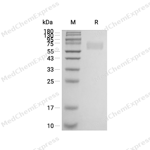
Purity - SDS-PAGE
-
Bioactivity - ELISA
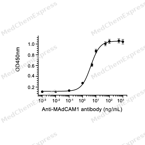
Bioactivity - ELISA


Technical Parameters
-
Species Human
-
Source HEK293
-
Tag C-Avi;C-6*His
-
Accession
Q4PKD0 (Q19-Q333)
-
Gene ID/
-
Molecular Construction
-
N-term
-
MAdCAM1 (Q19-Q333)
Accession # Q4PKD0 -
Avi-6*His
-
C-term
-
-
Protein Length
Partial
-
Conjugation
Biotin
-
Conjugate Method
The single lysine residue in Avitag is biotinylated through an enzymatic reaction.
-
Synonyms
MADCAM1; HMAdCAM-1; Mucosal Vascular Addressin Cell Adhesion Molecule 1; MAdCAM-1; Mucosal Addressin Cell Adhesion Molecule 1; Mucosal Addressin Cell Adhesion Molecule-1; MACAM1
-
AA Sequence
QSLQVKPLQVEPPEPVVAVALGASRQLTCRLACADRGASVQWRGLDTSLGAVQSDTGRSVLTVRNASLSAAGTRVCVGSCGGRTFQHTVQLLVYAFPDQLTVSPAALVPGDPEVACTAHKVTPVDPNALSFSLLVGGQELEGAQALGPEVQEEEEEPQGDEDVLFRVTERWRLPPLGTPVPPALYCQATMRLPGLELSHRQAIPVLHSPTSPEPPDTTSPESPDTTSPESPDTTSQEPPDTTSQEPPDTTSQEPPDTTSPEPPDKTSPEPAPQQGSTHTPRSPGSTRTRRPEISQAGHTQGKVIPTGSSKPAGDQ
-
Predicted Molecular Mass
35.8 kDa
-
Molecular Weight
Approximately 55-80 kDa, based on SDS-PAGE under reducing conditions.
-
Purity
≥ 95%, as determined by reducing SDS-PAGE.


Product Properties
Lyophilized powder.
Lyophilized from a 0.22 μm filtered solution of PBS, pH 7.4.
<1 EU/μg, determined by LAL method.
It is not recommended to reconstitute to a concentration less than 100 μg/mL in ddH2O. For long term storage it is recommended to add a carrier protein (0.1% BSA, 5% HSA, 10% FBS or 5% Trehalose).
Stored at -20°C for 2 years from date of receipt. After reconstitution, it is stable at 4°C for 1 week or -20°C for longer (with carrier protein). It is recommended to freeze aliquots at -20°C or -80°C for extended storage.
Room temperature in continental US; may vary elsewhere.


Documentation
-
Data Sheet (261 KB)
-
SDS (252 KB)
- English - EN (252 KB)
- Français - FR (252 KB)
- Deutsch - DE (252 KB)
- Norwegian - NO (252 KB)
- Español - ES (252 KB)
- Swedish - SV (252 KB)
- Italian - IT (252 KB)
- Korean - KR (252 KB)
- Portuguese - PT (252 KB)
-
Handling Instructions (2659 KB)

Calculators
Concentration (start) × Volume (start) = Concentration (final) × Volume (final)


 Powered by Bioz

