



Resazurin
Based on 11 publication(s) in Google Scholar
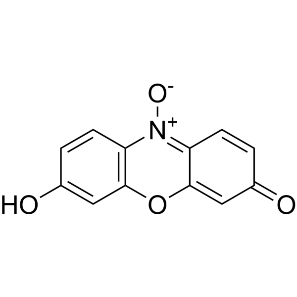
Resazurin (Diazoresorcinol) is a water-soluble, non-toxic, stable, membrane-permeable blue non-fluorescent dye (faintly fluorescent). Resazurin is used as a redox indicator, can be reduced to pink, highly fluorescent Resorufin (Ex=530-560 nm, Em=590 nm) in living cells. Resazurin can be used for the detection of cell viability, toxicity, proliferation, migration and invasion in cells (human, plant and animal, bacterial and fungal).
For research use only. We do not sell to patients.
- CAS No.: 550-82-3
- Formula: C12H7NO4
- Molecular Weight:229.19
-
Storage:
Please store the product under the recommended conditions in the Certificate of Analysis.

To place orders, for customer services and technical support, please contact: MedChemExpress USA
Tel: 609-228-6898 E-mail: [email protected] [email protected]


-
 Biological Activity
-
Chemical Information
- Protocols
- Purity & Documentation
- References
-
 Help & FAQs



Publications Citing Use of MedChemExpress (MCE) Resazurin
More- Nat Metab. 2021 Oct;3(10):1357-1371. [Abstract]
- Environ Sci Technol. 2025 Oct 21;59(41):21898-21909. [Abstract]
- J Immunother Cancer. 2026 May 27;14(5):e014533. [Abstract]
- Pharmaceuticals (Basel). 2024 Feb 2;17(2):197. [Abstract]
- Eur J Pharm Sci. 2026 Oct 1:225:107613.
- Surgery. 2024 Mar;175(3):726-734. [Abstract]
- bioRxiv. 2026 Jul 17.
- bioRxiv. 2026 Jun 29:2026.06.26.734853.
- bioRxiv. 2026 May 13:2026.05.13.724856. [Abstract]
- The Catholic University of America. 2026.
- bioRxiv. 2023 Apr 8:2023.04.07.536013. [Abstract]


Biological Activity
Description
In Vitro
Resazurin (Diazoresorcinol) is commonly used to measure the viability of bacterial and eukaryotic cells through its reduction to the fluorescent product resorufin. In TSBc medium, no viable bacteria were detected 24 hours after inoculation of F. tularensis LVS in the presence of Resazurin at a concentration of 44 μM. Reducing the concentration of Resazurin to 4.4 μM still resulted in a 10-fold reduction in the survival of F. tularensis LVS compared with medium alone. Both Resazurin treatments significantly reduced the number of viable F. tularensis LVS bacteria within 22 hours. Following infection of HEK293 cells for 22 hours, Resazurin treatment significantly reduced the number of viable F. tularensis LVS bacteria[3].
Guide (The following is our recommended protocol. This protocol is provided as a guideline only and should be modified according to your specific needs).
1. Solution Preparation[2][3]
1.1 Stock Solution Preparation
Solvent: DMSO or ddH2O
Concentration: 1 mg/mL (optimize according to the experiment).
Storage: Aliquot and store at -20°C or -80°C protected from light. Avoid repeated freeze-thaw cycles.
1.2 Working Solution Preparation
Dilute with PBS or serum-free culture medium to 1 μg/mL (optimize according to the experiment).
Note: The working solution should be prepared immediately before use and protected from light.
2. Cell Viability
1. Thaw the Resazurin solution in a 37°C water bath.
2. Plate cells in a 96-well plate and wash with PBS (avoid light exposure).
3. Remove the PBS wash solution, then add 500 μL of Resazurin solution (1 μg/mL).
4. Place the plate in an incubator for 30 minutes (incubation time depends on cell type and cell number).
5. Measure Resazurin fluorescence using a spectrophotometer (Ex=530-560 nm, Em=590 nm).
MedChemExpress (MCE) has not independently confirmed the accuracy of these methods. They are for reference only. Further protocols information, click here.
Emission (Em)
590
Excitation (Ex)
530/560


Chemical Information
-
CAS No. 550-82-3
-
Molecular Weight 229.19
-
Formula C12H7NO4
-
SMILES
O=C1C=CC2=[N+](C3=C(OC2=C1)C=C(C=C3)O)[O-]
-
Synonyms
Diazoresorcinol
-
Shipping
Room temperature in continental US; may vary elsewhere.
-
Storage
Please store the product under the recommended conditions in the Certificate of Analysis.


Publications (11)
-
Journal Impact Factor
-
Most Recent
-
Nat Metab
The alternative activity of nuclear PHGDH contributes to tumour growth under nutrient stress. [Abstract]2021 Oct;3(10):1357-1371. PMID: 34663976 -
Environ Sci Technol
Revealing Ferroptosis Induction by Bisphenol A and Bisphenol S through Distinct Protein Targets. [Abstract]2025 Oct 21;59(41):21898-21909. PMID: 41068997 -
J Immunother Cancer
Gasdermin D antagonizes immunosuppression in prostate cancer by inducing LAMC2 degradation to block M2 macrophage polarization. [Abstract]2026 May 27;14(5):e014533. PMID: 42203263 -
Pharmaceuticals (Basel)
Therapeutic Implications of Ceritinib in Cholangiocarcinoma beyond ALK Expression and Mutation. [Abstract]2024 Feb 2;17(2):197. PMID: 38399413 -
-
Surgery
2024 Mar;175(3):726-734. PMID: 37914574 -
-
-
bioRxiv
Physiological levels of 3-hydroxykynurenine alter mitochondrial function and morphology in neuronal cells. [Abstract]2026 May 13:2026.05.13.724856. PMID: 42182345 -
-
bioRxiv
Inhibition of serotonin biosynthesis in neuroendocrine neoplasm suppresses tumor growth in vivo. [Abstract]2023 Apr 8:2023.04.07.536013. PMID: 37066322

Protocols
-
Cell Cytotoxicity Assay
Cytotoxicity assays are usually based on the assessment of cell membrane damage, which can also be indirectly detected by measuring cell viability. Detection methods include MTT assay, CKK-8 assay, LDH assay and ATP assay, etc.
-
Mammalian live/dead viability and cytotoxicity staining
Live/dead viability and cytotoxicity staining assays are based on the simultaneous detection of intracellular esterase activity in metabolically active (viable) cells and membrane integrity loss in non-viable cells. In commonly used dual-staining approaches, membrane-permeant fluorogenic substrates are converted by intracellular esterases into fluorescent products in live cells, while impermeant DNA-binding dyes selectively enter cells with compromised plasma membranes and label nucleic acids in dead or dying cells, enabling discrimination between viable and non-viable populations by fluorescence microscopy or flow cytometry.
-
ROS/oxidative-stress fluorescent staining
ROS/oxidative-stress fluorescent staining uses cell-permeant fluorogenic probes that become fluorescent after oxidation inside cells or tissues; commonly used examples include DCFH-DA/DCFDA for broad cellular oxidant detection, DHE for superoxide-related signal detection, MitoSOX for mitochondrial superoxide-related signal detection, and CellROX probes for oxidative-stress-associated fluorescence readouts. The assay detects probe oxidation rather than a single ROS species unless the probe and analysis method have been chemically validated for that species. DCFH-DA enters cells, is deacetylated by intracellular esterases to DCFH, and produces fluorescent DCF after oxidation, so the readout is used as an operational measure of total cellular oxidative stress rather than a species-specific ROS measurement. DHE and MitoSOX can report superoxide-related oxidation, but red fluorescence alone can include non-specific ethidium-like oxidation products; HPLC or optimized spectral approaches are
-
Cell migration
Cell migration is a method that plays an important role in wound healing, cell differentiation, embryonic development, etc.
-
CCK-8/WST-8 Cell Proliferation Assay
The CCK-8/WST-8 assay is based on the reduction of the water-soluble tetrazolium salt WST-8 to a water-soluble formazan product by cellular dehydrogenases in metabolically active cells, where the generated formazan amount is proportional to the number of living cells and is quantified by measuring absorbance in the visible range, providing a colorimetric readout for cell viability and proliferation assessment. This class of tetrazolium-based assays improves upon earlier MTT-based systems by producing a water-soluble formazan, eliminating the need for organic solubilization steps and enabling direct spectrophotometric measurement in culture medium.
-
Cell invasion
Cell invasion is the ability of cells to migrate from one area to another via the extracellular matrix. Cell invasion is the response of normal and cancer cells to chemical and mechanical stimuli. Before migrating to a new region, the extracellular matrix is degraded by proteases within the cell. Cell invasion often occurs during wound repair, vascularization and inflammation, abnormal tissue invasion, and tumor cell metastasis.
-
MTT Cell Proliferation Assay
The MTT assay is a colorimetric endpoint assay for estimating viable cell number, cell growth, cytotoxicity, or cell activation in cultured mammalian cells. Living cells reduce the yellow tetrazolium salt MTT into purple/blue formazan, while dead cells do not generate the same signal; the resulting color can be quantified with a multiwell spectrophotometer. MTT reduction is commonly interpreted as a readout of metabolic activity that often correlates with viable cell number, but it should not be treated as a direct cell-counting method unless the assay is optimized for the cell type and experimental condition. Studies show that MTT reduction can involve mitochondrial and non-mitochondrial reducing systems, and formazan may accumulate in intracellular lipid droplets rather than simply marking mitochondria.
-
Cell Viability Determination by MTT Colorimetric Assay
The following protocol uses the MTT colorimetric assay as a classic literature-established method for assessing cell viability/metabolic activity in cultured mammalian cells. MTT[3-(4,5-dimethylthiazol-2-yl)-2,5-diphenyltetrazolium bromide] is reduced by metabolically active cells to a colored formazan product; the amount of formazan is quantified spectrophotometrically and provides an indirect measure of metabolically active viable cells. Importantly, MTT reduction reflects cellular oxidoreductase/metabolic activity rather than an absolute direct count of living cells, so changes in cellular metabolism can alter the signal independently of cell number.


Purity & Documentation
References
[1]. Silva FSG, et al. Determination of Metabolic Viability and Cell Mass Using a Tandem Resazurin/Sulforhodamine B Assay. Curr Protoc Toxicol. 2016 May 4;68:2.24.1-2.24.15. [Content Brief]
[2]. Rampersad SN. Multiple applications of Alamar Blue as an indicator of metabolic function and cellular health in cell viability bioassays. Sensors (Basel). 2012;12(9):12347-60. [Content Brief]
[3]. Schmitt DM, et al. The use of resazurin as a novel antimicrobial agent against Francisella tularensis. Front Cell Infect Microbiol. 2013 Dec 6;3:93. [Content Brief]

Calculators
Concentration (start) × Volume (start) = Concentration (final) × Volume (final)


Resazurin Related Classifications
HY-118540 Related Classifications
 Powered by Bioz