



S1QEL1.1

Based on 1 Customer Validation
S1QEL1.1 is a cell-permeable selective site IQ electron leak inhibitor and mitochondrial complex I site IQ superoxide/hydrogen peroxide production inhibitor (IC50 of 70.3 nM for IQr; IC50 of 64.0 nM for IQf), exerting cytoprotective effects by inhibiting site IQ-dependent ROS production during reverse and forward electron transport without affecting oxidative phosphorylation and normal electron transport. S1QEL1.1 can be used for research on ischemia-reperfusion injury.
For research use only. We do not sell to patients.
- Purity : 99.9%
- CAS No.: 897613-29-5
- Formula: C23H24N4O3S
- Molecular Weight:436.53
-
Storage:Powder -20°C, 3 years ; In solvent -80°C, 6 months , -20°C, 1 month


To place orders, for customer services and technical support, please contact: MedChemExpress USA
Tel: 609-228-6898 E-mail: [email protected] [email protected]


-
 Biological Activity
-
Chemical Information
-
Solvent & Solubility
- Protocols
- Purity & Documentation
- References
-
 Help & FAQs


-
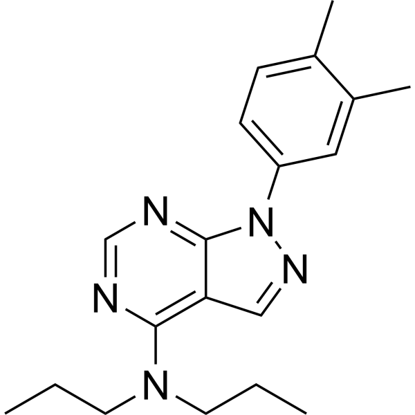
HY-110282
S3QEL-2Site IIIQo Superoxide Production Suppressor -
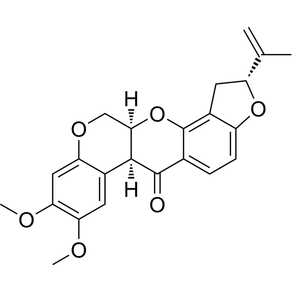
HY-B1756
RotenoneMitochondrial Complex I Inhibitor -

HY-117605R
N-Desmethyl Sildenafil (Standard)Sildenafil Metabolite -
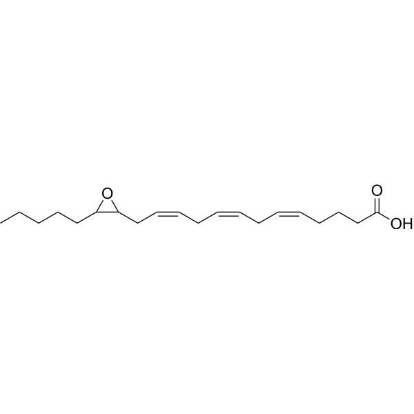
HY-150084
(±)14,15-Epoxyeicosatrienoic acidProliferation Inducer -
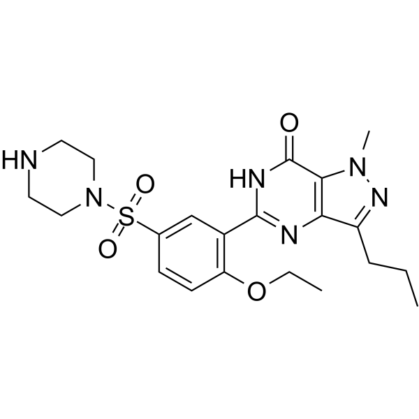
HY-117605
N-Desmethyl SildenafilSildenafil Metabolite -
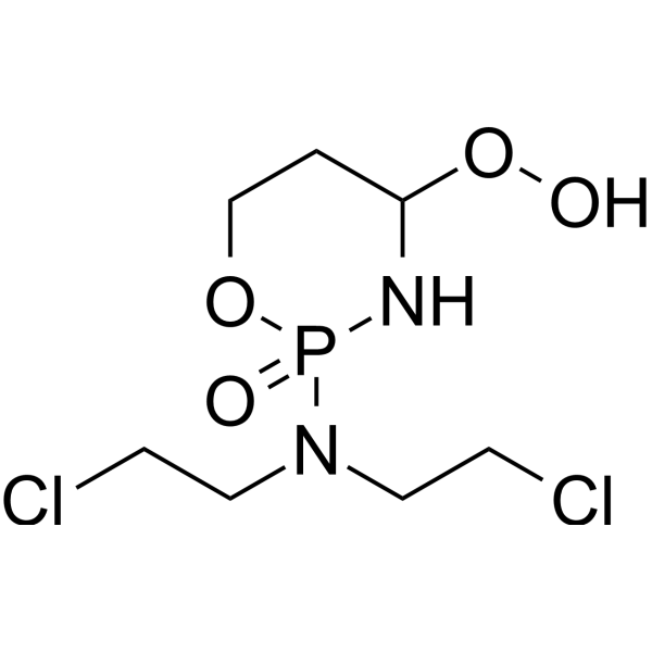
HY-117433
4-Hydroperoxy cyclophosphamideDrug Metabolite -

HY-W009512
CVT-2738Ranolazine Metabolite -
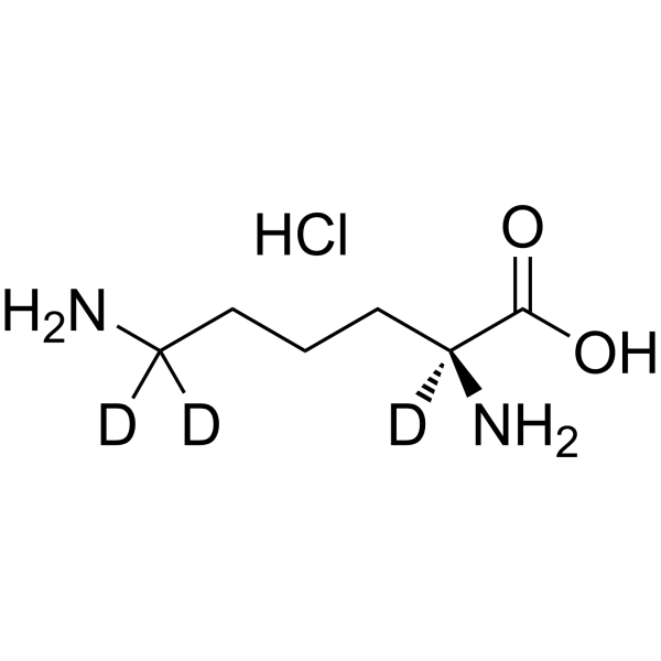
HY-N0469S
L-Lysine-d3 hydrochlorideStable Isotope -

HY-W009512R
CVT-2738 (Standard)Ranolazine Metabolite -

HY-127020
DeoxyenterocinBacterial Metabolite -

HY-113322
3-HydroxyquinineQuinidine Metabolite


Biological Activity
Description
IC50 & Target
[1]|
H2O2 70.3 nM (IC50, IQr) |
H2O2 64.0 nM (IC50, IQf) |
In Vitro
S1QEL1.1 (1 μM) inhibits superoxide/hydrogen peroxide production at site IQf during forward electron transfer in isolated rat skeletal muscle mitochondria[1].
S1QEL1.1 (titration) also inhibited superoxide/hydrogen peroxide production at sites IQr and IQf in isolated rat skeletal muscle mitochondria, with nominal IC50 values of 70.3 nM and 64.0 nM, respectively[1].
S1QEL1.1 (0.07 μM) is a potent inhibitor of superoxide-H2O2 production at site IQ in isolated rat skeletal muscle mitochondria, with an IC50 of 0.07 μM[2].
S1QEL1.1 (1.4-10 μM) is a selective inhibitor of superoxide-H2O2 production at site IQ in isolated rat skeletal muscle mitochondria, without affecting oxidative phosphorylation[2].
S1QEL1.1 (1.4-10 μM; 3-72 h) had no effect on oxidative phosphorylation or viability in HEK293 cells, even at high concentrations[2].
S1QEL1.1 (24 h) attenuated caspase activation in the ER stress H9c2 cardiomyocyte model[2].
MedChemExpress (MCE) has not independently confirmed the accuracy of these methods. They are for reference only. Further protocols information, click here.
In Vivo
S1QEL1.1 (800 nM; dietary) reduces RasV12-induced intestinal stem cell hyperplasia in Drosophila[2].
S1QEL1.1 (800 nM; dietary) had no effect on baseline stem cell division or differentiation in unstressed Drosophila intestines[2].
S1QEL1.1 (1.6 μM; infused via perfusion port; 5 min; at onset of reperfusion) improves functional recovery and reduces infarct size after ischemia in perfused mouse hearts[2].
S1QEL1.1 (0.08-1.6 μM; perfused; each dose for 5 min followed by 10 min wash-out) had no effect on baseline cardiac function in perfused mouse hearts at concentrations ranging from 0.08 μM to 1.6 μM[2].
MedChemExpress (MCE) has not independently confirmed the accuracy of these methods. They are for reference only.
-
Animal Model:Oregon Red (w;esgGal4,UAS-GFP; virgins crossed to Oregon Red males, 6-day old offspring)[2]
-
Dosage:800 nM
-
Administration:dietary
-
Result:Suppressed the tunicamycin-induced increase in the mitotic marker phospho-histone H3 by up to 45%.
-
Animal Model:Oregon Red (w;esgGal4,UAS-GFP; tubGal80ts virgins crossed to yw;UAS-RasV12 males, 4-day old offspring)[2]
-
Dosage:800 nM
-
Administration:dietary
-
Result:Decreased RasV12-induced hyperplasia in fly intestinal stem cells.
-
Animal Model:Oregon Red (MARCM82A:hsFlp,UAS-GFP;;tubGal4,FRT82,tubGal80 virgins crossed to w;FRT82 males, 4-day old offspring)[2]
-
Dosage:800 nM
-
Administration:dietary
-
Result:Did not affect stem cell division in unstressed conditions.
No differences in cell type distribution were observed; 90% of clones had an identifiable stem cell progenitor with and without S1QELs.
-
Animal Model:C57BL/6J (male, 8-12 weeks old)[2]
-
Dosage:1.6 μM
-
Administration:infused via perfusion port; 5 min; at onset of reperfusion
-
Result:Significantly improved the post-ischemic recovery of cardiac function (rate × pressure product).
Caused a small but significant decrease in infarct size.


Chemical Information
-
CAS No. 897613-29-5
-
Appearance Solid
-
Molecular Weight 436.53
-
Formula C23H24N4O3S
-
Color White to off-white
-
SMILES
O=C(NC1=CC=CC(NC(C)=O)=C1)C(NCCC2=C(C)N=C(C3=CC=C(C)C=C3)S2)=O
-
Shipping
Room temperature in continental US; may vary elsewhere.
-
Storage
Powder -20°C 3 years In solvent -80°C 6 months -20°C 1 month


Solvent & Solubility
In Vitro:
DMSO : 12.5 mg/mL (28.63 mM; ultrasonic and warming and heat to 60°C; Hygroscopic DMSO has a significant impact on the solubility of product, please use newly opened DMSO)
DMF : 3 mg/mL (6.87 mM; Need ultrasonic and warming)
Please refer to the solubility information to select the appropriate solvent. Once prepared, please aliquot and store the solution to prevent product inactivation from repeated freeze-thaw cycles.
Storage method and period of stock solution: -80°C, 6 months; -20°C, 1 month. When stored at -80°C, please use it within 6 months. When stored at -20°C, please use it within 1 month.
Please refer to the solubility information to select the appropriate solvent. Once prepared, please aliquot and store the solution to prevent product inactivation from repeated freeze-thaw cycles.
Storage method and period of stock solution: -80°C, 6 months; -20°C, 1 month. When stored at -80°C, please use it within 6 months. When stored at -20°C, please use it within 1 month.
Concentration (start) × Volume (start) = Concentration (final) × Volume (final)

Protocols
-
Kinase activity and phosphorylation assays
Kinase activity assays measure the ability of kinases to transfer phosphate groups from ATP to specific substrates, while phosphorylation assays detect the presence and levels of phosphorylated proteins. Common methods include radiolabeled ATP incorporation (e. g. ,), ADP release detection via bioluminescence (e. g. ,[3]), enzyme-linked immunosorbent assays (ELISA) for phospho-specific epitopes (e. g. ,[6]), and microtiter-based formats for high-throughput screening (e. g. ,[8]). The ADP-Glo assay quantifies kinase activity by measuring ADP produced during phosphorylation using a luciferase-based system. Radiometric assays involve autoradiography or scintillation counting after incorporation of 32P-labeled ATP into substrate proteins. ELISA-based approaches rely on phospho-specific antibodies to detect activated kinases in cell lysates or purified samples.
-
Western Blot
Western blotting (WB) is a commonly used experimental method in molecular biology, biochemistry, and immunogenetics for identifying and quantifying target proteins. It combines gel electrophoresis with immunoassay, enabling researchers to analyze protein expression, post-translational modifications, and molecular weight.
-
Cell Cytotoxicity Assay
Cytotoxicity assays are usually based on the assessment of cell membrane damage, which can also be indirectly detected by measuring cell viability. Detection methods include MTT assay, CKK-8 assay, LDH assay and ATP assay, etc.
-
ROS/oxidative-stress fluorescent staining
ROS/oxidative-stress fluorescent staining uses cell-permeant fluorogenic probes that become fluorescent after oxidation inside cells or tissues; commonly used examples include DCFH-DA/DCFDA for broad cellular oxidant detection, DHE for superoxide-related signal detection, MitoSOX for mitochondrial superoxide-related signal detection, and CellROX probes for oxidative-stress-associated fluorescence readouts. The assay detects probe oxidation rather than a single ROS species unless the probe and analysis method have been chemically validated for that species. DCFH-DA enters cells, is deacetylated by intracellular esterases to DCFH, and produces fluorescent DCF after oxidation, so the readout is used as an operational measure of total cellular oxidative stress rather than a species-specific ROS measurement. DHE and MitoSOX can report superoxide-related oxidation, but red fluorescence alone can include non-specific ethidium-like oxidation products; HPLC or optimized spectral approaches are
-
Protocol for Kinase activity and phosphorylation assays
Kinase activity assays measure transfer of phosphate from ATP to a protein or peptide substrate, generating phosphorylated substrate, ADP, or incorporated radiolabeled phosphate as the readout; phosphorylation assays measure site-specific phosphorylation in cells or tissues as a proxy for kinase-pathway activation, inhibition, or substrate regulation. Phosphorylation can be detected by phospho-specific Western blot, immunoprecipitation kinase assay, phospho-immunofluorescence, phospho-flow cytometry, luminescent ADP detection, radiolabeled ATP incorporation, or reporter-based pathway assays, and these readouts can be applied to cancer cells, primary neurons, mouse tumors, organoids, inflammatory macrophages, ferroptosis studies, and mitophagy studies when the kinase target is biologically relevant.


Purity & Documentation
-
Data Sheet (283 KB)
-
SDS (596 KB)
- English - EN (596 KB)
- Français - FR (596 KB)
- Deutsch - DE (596 KB)
- Norwegian - NO (596 KB)
- Español - ES (596 KB)
- Swedish - SV (596 KB)
- Italian - IT (596 KB)
- Korean - KR (596 KB)
- Portuguese - PT (596 KB)
-
Handling Instructions (2659 KB)
References


Complete Stock Solution Preparation Table
Please refer to the solubility information to select the appropriate solvent. Once prepared, please aliquot and store the solution to prevent product inactivation from repeated freeze-thaw cycles.
Storage method and period of stock solution: -80°C, 6 months; -20°C, 1 month. When stored at -80°C, please use it within 6 months. When stored at -20°C, please use it within 1 month.
| Optional Solvent | Concentration Solvent Mass | 1 mg | 5 mg | 10 mg | 25 mg |
|---|---|---|---|---|---|
| DMF / DMSO | 1 mM | 2.2908 mL | 11.4540 mL | 22.9079 mL | 57.2698 mL |
| 5 mM | 0.4582 mL | 2.2908 mL | 4.5816 mL | 11.4540 mL | |
| DMSO | 10 mM | 0.2291 mL | 1.1454 mL | 2.2908 mL | 5.7270 mL |
| 15 mM | 0.1527 mL | 0.7636 mL | 1.5272 mL | 3.8180 mL | |
| 20 mM | 0.1145 mL | 0.5727 mL | 1.1454 mL | 2.8635 mL | |
| 25 mM | 0.0916 mL | 0.4582 mL | 0.9163 mL | 2.2908 mL |


S1QEL1.1 Related Classifications
HY-129115 Related Classifications
 Powered by Bioz
Keywords
- S1QEL1.1
- 897613-29-5
- Reactive Oxygen Species (ROS)
- site IQ electron leak
- superoxide/hydrogen peroxide production
- primary astrocytes
- cardiomyocyte ER stress
- oxidative phosphorylation
- rat skeletal muscle mitochondria
- HEK293
- mitochondrial complex I
- ischemia-reperfusion injury
- Drosophila intestine
- Inhibitor
- inhibitor
- inhibit