



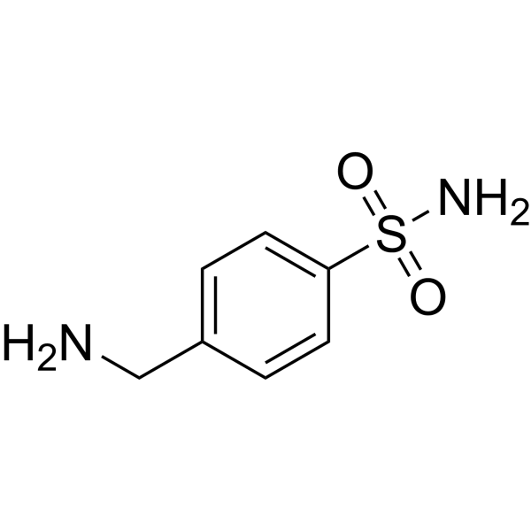
Mafenide

Based on 1 Customer Validation
Mafenide is a potent sulfonamide antimicrobial agent. Mafenide exhibits antibacterial activity against Staphylococcus aureus and Pseudomonas aeruginosa. Mafenide also exhibits antifungal activity against filamentous fungi (e.g., Lichtheimia and Aspergillus flavus). Mafenide can be used in the research of skin grafts on burn wounds, post-traumatic invasive fungal infections, and bacterially contaminated wounds.
For research use only. We do not sell to patients.
- Purity : 99.96%
- CAS No.: 138-39-6
- Formula: C7H10N2O2S
- Molecular Weight:186.23
-
Storage:Powder -20°C, 3 years , 4°C, 2 years ; In solvent -80°C, 6 months , -20°C, 1 month

To place orders, for customer services and technical support, please contact: MedChemExpress USA
Tel: 609-228-6898 E-mail: [email protected] [email protected]


-
 Biological Activity
-
Chemical Information
-
Solvent & Solubility
- Protocols
- Purity & Documentation
- References
-
 Help & FAQs




Biological Activity
Description
In Vitro
Mafenide (5%) maintains antimicrobial activity with a zone of inhibition >2 mm against Staphylococcus aureus and Pseudomonas aeruginosa in vitro[1].
Mafenide (2.5%-7.5%; 1-24 h) exhibits dose-dependent antifungal activity against filamentous fungi (e.g., Lichtheimia spp., Aspergillus flavus)[3].
MedChemExpress (MCE) has not independently confirmed the accuracy of these methods. They are for reference only.
-
Cell Line:Human keratinocytes (HEK-001; ATCC CRL-2404), human dermal fibroblasts (PromoCell), human osteoblasts (PromoCell)
-
Concentration:2.5%, 5%, 7.5%
-
Incubation Time:0.5 h, 1 h, 3 h, 24 h
-
Result:Showed a viability range of 30%-78% (fibroblasts).
Showed a viability range of 31%-77% (keratinocytes).
Showed a viability range of 22%-81% (osteoblasts).
In Vivo
MedChemExpress (MCE) has not independently confirmed the accuracy of these methods. They are for reference only.
-
Animal Model:Male Wistar albino rats (mean weight 200 g) with Pseudomonas aeruginosa-contaminated full-thickness skin graft (FTSG) model (2.5×2.5 cm FTSG harvested from back, wound bed inoculated with 107 CFU/mL Pseudomonas aeruginosa in 0.6 mL buffer solution)[4]
-
Dosage:2.5% Mafenide Acetate solution
-
Administration:Topical application via soaked dressings (hydrofiber or gauze); group 3 (hydrofiber + MA): dressing changed every 2 days; group 4 (gauze + MA): dressing changed twice a day; total cycle of 14 days
-
Result:Showed a mean graft survival rate of 50.1% in group 3 and 66.3% in group 4.
Showed detachment of the dermal-epidermal junction, collagen disorganization, decreased fibroblast number, and decreased capillary count.
Clinical Trial
| NCT Number | Sponsor | Condition | Start Date |
Phase

|
|---|---|---|---|---|
| NCT01329991 | Plexxikon| | 2011-05 | PHASE1 |


Chemical Information
-
CAS No. 138-39-6
-
Appearance Solid
-
Molecular Weight 186.23
-
Formula C7H10N2O2S
-
Color White to off-white
-
SMILES
O=S(C1=CC=C(CN)C=C1)(N)=O
-
Shipping
Room temperature in continental US; may vary elsewhere.
-
Storage
Powder -20°C 3 years 4°C 2 years In solvent -80°C 6 months -20°C 1 month


Solvent & Solubility
In Vitro:
DMSO : 30.3 mg/mL (162.70 mM; Need ultrasonic; Hygroscopic DMSO has a significant impact on the solubility of product, please use newly opened DMSO)
Please refer to the solubility information to select the appropriate solvent. Once prepared, please aliquot and store the solution to prevent product inactivation from repeated freeze-thaw cycles.
Storage method and period of stock solution: -80°C, 6 months; -20°C, 1 month. When stored at -80°C, please use it within 6 months. When stored at -20°C, please use it within 1 month.
Please refer to the solubility information to select the appropriate solvent. Once prepared, please aliquot and store the solution to prevent product inactivation from repeated freeze-thaw cycles.
Storage method and period of stock solution: -80°C, 6 months; -20°C, 1 month. When stored at -80°C, please use it within 6 months. When stored at -20°C, please use it within 1 month.
Concentration (start) × Volume (start) = Concentration (final) × Volume (final)


Purity & Documentation
-
Data Sheet (274 KB)
-
SDS (393 KB)
- English - EN (393 KB)
- Français - FR (393 KB)
- Deutsch - DE (393 KB)
- Norwegian - NO (393 KB)
- Español - ES (393 KB)
- Swedish - SV (393 KB)
- Italian - IT (393 KB)
- Korean - KR (393 KB)
- Portuguese - PT (393 KB)
-
Handling Instructions (2659 KB)
References
[1]. Ashkan Afshari, et al. The Effective Duration of Antimicrobial Activity of Mafenide Acetate After Reconstitution. J Burn Care Res. 2018 Aug 17;39(5):736-738. [Content Brief]
[2]. Haynes, B.W., Jr., Mafenide acetate in burn treatment. N Engl J Med, 1971. 284(23): p. 1324. [Content Brief]
[3]. Barsoumian A, et al. In vitro toxicity and activity of Dakin's solution, mafenide acetate, and amphotericin B on filamentous fungi and human cells. J Orthop Trauma. 2013 Aug;27(8):428-36. [Content Brief]
[4]. Baver Acaban M, et al. The effects of topical mafenide acetate application on skin graft survival in bacterial contaminated wounds. Burns. 2024 Mar;50(2):433-443. [Content Brief]


Complete Stock Solution Preparation Table
Please refer to the solubility information to select the appropriate solvent. Once prepared, please aliquot and store the solution to prevent product inactivation from repeated freeze-thaw cycles.
Storage method and period of stock solution: -80°C, 6 months; -20°C, 1 month. When stored at -80°C, please use it within 6 months. When stored at -20°C, please use it within 1 month.
| Optional Solvent | Concentration Solvent Mass | 1 mg | 5 mg | 10 mg | 25 mg |
|---|---|---|---|---|---|
| DMSO | 1 mM | 5.3697 mL | 26.8485 mL | 53.6970 mL | 134.2426 mL |
| 5 mM | 1.0739 mL | 5.3697 mL | 10.7394 mL | 26.8485 mL | |
| 10 mM | 0.5370 mL | 2.6849 mL | 5.3697 mL | 13.4243 mL | |
| 15 mM | 0.3580 mL | 1.7899 mL | 3.5798 mL | 8.9495 mL | |
| 20 mM | 0.2685 mL | 1.3424 mL | 2.6849 mL | 6.7121 mL | |
| 25 mM | 0.2148 mL | 1.0739 mL | 2.1479 mL | 5.3697 mL | |
| 30 mM | 0.1790 mL | 0.8950 mL | 1.7899 mL | 4.4748 mL | |
| 40 mM | 0.1342 mL | 0.6712 mL | 1.3424 mL | 3.3561 mL | |
| 50 mM | 0.1074 mL | 0.5370 mL | 1.0739 mL | 2.6849 mL | |
| 60 mM | 0.0895 mL | 0.4475 mL | 0.8950 mL | 2.2374 mL | |
| 80 mM | 0.0671 mL | 0.3356 mL | 0.6712 mL | 1.6780 mL | |
| 100 mM | 0.0537 mL | 0.2685 mL | 0.5370 mL | 1.3424 mL |


Mafenide Related Classifications
HY-B0614 Related Classifications
 Powered by Bioz