



MSC 2032964A
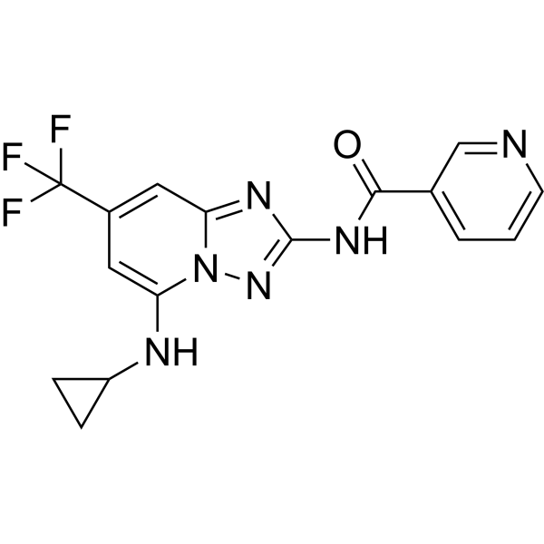
MSC 2032964A is an orally active, selective inhibitor for apoptosis signal-regulating kinase 1(ASK1) with IC50 of 96 nM. MSC 2032964A preserves the visual responses in EAE mice model and exhibits potency in ameliorating the neuroinflammation.
For research use only. We do not sell to patients.
- CAS No.: 1124381-43-6
- Formula: C16H13F3N6O
- Molecular Weight:362.31
-
Storage:
Please store the product under the recommended conditions in the Certificate of Analysis.

To place orders, for customer services and technical support, please contact: MedChemExpress USA
Tel: 609-228-6898 E-mail: [email protected] [email protected]


-
 Biological Activity
-
Chemical Information
- Protocols
- Purity & Documentation
- References
-
 Help & FAQs


-
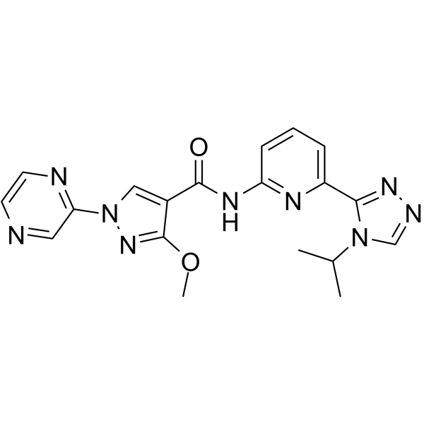
HY-133554
ASK1-IN-1ASK1 Inhibitor -
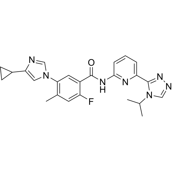
HY-18938
SelonsertibASK1 Inhibitor -
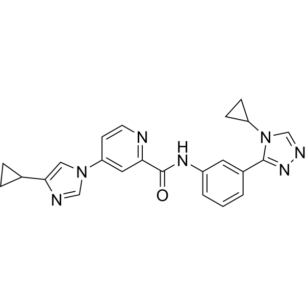
HY-100844
GS-444217ASK1 Inhibitor -
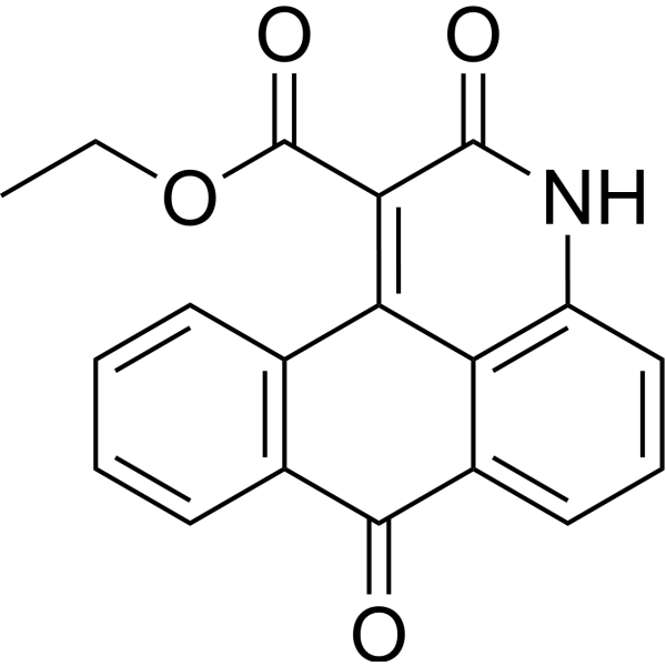
HY-19566
NQDI-1ASK1 Inhibitor -
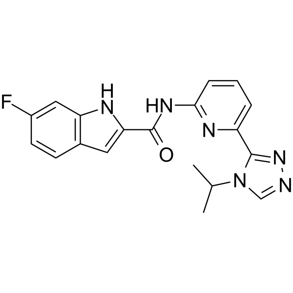
HY-131969
ASK1-IN-2ASK1 Inhibitor -
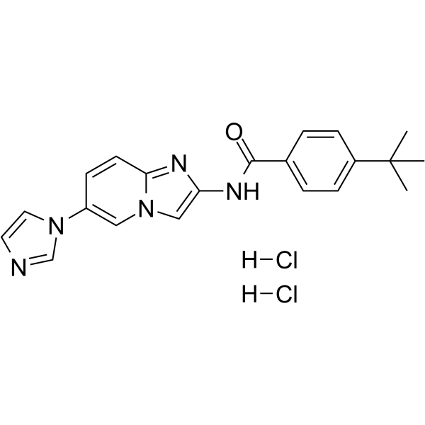
HY-103258
TC ASK 10ASK1 Inhibitor -

HY-100844R
GS-444217 (Standard)ASK1 Inhibitor -

HY-103258R
TC ASK 10 (Standard)Reference Standard -
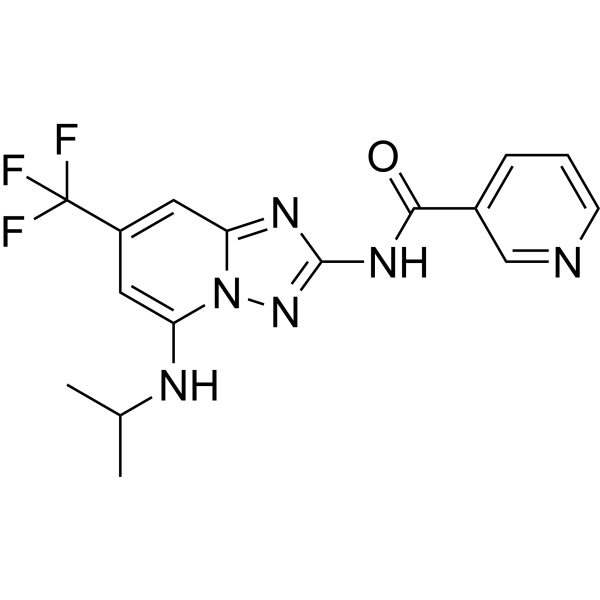
HY-18297
ASK1-IN-5ASK1 Inhibitor
All MAP3K Isoforms
More

Biological Activity
Description
IC50 & Target
|
ASK1 96 nM (IC50) |
Cellular Effect
|
Cell Line

|
Type | Value | Description | References |
|---|---|---|---|---|
| HEK-293T | IC50 |
13 nM
Compound: 3; MSC2032964A
|
Inhibition of V5 tagged human ASK1 expressed in HEK-293T cells assessed as reduction in T838 autophosphorylation incubated for 1 hr
Inhibition of V5 tagged human ASK1 expressed in HEK-293T cells assessed as reduction in T838 autophosphorylation incubated for 1 hr
|
[PMID: 31710475] |
In Vitro
MSC 2032964A (10 μM) reveals a good metabolic stability with microsomal clearance <4 ml/min/mg, and a good apparent permeability[1].
MSC 2032964A (10 μM) inhibits Lipopolysaccharide (LPS, HY-D1056)-induced ASK1 and p38 phosphorylation in mouse astrocytes[1].
MedChemExpress (MCE) has not independently confirmed the accuracy of these methods. They are for reference only. Further protocols information, click here.
-
Cell Line:mouse astrocytes
-
Concentration:10 μM
-
Incubation Time:90 min
-
Result:Inhibited phosphorylation of ASK1 and p38.
In Vivo
MSC 2032964A (30 mg/kg, p.o., once daily for 40 days) attenuates neurological symptoms in both spinal cord and optic nerve lesions, and thus ameliorates the MOG (35-55) (HY-P1240)-induced autoimmune encephalomyelitis in C57BL/6J mice model[1].
MedChemExpress (MCE) has not independently confirmed the accuracy of these methods. They are for reference only.
-
Animal Model:MOG (35-55)-induced autoimmune encephalomyelitis in C57BL/6J mice[1]
-
Dosage:30 mg/kg
-
Administration:p.o., once daily for 40 days
-
Result:Reduced demyelination in optic nerve, decreased astrocyte and microglia activation in spinal cord.


Chemical Information
-
CAS No. 1124381-43-6
-
Molecular Weight 362.31
-
Formula C16H13F3N6O
-
SMILES
O=C(C1=CC=CN=C1)NC2=NN3C(NC4CC4)=CC(C(F)(F)F)=CC3=N2
-
Shipping
Room temperature in continental US; may vary elsewhere.
-
Storage
Please store the product under the recommended conditions in the Certificate of Analysis.

Protocols
-
Apoptosis
Apoptosis, also called programmed cell death, is generally characterized by distinct morphological characteristics.
-
TUNEL staining for apoptotic DNA fragmentation
TUNEL staining detects DNA strand breaks by using terminal deoxynucleotidyl transferase to add labeled nucleotides to exposed 3′-OH DNA termini, generating either microscopic staining in fixed cells or tissue sections, or fluorescence/cytometric signal in cell suspensions. TUNEL positivity reflects DNA fragmentation but should not be interpreted alone as definitive apoptosis, because TUNEL can also label necrotic, autolytic, mechanically damaged, or DNA-repair-associated DNA breaks.
-
Cell Cytotoxicity Assay
Cytotoxicity assays are usually based on the assessment of cell membrane damage, which can also be indirectly detected by measuring cell viability. Detection methods include MTT assay, CKK-8 assay, LDH assay and ATP assay, etc.
-
Annexin V plus membrane-impermeant dye apoptosis staining
Annexin V-based apoptosis assays rely on the detection of phosphatidylserine (PS) externalization from the inner leaflet of the plasma membrane to the outer leaflet, an early biochemical hallmark of apoptosis. Fluorescently labeled Annexin V binds PS in a calcium-dependent manner, enabling identification of early apoptotic cells by flow cytometry or fluorescence microscopy. When combined with a membrane-impermeant DNA-binding dye (e. g. , propidium iodide), this approach allows discrimination between viable (Annexin V−/dye−), early apoptotic (Annexin V+/dye−), and late apoptotic or necrotic (Annexin V+/dye+) cell populations by assessing membrane integrity and PS exposure.
-
Apoptosis Solutions
Apoptosis is a regulated, generally non-lytic cell-death pathway that removes unwanted, damaged, infected, or abnormal cells through coordinated morphological changes, caspase activation, DNA fragmentation, and membrane remodeling. The intrinsic apoptosis pathway is controlled mainly by mitochondrial outer membrane permeabilization, BCL-2 family proteins, cytochrome c release, apoptosome formation, caspase-9 activation, and downstream executioner caspase-3/7 activation. The extrinsic apoptosis pathway is initiated by death receptors such as Fas, TNFR, and TRAIL receptors, which recruit adaptor proteins and activate caspase-8 before engaging executioner caspases or mitochondrial amplification through BID cleavage. Apoptosis is linked to many phenotypes, including cancer cell killing, tissue homeostasis, immune regulation, neurodegeneration, infection response, and treatment-induced cytotoxicity; unresolved questions include how apoptosis interacts with necroptosis, pyroptosis, ferroptos
-
Research Protocol for Neurological Diseases
PINK1/Parkin-mediated mitophagy pathway is a mitochondrial quality-control signaling axis in which mitochondrial depolarization stabilizes PINK1 on damaged mitochondria, activates Parkin recruitment and E3 ubiquitin ligase activity, promotes ubiquitination of outer mitochondrial membrane proteins, recruits selective autophagy adaptors, and drives lysosomal degradation of damaged mitochondria. In neurological disease research, this pathway is experimentally important because neurons, especially dopaminergic neurons, are highly dependent on mitochondrial integrity, and defective mitochondrial turnover can lead to mitochondrial dysfunction, oxidative stress, impaired neuronal survival, α-synuclein accumulation, and neuroinflammatory damage-associated signals. The genetic disease link is strongest in Parkinson’s disease because mutations in PRKN/parkin cause autosomal recessive juvenile parkinsonism, mutations in PINK1 cause hereditary early-onset Parkinson’s disease, and Drosophila studie


Purity & Documentation
References

Calculators
Concentration (start) × Volume (start) = Concentration (final) × Volume (final)


MSC 2032964A Related Classifications
HY-110262 Related Classifications
 Powered by Bioz