192 Results for "

visualize

" in MedChemExpress (MCE) Product Catalog:
Products (192)

192 Results for "visualize" in MCE Product Catalog:

Cat. No.: HY-P10978
CAS No.: 3137461-57-2
Target:  

PD-1/PD-L1

Research Areas:  

Cancer

NK224 is a peptide-based radiotracer targeting human PD-L1, with dual-radionuclide (68Ga and 18F) labeling compatibility enabled by the NOTA chelator. NK224 exhibits high binding affinity to PD-L1, with an IC50 value of 2.45 nM. NK224 visualizes tumor heterogeneity and dynamically monitors PD-L1 target occupancy during immunotherapy. NK224 can be used for the study of non-small cell lung cancer (NSCLC) .
loading...
    loading...
Cat. No.: HY-P11850
CAS No.: 1192023-11-2
Research Areas:  

Cancer

PTP peptide is a specific Plectin-1 binder. PTP peptide can serve as a component of imaging agents; when conjugated with magnetofluorescent nanoparticles, it enables the detection of small pancreatic ductal adenocarcinomas and precancerous lesions via in vivo confocal microscopy and MRI. When conjugated with imaging agents, PTP peptide specifically targets pancreatic ductal adenocarcinoma cells, and can be used for tumor visualization and compound delivery. PTP peptide is applicable to research related to pancreatic ductal adenocarcinoma .
loading...
    loading...
Cat. No.: HY-P11088F
VCAM1 binding peptide, FITC labeled acetate is a FITC-labeled VCAM1 binding peptide acetate (HY-P11088A). VCAM1 binding peptide acetate is a VCAM1 binder with internalization activity. VCAM1 binding peptide acetate serves as a component of biosensing systems for visualizing in vitro VCAM1 endocytic pathways. VCAM1 binding peptide acetate forms the research and development basis for MacroP and NAMP, PET radiotracers targeting VCAM1. VCAM1 binding peptide acetate is applicable to studies related to atherosclerosis .
loading...
    loading...
Cat. No.: HY-P11838
Research Areas:  

Cancer

DOTA-TH10 is a CSPG4-targeted probe that enables targeted enrichment in tumors with overexpressed CSPG4. 68Ga-radiolabeled DOTA-TH10 specifically binds to CSPG4, allowing visualization of CSPG4 expression. 68Ga-radiolabeled DOTA-TH10 exhibits significant tumor uptake capacity in pancreatic cancer and gastric cancer models. DOTA-TH10 can be used for research on pancreatic cancer and gastric cancer .
loading...
    loading...
Cat. No.: HY-P11858
Research Areas:  

Cancer

DP1 peptide is a high-affinity ROR1-binding peptide with a Kd value of 74.9 nM. When labeled with 68Ga, DP1 peptide serves as a PET imaging agent, which features rapid tumor uptake, favorable target-to-nontarget ratios in various tumor models, and renal clearance, enabling non-invasive quantitative visualization of ROR1 expression. DP1 peptide can be used in research related to melanoma, hepatocellular carcinoma, colorectal cancer, and non-small cell lung cancer .
loading...
    loading...
Cat. No.: HY-D3074
Research Areas:  

Others

TCM-4COOH is an AIE-active fluorescent probe for calcium ions. TCM-4COOH relies on aggregation-induced emission activity, and its carboxyl chelating group can bind to Ca 2+ and Mg 2+ to form insoluble aggregates, triggering fluorescence enhancement via restricting intramolecular motion (Ex/Em = 477/603 nm). TCM-4COOH shows pH independence in physiological environments (pH 4-9), can be strongly activated at Ca 2+ concentrations ranging from low nM to mM, and has a stronger binding ability to Ca 2+ than to Mg 2+. TCM-4COOH, as its acetoxymethyl ester derivative TCM-AM, can penetrate cell membranes for intracellular detection. TCM-4COOH can be used for imaging of cellular calcium overload, detection of bone microcracks, and studies related to calcium-rich biological environments .
loading...
    loading...
Cat. No.: HY-128536
CAS No.: 2436544-28-2
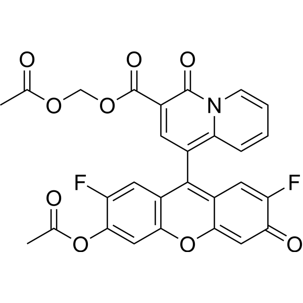
KMG-104AM is a Mg 2+ fluorescent probe. KMG-104AM is a membrane-permeable ester-modified derivative of KMG-104, which serves as a reporter and imaging agent. KMG-104AM can track the increase in intracellular free magnesium ion concentration induced by mitochondrial uncoupling. KMG-104AM enables visualization of the three-dimensional distribution of intracellular magnesium ion concentration. KMG-104AM is applicable to research related to the functions of intracellular magnesium ions .
loading...
    loading...
Cat. No.: HY-145746
CAS No.: 1782950-80-4
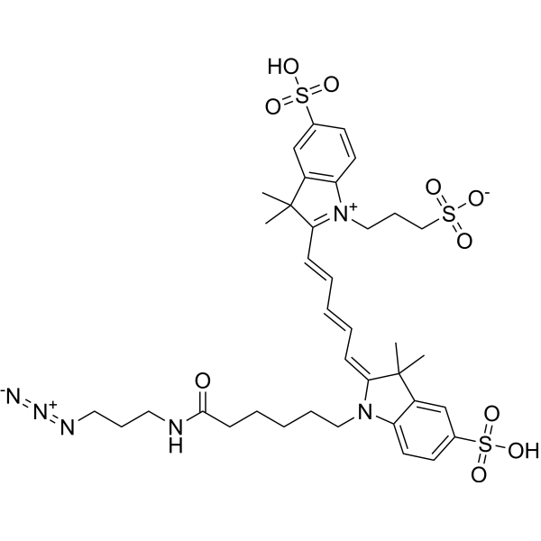
Sulfo-Cy5 azide is a near-infrared fluorescent probe with favorable click chemistry reactivity. Sulfo-Cy5 azide enables fluorescence imaging, tissue and cellular visualization of PD-L1 in tumors, and site-specific modification of anti-PD-L1 antibodies. Sulfo-Cy5 azide has been employed for RNA labeling and imaging. Sulfo-Cy5 azide can be conjugated to targeting agents for fluorescence imaging in atherosclerosis and breast cancer models (Ex/Em = 645/670 nm) .
loading...
    loading...
Cat. No.: HY-156124
CAS No.: 2860554-32-9
Target:  

Sigma Receptor

Research Areas:  

Cancer

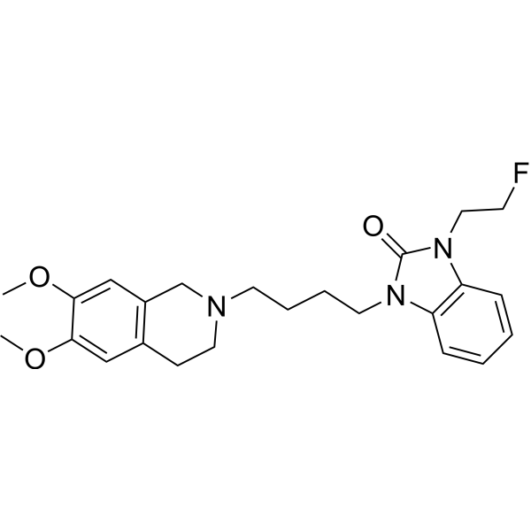
Sigma-2 Radioligand 1 (compound 1) is a Sigma-2 selective ligand. Sigma-2 Radioligand 1 has good biodistribution in mice and good in vivo activity in rats. [18F] Modified Sigma-2 Radioligand 1 for visualization of tumors in micro-PET/CT imaging, exhibiting high tumor uptake and tumor-to-background ratio. Experiments show that Sigma-2 Radioligand 1 binds highly specifically in U87MG glioma xenografts .
loading...
    loading...
Cat. No.: HY-D2004
CAS No.: 1430816-27-5
ATTO 488 streptavidin is a fluorescent reagent that specifically targets and binds to biotin (biotin), formed by the conjugation of ATTO 488 with streptavidin (HY-P3152). ATTO 488 streptavidin enables visualization of the activity of streptavidin immobilized on the surface of polymeric nanoparticles, or acts as a fluorescent probe to detect the selective binding and internalization process of anti-HB-EGF/NA with cells expressing HB-EGF (with no such effect on cells that do not express this receptor). ATTO 488 streptavidin effectively verifies the function of streptavidin conjugated to the surface of nanoparticles and is suitable for research related to atherosclerotic cardiovascular diseases .
loading...
    loading...
Cat. No.: HY-D2729
CAS No.: 3012597-48-4
Cy3-NO2-Tre is a nitroreductase-responsive cyanine-based fluorescent probe that specifically labels Mycobacterium tuberculosis (Mtb). Cy3-NO2-tre generated fluorescence after activation by a specific nitroreductase, Rv3368c, which is conserved in the Mycobacteriaceae. Cy3-NO2-tre effectively imaged mycobacteria within infected host cells, tracked the infection process, and visualized Mycobacterium smegmatis being endocytosed by macrophages (Ex/Em= 540-550/575-625 nm) .
loading...
    loading...
Cat. No.: HY-D3007
LD-CK is a fluorescent probe based on a coumarin-chromone structure, specifically designed to visualize lipid droplet polarity changes and endowed with multimodal imaging capability. LD-CK contains a high-performance lipophilic coumarin moiety, which enables it to specifically target lipid droplets upon cellular entry while minimizing background fluorescence to the greatest extent. LD-CK has an excitation wavelength (Ex) of 488 nm and an emission wavelength (Em) of 540 nm (in low-polarity environments such as toluene) or 640 nm (in high-polarity environments such as glycerol), with its emission peak undergoing a redshift as solvent polarity increases .
loading...
    loading...
Cat. No.: HY-D3251
LCP is a polar-responsive fluorescent probe for the cellular microenvironment. LCP responds to polarity changes in the cellular microenvironment via fluorescence resonance energy transfer, emitting blue fluorescence in low-polarity environments (Ex/Em = 420 nm/550 nm) and red fluorescence (Ex/Em = 500 nm/662 nm) in high-polarity environments. LCP enables dual-color visualization of dynamic changes in lysosomes and cytoplasmic membranes during cell apoptosis, and monitors cell viability through localization and emission color changes. LCP can be used in cancer research .
loading...
    loading...
Cat. No.: HY-K0611

MCE Modified Van Gieson Staining Kit employs Celestine Blue and Mayer’s hematoxylin for nuclear staining, providing clearer and more stable nuclear visualization while facilitating longer preservation of stained sections. Ponceau S is used for collagen fiber staining, offering stable coloration and strong resistance to fading. This method enables effective differentiation between collagen fibers and muscle fibers, and can assist in distinguishing collagen fiber–derived tumors from myogenic tumors to a certain extent. It is also suitable for observing tissue or organ injury, repair processes, and the degree of fibrosis.

loading...
    loading...
Cat. No.: HY-D3162
CAS No.: 2390002-31-8
Research Areas:  

Cancer

HMBI is a fluorescent probe that enables dual-color visualization of both the nucleus and mitochondria and monitors apoptosis. HMBI emits green fluorescence in the nucleus with Ex/Em=405/510-560 nm, and red fluorescence in mitochondria with Ex/Em=543/560-660 nm. The localization and emission properties of HMBI depend on subcellular pH and mitochondrial membrane potential. HMBI can serve as a feed additive for in vitro rumen fermentation; it is partially degraded, absorbed or converted into HMB by microorganisms in the system, so as to support microbial protein synthesis, alter the concentration of rumen metabolites and adjust the composition of bacterial communities .
loading...
    loading...
Cat. No.: HY-D3188
CAS No.: 2730023-28-4
gGlu-2OMe SiR600 is a γ-glutamyl transpeptidase (GGT) responsive fluorescent probe. gGlu-2OMe SiR600 can be converted into a highly fluorescent molecule via reaction with GGT, and its initial fluorescence is quenched through a photoinduced electron transfer (PeT) mechanism. gGlu-2OMe SiR600 exhibits fluorescence activation in malignant breast cancer and benign breast fibroadenoma tissues, enabling lesion visualization. gGlu-2OMe SiR600 can be used for research related to breast cancer and fibroadenoma .
loading...
    loading...
Cat. No.: HY-D3195
CAS No.: 2028316-19-8
CDr15 is a deep-red fluorescent probe (Ex=733 nm) that can selectively intercalate into and label bacterial extracellular DNA (eDNA). CDr15 exhibits extremely high specificity for biofilm imaging and cannot effectively bind to mammalian nuclear DNA. CDr15 enables real-time visualization of the microcolony structure and developmental process of three-dimensional *Pseudomonas aeruginosa* biofilms, and accurately localizes biofilm-forming regions of microorganisms in a mouse corneal infection model. With low background interference signals, CDr15 serves as an ideal diagnostic tool for research fields including bacterial biofilms and corneal infections .
loading...
    loading...
Cat. No.: HY-P11486
NOTA-RHAU18 is a conjugate of the chelator NOTA and the peptide RHAU18. NOTA-RHAU18 can be used to synthesize the radiolabeled peptide probe [ 18F]AlF-NOTA-RHAU18. [ 18F]AlF-NOTA-RHAU18 targets mitochondrial DNA G4. [ 18F]AlF-NOTA-RHAU18 can be used to visualize and detect solid tumors in homozygous mice. [ 18F]AlF-NOTA-RHAU18 can also be used in PET imaging studies .
loading...
    loading...
Cat. No.: HY-P12120
CAS No.: 499773-67-0
Synonyms: KCCYSL
Research Areas:  

Cancer

ErbB-2-binding peptide-2 (KCCYSL) is a peptide targeting ErbB-2/HER2, which specifically binds to the extracellular domain of HER2 and accumulates in HER2-expressing cancer cells. When radiolabeled, ErbB-2-binding peptide-2 serves as a SPECT/CT and PET imaging agent, enabling the visualization of HER2-expressing tumor xenografts. ErbB-2-binding peptide-2 is applicable to research related to ovarian cancer and breast cancer .
loading...
    loading...
Cat. No.: HY-103336
CAS No.: 1186195-59-4
Synonyms: T1117
Research Areas:  

Cancer

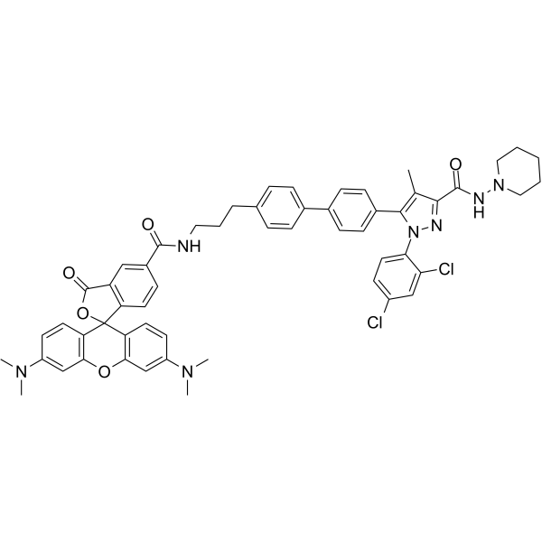
Tocrifluor 1117 is a selective GPR55 ligand and fluorescent probe. Tocrifluor 1117 shows extremely low affinity for mouse CB1 receptors (Ki >1000 nM) and only weakly displaces CB1 agonists. Tocrifluor 1117 specifically activates GPR55 to trigger intracellular calcium oscillations, and binds to vascular smooth muscle, endothelial and adventitial cells; all these binding effects are inhibited by pretreatment with unlabeled AM251 or O1602. Tocrifluor 1117 can clearly visualize the distribution of GPR55 in vascular tissues via confocal microscopy, while its uptake in cancer cells is completely dependent on GPR55 expression .
loading...
    loading...

