



Sulfo-Cy5 azide

Based on 1 Customer Validation
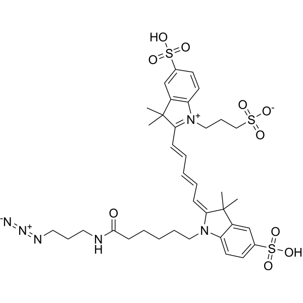
Sulfo-Cy5 azide is a near-infrared fluorescent probe with favorable click chemistry reactivity. Sulfo-Cy5 azide enables fluorescence imaging, tissue and cellular visualization of PD-L1 in tumors, and site-specific modification of anti-PD-L1 antibodies. Sulfo-Cy5 azide has been employed for RNA labeling and imaging. Sulfo-Cy5 azide can be conjugated to targeting agents for fluorescence imaging in atherosclerosis and breast cancer models (Ex/Em = 645/670 nm).
For research use only. We do not sell to patients.
- Purity : 98.97%
- CAS No.: 1782950-80-4
- Formula: C37H48N6O10S3
- Molecular Weight:833.01
-
Storage:
4°C, protect from light
* In solvent : -80°C, 6 months; -20°C, 1 month (protect from light)

To place orders, for customer services and technical support, please contact: MedChemExpress USA
Tel: 609-228-6898 E-mail: [email protected] [email protected]


-
 Biological Activity
-
Chemical Information
-
Solvent & Solubility
- Protocols
- Purity & Documentation
- References
-
 Help & FAQs





Biological Activity
Description
In Vitro
Sulfo-Cy5 azide covalently conjugates to alkyne-modified ADP-ribose units on ARTD1 and histone H1.2 via click chemistry, enabling fluorescent detection of ADP-ribosylation events catalyzed by ARTD1 with alkyne-containing NAD+ analogues[1].
Sulfo-Cy5 azide covalently conjugates to alkyne-functionalized TMV coat proteins via CuAAC[2].
MedChemExpress (MCE) has not independently confirmed the accuracy of these methods. They are for reference only. Further protocols information, click here.
Emission (Em)
670
Excitation (Ex)
645


Chemical Information
-
CAS No. 1782950-80-4
-
Appearance Solid
-
Molecular Weight 833.01
-
Formula C37H48N6O10S3
-
Color Brown to dark brown
-
SMILES
[N-]=[N+]=NCCCNC(CCCCCN1C2=CC=C(S(=O)(O)=O)C=C2C(C)(C)/C1=C\C=C\C=C\C3=[N+](CCCS(=O)([O-])=O)C4=C(C3(C)C)C=C(S(=O)(O)=O)C=C4)=O
-
Shipping
Room temperature in continental US; may vary elsewhere.
-
Storage
4°C, protect from light
* In solvent : -80°C, 6 months; -20°C, 1 month (protect from light)


Solvent & Solubility
In Vitro:
DMSO : 5 mg/mL (6.00 mM; Need ultrasonic; Hygroscopic DMSO has a significant impact on the solubility of product, please use newly opened DMSO)
Please refer to the solubility information to select the appropriate solvent. Once prepared, please aliquot and store the solution to prevent product inactivation from repeated freeze-thaw cycles.
Storage method and period of stock solution: -80°C, 6 months; -20°C, 1 month (protect from light). When stored at -80°C, please use it within 6 months. When stored at -20°C, please use it within 1 month.
Please refer to the solubility information to select the appropriate solvent. Once prepared, please aliquot and store the solution to prevent product inactivation from repeated freeze-thaw cycles.
Storage method and period of stock solution: -80°C, 6 months; -20°C, 1 month (protect from light). When stored at -80°C, please use it within 6 months. When stored at -20°C, please use it within 1 month.
Concentration (start) × Volume (start) = Concentration (final) × Volume (final)

Protocols
-
EdU Incorporation Assay (Click Chemistry-Based DNA Synthesis Measurement)
The EdU incorporation assay measures DNA synthesis by adding the thymidine analog 5-ethynyl-2′-deoxyuridine to cells or tissues, where it is incorporated into newly synthesized DNA during S phase. Incorporated EdU is detected by copper-catalyzed azide-alkyne cycloaddition, in which a fluorescent azide covalently reacts with the ethynyl group on EdU, allowing S-phase cells to be detected by fluorescence microscopy, flow cytometry, or high-content imaging. EdU detection does not require DNA denaturation or anti-BrdU antibody access, which preserves sample structure and improves compatibility with immunostaining and multiparameter cytometry compared with BrdU-based detection. EdU can be cytotoxic in a cell-type- and exposure-dependent manner, so pulse duration, concentration, and continuous-labeling designs should be validated for each cell type.
-
Research Protocol for Cardiovascular Diseases
Cardiovascular disease can be modeled as maladaptive cardiac remodeling, where ischemic injury or pressure overload activates inflammatory signaling, fibroblast activation, extracellular-matrix deposition, cardiomyocyte hypertrophy, vascular remodeling, and progressive ventricular dysfunction. The TGF-β/SMAD axis is a central profibrotic pathway after myocardial injury and pressure overload, while innate immune and cytokine pathways regulate leukocyte recruitment, scar formation, and adverse remodeling. Key unresolved questions include which inflammatory signals are reparative versus harmful, when fibrosis is protective versus maladaptive, and whether pathway inhibition improves function without weakening necessary infarct healing or compensatory remodeling.
-
Bioluminescent/Fluorescent Imaging Xenograft
Bioluminescent and fluorescent imaging xenograft models use tumor cells engineered to express optical reporters so tumor engraftment, growth, dissemination, and treatment response can be monitored longitudinally in living animals and validated ex vivo. Bioluminescence imaging usually measures luciferase activity after substrate administration and is commonly used as a surrogate for viable reporter-expressing tumor burden, while fluorescence imaging measures reporter or probe emission and can support tumor localization, ex vivo confirmation, or complementary multimodal analysis.
-
Breast Cancer Modeling
Breast cancer is a heterogeneous cancer, and it has been distinguished into four subtypes: luminal A, luminal B, HER2-positive and basal-like. Molecular mutations, epigenetic alterations, hormone exposure and immune microenvironment are related to the progression of breast cancer.


Purity & Documentation
-
Data Sheet (276 KB)
-
SDS (393 KB)
- English - EN (393 KB)
- Français - FR (393 KB)
- Deutsch - DE (393 KB)
- Norwegian - NO (393 KB)
- Español - ES (393 KB)
- Swedish - SV (393 KB)
- Italian - IT (393 KB)
- Korean - KR (393 KB)
- Portuguese - PT (393 KB)
-
Handling Instructions (2659 KB)
References
[1]. Wang Y, et al. Chain-terminating and clickable NAD⁺ analogues for labeling the target proteins of ADP-ribosyltransferases. Angew Chem Int Ed Engl. 2014;53(31):8159-8162. [Content Brief]
[2]. Bruckman MA, et al. Dual-modal magnetic resonance and fluorescence imaging of atherosclerotic plaques in vivo using VCAM-1 targeted tobacco mosaic virus. Nano Lett. 2014;14(3):1551-1558. [Content Brief]
[3]. Hagemans IM, et al. Multiscale imaging of therapeutic anti-PD-L1 antibody localization using molecularly defined imaging agents. J Nanobiotechnology. 2022;20(1):64. Published 2022 Feb 2. [Content Brief]


Complete Stock Solution Preparation Table
Please refer to the solubility information to select the appropriate solvent. Once prepared, please aliquot and store the solution to prevent product inactivation from repeated freeze-thaw cycles.
Storage method and period of stock solution: -80°C, 6 months; -20°C, 1 month (protect from light). When stored at -80°C, please use it within 6 months. When stored at -20°C, please use it within 1 month.
| Optional Solvent | Concentration Solvent Mass | 1 mg | 5 mg | 10 mg | 25 mg |
|---|---|---|---|---|---|
| DMSO | 1 mM | 1.2005 mL | 6.0023 mL | 12.0047 mL | 30.0116 mL |
| 5 mM | 0.2401 mL | 1.2005 mL | 2.4009 mL | 6.0023 mL |


Sulfo-Cy5 azide Related Classifications
HY-145746 Related Classifications
 Powered by Bioz