3 Results for "

MitoTracker Deep Red

" in MedChemExpress (MCE) Product Catalog:
Products (3)

3 Results for "MitoTracker Deep Red" in MCE Product Catalog:

18
18 Cited Publications
Cat. No.: HY-D1783
CAS No.: 873315-86-7
Synonyms: MTDR FM
Target:  

Fluorescent Dye

Research Areas:  

Cancer

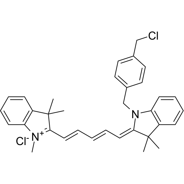
MitoTracker Deep Red (MTDR) FM fluorescent dye that selectively accumulates in the mitochondrial matrix. MitoTracker Deep Red FM covalently binds mitochondrial proteins by reacting with free mercaptan of cysteine residues, allowing staining of mitochondrial membrane potential independent of membrane potential. Excitation/emission wavelength 644/665 nm . MitoTracker Deep Red dyes have an excitation/emission wavelength of 633/650-750 nm .
The Ex/Em of MitoTracker Deep Red (MTDR) FM is 644/665 nm.
loading...
    loading...
Cat. No.: HY-DY1105
CAS No.: 873315-86-7
Synonyms: MTDR FM (solution)
Target:  

Fluorescent Dye

Research Areas:  

Others

MitoTracker Deep Red FM (solution) (MTDR FM (solution)) selectively accumulates in the mitochondrial matrix. MitoTracker Deep Red FM covalently binds mitochondrial proteins by reacting with free mercaptan of cysteine residues, allowing staining of mitochondrial membrane potential independent of membrane potential. The Ex/Em of MitoTracker Deep Red (MTDR) FM is 644/665 nm.
Solvent and concentration: DMSO: 1 mM
loading...
    loading...
Cat. No.: HY-N16300
CAS No.: 3091879-75-0
Target:  

Fluorescent Dye

Research Areas:  

Others

Mito-laurdan (bromide) is a Fluorescent dye for mitochondrial membrane fluidity and packing measurement. It depends on membrane potential for targeting: it contains a cationic triphenylphosphonium moiety that accumulates at the inner mitochondrial membrane due to the membrane's negative potential, connected via a 3-carbon linker. As a solvatochromic dye derived from Laurdan (HY-D0080), it retains the characteristic mechanism where fluorescence emission shifts based on membrane packing: in more fluid, loosely packed membranes, greater water penetration leads to increased dipolar relaxation and a bathochromic red shift in emission, while more ordered membranes show a blue-shifted emission; these shifts are quantified using generalized polarization, calculated from emission intensities in ordered and disordered spectral windows. Its excitation wavelength is 352 nm, with emission detected in ordered (409-463 nm) and disordered (473-516 nm) spectral windows. It localizes specifically to mitochondria in HeLa cells, showing high signal overlap with MitoTracker Deep Red, and can detect changes in inner mitochondrial membrane fluidity .
loading...
    loading...

