



Simtuzumab

Based on 1 Customer Validation
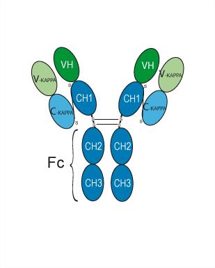
Simtuzumab (AB 0024; GS 6624) is a monoclonal antibody directed against Lysyl oxidase like-2 (LOXL2). Simtuzumab non-competitively blocks collagen cross-linking, reduces LOXL2 protein expression and attenuates extracellular matrix changes. Simtuzumab reduces myocardial fibrosis and prevents cardiac dysfunction. Simtuzumab lowers Myh7 and Nppa gene expression, reduces contraction heterogeneity, and cuts COL1A1 deposition. Simtuzumab can be used for the research of LMNA mutation-induced dilated cardiomyopathy, idiopathic pulmonary fibrosis, and primary sclerosing cholangitis.
For research use only. We do not sell to patients.
- Purity : 99.35%
- CAS No.: 1318075-13-6
- Molecular Weight:145.5 kDa
-
Storage:
Please store the product under the recommended conditions in the Certificate of Analysis.


To place orders, for customer services and technical support, please contact: MedChemExpress USA
Tel: 609-228-6898 E-mail: [email protected] [email protected]


-
 Biological Activity
-
Chemical Information
- Protocols
- Purity & Documentation
- References
-
 Help & FAQs


-

HY-P99288
FG-3019Anti-Human CTGF Recombinant Antibody -
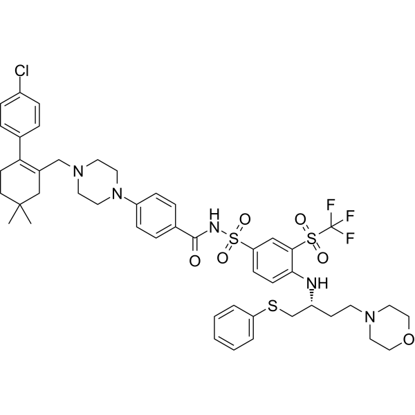
HY-10087
NavitoclaxBcl-2 Inhibitor -

HY-Y0882
Hydroxylamine hydrochlorideMAO Inhibitor -
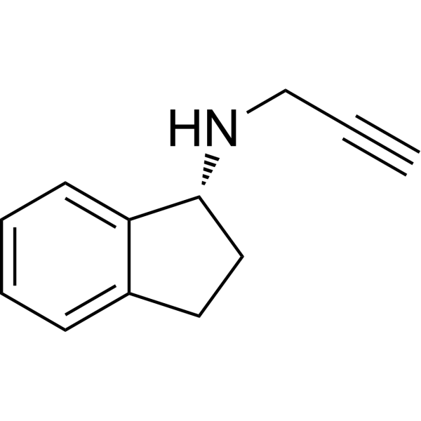
HY-14605A
RasagilineMAO Inhibitor -
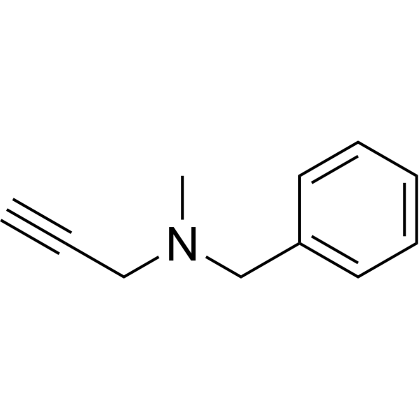
HY-A0091A
PargylineMonoamine Oxidase Inhibitor -
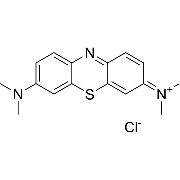
HY-14536
Methylene BluesGC Inhibitor -
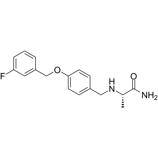
HY-70057
SafinamideMAO-B Inhibitor -
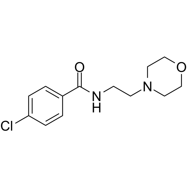
HY-B0534
MoclobemideMAOI Antidepressant -
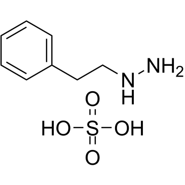
HY-B1018A
Phenelzine sulfateMAO Inhibitor -
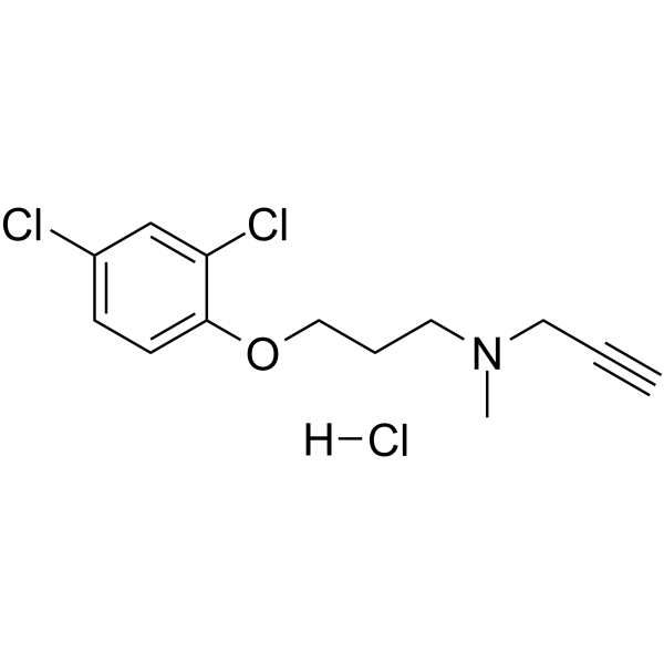
HY-14197A
Clorgyline hydrochlorideMAO-A Inhibitor -

HY-P9902
PembrolizumabPD-1 Inhibitor -
HY-N2554
OsthenolhMAO-A Inhibitor


Biological Activity
Description
Isotype
Human IgG4 kappa
Recommend Isotype Controls
Species Reactivity
Human
IC50 & Target
LOXL2
In Vitro
Simtuzumab (1 µM; 15 days) reduces contraction heterogeneity, contraction time, diastolic time, and COL1A1 deposition area in LMNAH222P hiPSC-CMs, without altering contraction amplitude[1].
Simtuzumab (1-50 μg/mL; 72 h) enhances myofibroblast differentiation in primary IPF human lung fibroblasts at 50 μg/mL, particularly with IL-13 stimulation, and induces mild differentiation in primary normal human lung fibroblasts at the same dose without additional stimuli[2].
Simtuzumab (50 μg/mL; 7 day) does not reduce or alter collagen 1 synthesis in primary normal or IPF human lung fibroblasts, even with pro-fibrotic stimuli[2].
Simtuzumab (50 μg/mL; 96 h) increases the invasive migration of primary normal and IPF human lung fibroblasts in a scratch wound assay[2].
Simtuzumab (50 μg/mL; 26 days, replenished twice weekly) accelerates the outgrowth of primary fibroblasts from mixed normal and IPF human lung explant cells and increases the expression of fibrosis-related genes in these cultures[2].
Simtuzumab (50 μg/mL; 24 h) increases αSMA and Collagen 3 transcript expression in SSEA4+-enriched mesenchymal progenitors from normal human lung samples[2].
Simtuzumab (50 μg/mL; 10 days, replenished every 3 days) increases LOXL2 and PDGFRA transcript expression and induces mesenchymal-like morphological changes in mixed primary cells from normal and IPF human lung explants[2].
Simtuzumab inhibits human LOXL2 enzymatic activity, blocks fibroblast activation, and reduces LOXL2-mediated extracellular matrix modifications in in vitro assays[4].
MedChemExpress (MCE) has not independently confirmed the accuracy of these methods. They are for reference only. Further protocols information, click here.
-
Cell Line:primary IPF human lung fibroblasts
-
Concentration:1 μg/mL; 10 μg/mL; 50 μg/mL
-
Incubation Time:72 h
-
Result:Enhanced myofibroblast differentiation at 50 μg/mL with IL-13 stimulation.
Induced mild differentiation in primary normal human lung fibroblasts at 50 μg/mL without additional stimuli.
-
Cell Line:primary normal and IPF human lung fibroblasts
-
Concentration:50 μg/mL
-
Incubation Time:7 days
-
Result:Did not reduce or alter collagen 1 synthesis.
-
Cell Line:primary normal and IPF human lung fibroblasts
-
Concentration:50 μg/mL
-
Incubation Time:96 h
-
Result:Increased the invasive migration.
In Vivo
Simtuzumab (15 mg/kg; i.v.; twice a week; 35 days (preventative); 28 days (therapeutic)) fails to modulate lung fibrosis in a humanized NSG mouse model of idiopathic pulmonary fibrosis, with no significant differences in key fibrotic endpoints compared to IgG control, though it increases lung hydroxyproline levels relative to naive or cell-only groups and elevates SCA1+ mesenchymal progenitors in the preventative regimen[2].
MedChemExpress (MCE) has not independently confirmed the accuracy of these methods. They are for reference only.
-
Animal Model:LmnaH222P/H222P (male, 4 months old at treatment initiation, homozygous genetic model with established cardiac disease phenotype)[1]
-
Dosage:5 mg/kg
-
Administration:i.p.; 3× weekly; 1 month
-
Result:Reduced Loxl2 protein expression in mouse hearts.
Decreased myocardial fibrosis as shown by Sirius red staining.
Stabilized left ventricular end-diastolic diameter (LVEDd) at ~3.5 mm from 4 to 5 months, compared to an increase from 3.5 mm to 4.1 mm in untreated mice.
Preserved ejection fraction (EF) from 70.9% at 4 months to 67.1% at 5 months, compared to a decline from 67.8% to 50.0% in untreated mice.
Maintained fractional shortening from 39.6% at 4 months to 36.5% at 5 months, compared to a decline from 37.2% to 25.1% in untreated mice.
Lowered mRNA expression of cardiac stress markers Myh7 and Nppa in mouse hearts.
-
Animal Model:NOD Cg-PrkdcSCID IL2rgTm1wil Szi (NSG) (6- to 8-week-old, female, pathogen-free, humanized model induced by intravenous injection of ~1×10⁶ IPF lung cells)[2]
-
Dosage:15 mg/kg
-
Administration:i.v.; twice a week; 35 days (preventative); 28 days (therapeutic)
-
Result:Significantly increased lung hydroxyproline levels compared to mice that received IPF cells alone in the preventative treatment group, but showed no difference compared to IgG-treated mice.
Significantly increased total lung hydroxyproline levels compared to naive mice in the therapeutic treatment group, but showed no difference compared to IgG-treated mice.
Significantly increased SCA1+ mesenchymal progenitors in bronchoalveolar lavage fluid compared to IgG-treated mice at day 35 of the preventative regimen.
Showed no significant differences compared to IgG-treated groups in fibrosis-related lung transcripts, αSMA+ cell counts, collagen intensity, or histological appearance at day 63.
Clinical Trial
| NCT Number | Sponsor | Condition | Start Date |
Phase

|
|---|---|---|---|---|
| NCT01329991 | Plexxikon| | 2011-05 | PHASE1 |
Gene ID
Accession
Conjugated
Unconjugated
Reconsititution
The product can be reconstituted/diluted with sterile PBS or saline.
Format
-
Human IgG4 kappa
Application
ELISA, FACS, Functional assay
Verified Bioactivity
-
 Immobilized LOXL2 Protein, Human (HEK293, His, HY-P74770) can bind Simtuzumab. The EC50 for this effect is 9.679 ng/mL.
 -
 Flow cytometric analysis of 1X10⁶ A549 cells with Simtuzumab (HY-P99047, red). Cells were fixed with 4% paraformaldehyde. Then stained with the primary antibody at 1/200 dilution for an hour at 4℃. AF 488-conjugated AffiniPure Goat Anti-Human IgG H&L (HY-P83776) was used as the secondary antibody at 1/1,000 dilution for 30 minutes at 4℃. Human IgG4 kappa (HY-P99003, blue) was used as the isotype control.


Chemical Information
-
CAS No. 1318075-13-6
-
Appearance Liquid
-
Molecular Weight 145.5 kDa
-
Color Colorless to light yellow
-
SMILES
[Simtuzumab]
-
Synonyms
AB 0024; GS 6624
-
Shipping
Shipping with dry ice.
-
Formulation
Please refer to the lot-specific COA for specific buffer information.
-
Storage
Please store the product under the recommended conditions in the Certificate of Analysis.

Protocols
-
Fibrosis/Collagen Morphometry
Fibrosis and collagen morphometry is based on the quantitative visualization of fibrillar collagen deposition in tissue sections using histochemical stains such as Sirius Red (Picrosirius Red) or Masson's trichrome, followed by image-based or polarization-enhanced analysis to estimate collagen proportional area as a surrogate of extracellular matrix accumulation during fibrotic remodeling. Sirius Red combined with polarized light microscopy enhances detection of collagen fibers due to birefringence properties, enabling more specific visualization of collagen type I and III fibrils compared to conventional bright-field histology, while whole-section or region-restricted digital morphometry reduces field-selection bias in fibrosis assessment. Alternative quantitative approaches include second harmonic generation (SHG) and two-photon excited fluorescence microscopy, which enable label-free detection of fibrillar collagen and have been validated against histological staining and biochemica
-
RT-PCR
Reverse transcription technology uses RNA as a template to synthesize DNA. RT-PCR is simple, specific and sensitive, and can be used to detect gene expression levels and expression differences in cells; detect RNA virus content; clone cDNA sequences of specific genes.
-
RNA extraction experimental
By lysing cells, releasing RNA, and removing impurities such as proteins and DNA, high-purity RNA products are finally obtained. The commonly used traditional method is the guanidine isothiocyanate/phenol/chloroform method (Trizol), which is suitable for a variety of animal materials including animal tissues, microorganisms, cultured cells, etc., and most plant materials.
-
Western Blot
Western blotting (WB) is a commonly used experimental method in molecular biology, biochemistry, and immunogenetics for identifying and quantifying target proteins. It combines gel electrophoresis with immunoassay, enabling researchers to analyze protein expression, post-translational modifications, and molecular weight.
-
Collagen: Sirius Red Staining
Sirius Red or picrosirius red staining is a histochemical method for visualizing collagen-rich extracellular matrix in tissue sections, and collagen fibers are detected as red-stained structures under bright-field microscopy with enhanced birefringence under polarized light. Picrosirius red is useful for assessing total collagen organization, distribution, and fibrosis burden, but polarized color should not be interpreted as a definitive collagen type I versus type III readout because color is affected by fiber orientation, thickness, and packing.
-
ECM-Embedded Organoid (Matrigel/Dome) Culture
ECM-embedded organoid dome culture embeds epithelial stem cells, crypts, organoid fragments, or tumor-derived epithelial cells in a basement-membrane-like hydrogel such as Matrigel, allowing 3D growth, self-organization, lumen formation, budding or cystic morphogenesis, and lineage maintenance under defined niche-factor-containing medium. The primary readouts are organoid establishment efficiency, growth, morphology, passaging capacity, lineage-marker expression, and, when fluorescently labeled lines are used, microscopy- or flow-cytometry-based quantification of population behavior in 3D culture.
-
Connective Tissue: Masson's Trichrome/Collagen Trichrome Staining
Masson’s Trichrome (collagen/trichrome staining) is a histological technique that differentially stains tissue compartments using sequential acidic dyes to distinguish collagen from muscle and cytoplasmic components based on dye affinity and tissue permeability differences, enabling visualization of fibrosis and connective tissue architecture in histological sections. The classical formulation typically uses Weigert's iron hematoxylin for nuclear staining, Biebrich scarlet-acid fuchsin for cytoplasm and muscle, and aniline blue (or light green variants) for collagen, producing a characteristic blue/green collagen signal contrasted against red cytoplasm and dark nuclei. The staining principle relies on selective displacement of smaller dye molecules by larger anionic dyes in collagen-rich regions under controlled acidified conditions, which enhances collagen-specific dye retention. This property makes the method widely used for fibrosis assessment in organs such as heart, liver, lung, a
-
Matrigel/ECM Transwell Invasion Assay
The Matrigel/ECM Transwell invasion assay measures the ability of cells to move toward a chemoattractant while crossing an extracellular-matrix barrier placed on a porous membrane; therefore, the readout reflects both chemotactic motility and matrix invasion rather than migration alone. Matrigel is a basement-membrane-rich matrix derived from Engelbreth-Holm-Swarm mouse sarcoma and has been used as a reconstituted basement membrane barrier in chemoinvasion assays. The assay readout is generated by quantifying cells that reach the underside of the insert membrane or lower compartment after incubation, commonly by staining and counting invaded cells or by fluorescence-based quantification.
-
Real Time qPCR (Q-PCR)
Real-time quantitative PCR (qPCR) quantifies an amplifiable nucleic-acid target by monitoring fluorescence during PCR cycling rather than measuring product only after amplification. The increase in fluorescence tracks accumulation of PCR product, and the quantification cycle (Cq; historically also Ct/CP) is related to the initial amount of target: samples containing more starting target generally reach the defined fluorescence threshold in fewer cycles.
-
Genotoxicity/Mutagenicity Study
The bacterial reverse mutation assay detects point mutations that restore amino-acid prototrophy in auxotrophic Salmonella typhimurium or Escherichia coli tester strains; after exposure to a test article, mutagenic activity is read out as an increased number of revertant colonies on minimal agar compared with the vehicle control. The assay uses tester strains with different mutation targets so that base-substitution and frameshift mutagens can be detected, and testing is performed with and without exogenous mammalian metabolic activation because some chemicals require biotransformation to become mutagenic.
-
Protocol For Protein Expression And Purification
Recombinant protein expression in Escherichia coli followed by purification of a His-tagged soluble protein by immobilized metal affinity chromatography (IMAC), with optional MBP fusion and TEV tag removal when the construct includes these elements. The biological readout is production of the encoded target protein, detected as an inducible band at the expected molecular mass by SDS-PAGE and quantified by total protein assay or chromatographic absorbance; the purification readout is enrichment of the target protein in elution fractions after selective binding of polyhistidine residues to immobilized Ni2+/metal-chelate resin and elution by imidazole-containing buffer. Expression is driven by an inducible bacterial expression system, commonly T7/lac-based, in which IPTG or lactose/auto-induction activates transcription and translation of the cloned gene; lower induction temperature, lower inducer concentration, induction timing, and solubility-enhancing fusion tags can influence the frac
-
Matrigel Transwell/Boyden Chamber Invasion Assay
Matrigel Transwell/Boyden chamber invasion assay measures the ability of cells to degrade or traverse an extracellular matrix-coated porous membrane and move from an upper chamber toward a chemoattractant in a lower chamber. Invasion is distinguished from migration by coating the membrane with Matrigel or basement membrane matrix; uncoated inserts measure migration, while coated inserts require cells to cross an ECM barrier before reaching the underside of the membrane.


Purity & Documentation
-
Data Sheet (279 KB)
-
SDS (251 KB)
- English - EN (251 KB)
- Français - FR (251 KB)
- Deutsch - DE (251 KB)
- Norwegian - NO (251 KB)
- Español - ES (251 KB)
- Swedish - SV (251 KB)
- Italian - IT (251 KB)
- Korean - KR (251 KB)
- Portuguese - PT (251 KB)
-
Inhibitory Antibodies User Guide (603 KB)
References
[1]. Kervella M, et al. Simtuzumab Attenuates Loxl2-Mediated Extracellular Matrix Remodeling and Preserves Cardiac Function in LMNA Mutation-Induced Dilated Cardiomyopathy. Circ Heart Fail. 2026;19(4):e013806. [Content Brief]
[2]. Espindola MS, et al. Translational Studies Reveal the Divergent Effects of Simtuzumab Targeting LOXL2 in Idiopathic Pulmonary Fibrosis. Fibrosis (Hong Kong). 2023;1(2):10007. [Content Brief]
[3]. Harrison SA, et al. Simtuzumab Is Ineffective for Patients With Bridging Fibrosis or Compensated Cirrhosis Caused by Nonalcoholic Steatohepatitis. Gastroenterology. 2018;155(4):1140-1153. [Content Brief]
[4]. Verstovsek S, et al. A phase 2 study of simtuzumab in patients with primary, post-polycythaemia vera or post-essential thrombocythaemia myelofibrosis. Br J Haematol. 2017;176(6):939-949. [Content Brief]
[5]. Chen W, et al. Lysyl Oxidase (LOX) Family Members: Rationale and Their Potential as Therapeutic Targets for Liver Fibrosis. Hepatology. 2020;72(2):729-741. [Content Brief]

Calculators
Concentration (start) × Volume (start) = Concentration (final) × Volume (final)


Simtuzumab Related Classifications
HY-P99047 Related Classifications
 Powered by Bioz
Keywords
- Simtuzumab
- 1318075-13-6
- AB 0024
- GS 6624
- AB0024
- AB-0024
- GS6624
- GS 6624
- GS-6624
- Monoamine Oxidase
- collagen cross-linking
- myocardial fibrosis
- LMNA mutation-induced dilated cardiomyopathy
- idiopathic pulmonary fibrosis
- hiPSC-CMs
- mouse LOXL2
- human LOXL2
- primary sclerosing cholangitis
- LOXL2
- fibroblast activation
- Inhibitor
- inhibitor
- inhibit