



Siramesine
Based on 6 publication(s) in Google Scholar
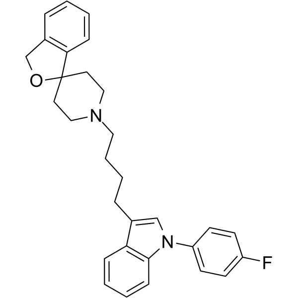
Siramesine (Lu 28-179) is a potent sigma-2 receptor agonist. Siramesine has a subnanomolar affinity for sigma-2 receptors (IC50=0.12 nM) and exhibits a 140-fold selectivity for sigma-2 receptors over sigma-1 receptors (IC50=17 nM). Siramesine triggers cell death through destabilisation of mitochondria, but not lysosomes. Anti-cancer activity.
For research use only. We do not sell to patients.
- Purity : 98.98%
- CAS No.: 147817-50-3
- Formula: C30H31FN2O
- Molecular Weight:454.58
-
Storage:Pure form -20°C, 3 years , 4°C, 2 years ; In solvent -80°C, 6 months , -20°C, 1 month

To place orders, for customer services and technical support, please contact: MedChemExpress USA
Tel: 609-228-6898 E-mail: [email protected] [email protected]


-
 Biological Activity
-
Chemical Information
- Protocols
- Purity & Documentation
- References
-
 Help & FAQs


-
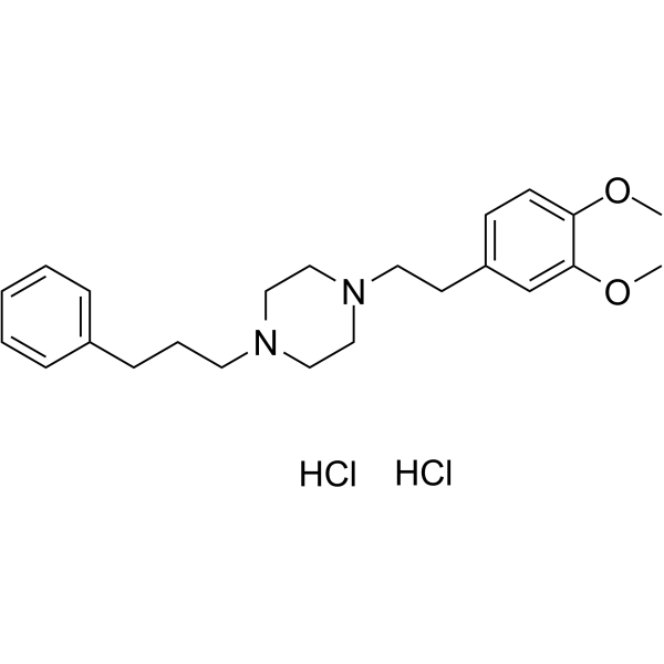
HY-13510
Cutamesine dihydrochlorideσ1R Agonist -
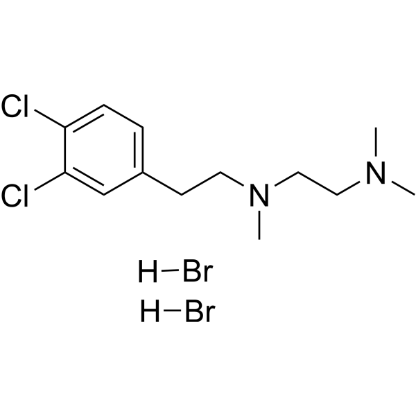
HY-16996A
BD-1047 dihydrobromideSigma-1 Receptor Antagonist -
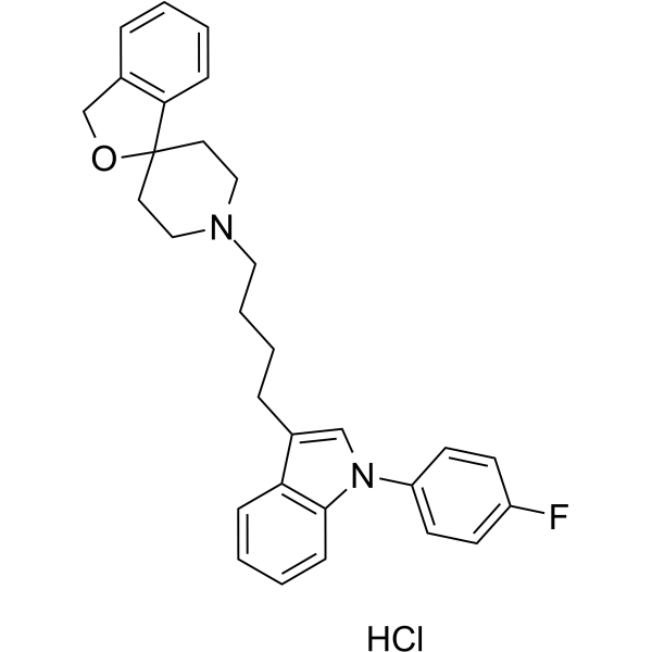
HY-14221A
Siramesine hydrochlorideSigma-2 Receptor Agonist -
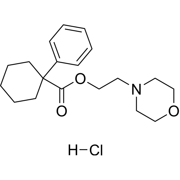
HY-18100A
PRE-084 hydrochlorideS1R Agonist -
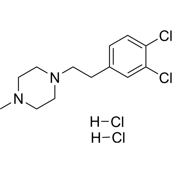
HY-18101A
BD-1063 dihydrochlorideσ-1R Antagonist -
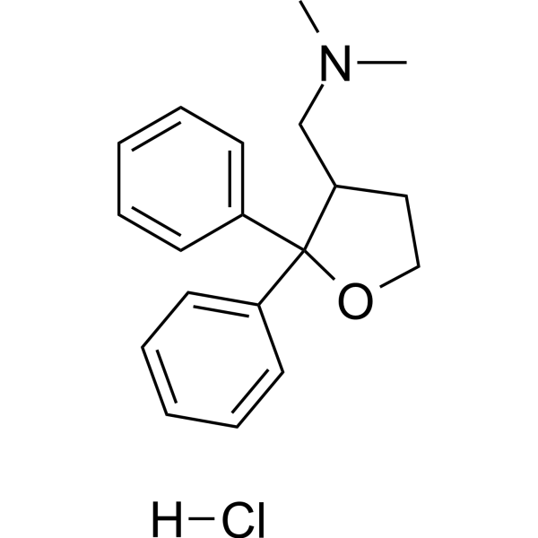
HY-101864
Blarcamesine hydrochlorideSigma-1 Receptor Agonist -
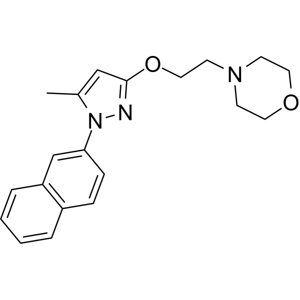
HY-18099
S1RAσ1R Antagonist -
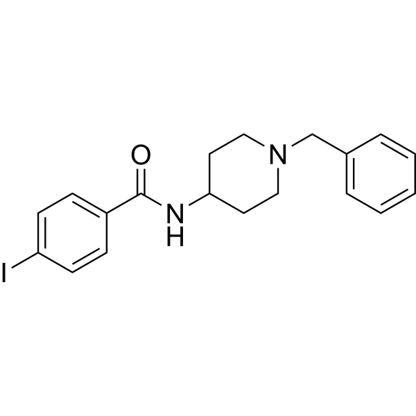
HY-100155
4-IBPσ1 Receptor Agonist -
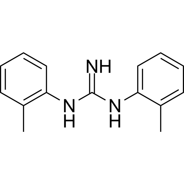
HY-14218
DitolylguanidineSigma Receptor Agonist -
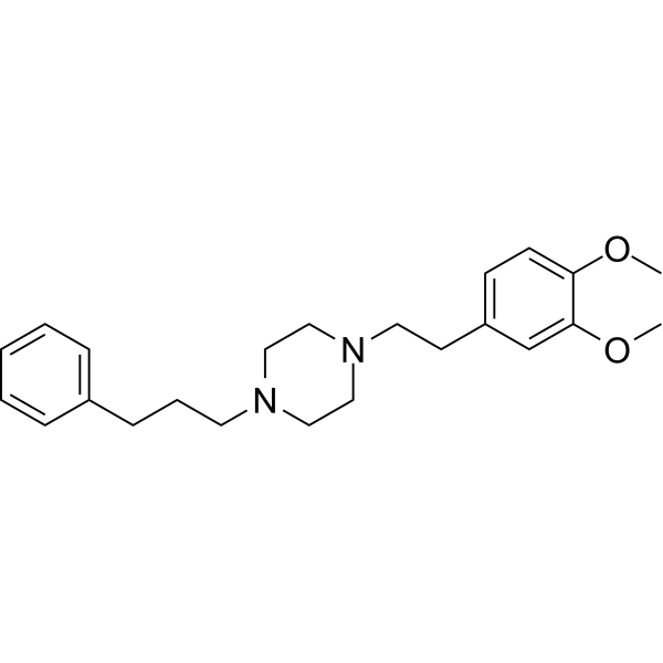
HY-14813
Cutamesineσ1R Agonist -

HY-14218R
Ditolylguanidine (Standard)Sigma Receptor Agonist -
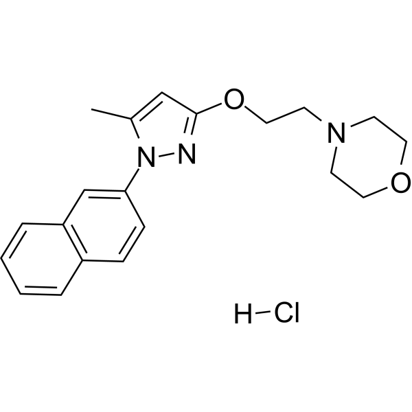
HY-18099A
S1RA hydrochlorideσ1R Antagonist
Publications Citing Use of MedChemExpress (MCE) Siramesine
MoreAll Sigma Receptor Isoforms
More

Biological Activity
Description
IC50 & Target
|
Sigma 1 Receptor |
Cellular Effect
|
Cell Line

|
Type | Value | Description | References |
|---|---|---|---|---|
| C6 | EC50 |
43.1 μM
Compound: Lu-28-179
|
Antiproliferative activity against rat C6 cells assessed as reduction in cell viability after 24 hrs by MTT assay
Antiproliferative activity against rat C6 cells assessed as reduction in cell viability after 24 hrs by MTT assay
|
10.1039/C5MD00079C |
| DU-145 | EC50 |
13.9 μM
Compound: Lu-28-179
|
Antiproliferative activity against human DU145 cells assessed as reduction in cell viability after 24 hrs by MTT assay
Antiproliferative activity against human DU145 cells assessed as reduction in cell viability after 24 hrs by MTT assay
|
10.1039/C5MD00079C |
| EMT6 | EC50 |
14.9 μM
Compound: 6
|
Antagonist activity at sigma-2 receptor in mouse EMT6 cells assessed as inhibition of cell proliferation after 24 hrs by MTS assay
Antagonist activity at sigma-2 receptor in mouse EMT6 cells assessed as inhibition of cell proliferation after 24 hrs by MTS assay
|
[PMID: 24821398] |
| EMT6 | EC50 |
5.3 μM
Compound: 6
|
Antagonist activity at sigma-2 receptor in mouse EMT6 cells assessed as inhibition of cell proliferation after 48 hrs by MTS assay
Antagonist activity at sigma-2 receptor in mouse EMT6 cells assessed as inhibition of cell proliferation after 48 hrs by MTS assay
|
[PMID: 24821398] |
| MCF7 | EC50 |
12.3 μM
Compound: Lu-28-179
|
Antiproliferative activity against human MCF7 cells assessed as reduction in cell viability after 48 hrs by MTT assay
Antiproliferative activity against human MCF7 cells assessed as reduction in cell viability after 48 hrs by MTT assay
|
10.1039/C5MD00079C |
| MCF7 | EC50 |
23.6 μM
Compound: Lu-28-179
|
Antiproliferative activity against human MCF7 cells assessed as reduction in cell viability after 24 hrs by MTT assay
Antiproliferative activity against human MCF7 cells assessed as reduction in cell viability after 24 hrs by MTT assay
|
10.1039/C5MD00079C |
| MCF7 | IC50 |
13 μM
Compound: Srms
|
Cytotoxicity against ER/PR-positive HER2-negative human MCF7 cells harbouring p53 R175H mutant assessed as inhibition of cell growth incubated for 48 hrs by resazurin dye based assay
Cytotoxicity against ER/PR-positive HER2-negative human MCF7 cells harbouring p53 R175H mutant assessed as inhibition of cell growth incubated for 48 hrs by resazurin dye based assay
|
[PMID: 35724925] |
| SK-BR-3 | IC50 |
14 μM
Compound: Srms
|
Cytotoxicity against ER/PR-negative HER2-positive human SK-BR-3 cells harbouring wild type p53 assessed as inhibition of cell growth incubated for 48 hrs by resazurin dye based assay
Cytotoxicity against ER/PR-negative HER2-positive human SK-BR-3 cells harbouring wild type p53 assessed as inhibition of cell growth incubated for 48 hrs by resazurin dye based assay
|
[PMID: 35724925] |
In Vitro
Siramesine displays the binding affinities: IC50 (sigma 1)=17 nM, IC50 (sigma 2)=0.12 nM, IC50 (5-HT1A)=21000 nM, IC50 (5-HT1A)=2000 nM, IC50 (D2)=800 nM, IC50 (alpha 1)=330 nM[1].
Siramesine (0-50μM; 8 hours) induces cell death in various cell lines (HaCaT, Hsc-4, HeLa and MCF-7, neuroblastoma cell line SH-SY5Y and glioblastoma cell line U-87MG)[2].
Siramesine (0-40 μM; 2-48 hours) activates caspases in HaCaT and in U-87MG cells[2].
MedChemExpress (MCE) has not independently confirmed the accuracy of these methods. They are for reference only. Further protocols information, click here.


Chemical Information
-
CAS No. 147817-50-3
-
Appearance Oil
-
Molecular Weight 454.58
-
Formula C30H31FN2O
-
Color Colorless to light yellow
-
SMILES
FC1=CC=C(N2C=C(CCCCN3CCC4(CC3)OCC5=C4C=CC=C5)C6=C2C=CC=C6)C=C1
-
Synonyms
Lu 28-179
-
Shipping
Room temperature in continental US; may vary elsewhere.
-
Storage
Pure form -20°C 3 years 4°C 2 years In solvent -80°C 6 months -20°C 1 month


Publications (6)
-
Journal Impact Factor
-
Most Recent
-
J Am Chem Soc
Fluorinated Ribonucleocarbohydrate Nanoparticles Allow Ultraefficient mRNA Delivery and Protein Expression in Tumor-Associated Myeloid Cells. [Abstract]2025 Mar 26. PMID: 40135499 -
Adv Sci (Weinh)
2023 May;10(13):e2300311. PMID: 36905240 -
Sci Adv
Quantitative prediction of siRNA complexation by ionizable drugs enables their codelivery in nanoparticles. [Abstract]2026 Jun 12;12(24):eaed2731. PMID: 42284421 -
J Pharmacol Exp Ther
Characterization of CM572, a Selective Irreversible Partial Agonist of the Sigma-2 Receptor with Antitumor Activity. [Abstract]2015 Aug;354(2):203-12. PMID: 26034081 -
-

Protocols
-
Cell Cytotoxicity Assay
Cytotoxicity assays are usually based on the assessment of cell membrane damage, which can also be indirectly detected by measuring cell viability. Detection methods include MTT assay, CKK-8 assay, LDH assay and ATP assay, etc.
-
Mammalian live/dead viability and cytotoxicity staining
Live/dead viability and cytotoxicity staining assays are based on the simultaneous detection of intracellular esterase activity in metabolically active (viable) cells and membrane integrity loss in non-viable cells. In commonly used dual-staining approaches, membrane-permeant fluorogenic substrates are converted by intracellular esterases into fluorescent products in live cells, while impermeant DNA-binding dyes selectively enter cells with compromised plasma membranes and label nucleic acids in dead or dying cells, enabling discrimination between viable and non-viable populations by fluorescence microscopy or flow cytometry.
-
Apoptosis Solutions
Apoptosis is a regulated, generally non-lytic cell-death pathway that removes unwanted, damaged, infected, or abnormal cells through coordinated morphological changes, caspase activation, DNA fragmentation, and membrane remodeling. The intrinsic apoptosis pathway is controlled mainly by mitochondrial outer membrane permeabilization, BCL-2 family proteins, cytochrome c release, apoptosome formation, caspase-9 activation, and downstream executioner caspase-3/7 activation. The extrinsic apoptosis pathway is initiated by death receptors such as Fas, TNFR, and TRAIL receptors, which recruit adaptor proteins and activate caspase-8 before engaging executioner caspases or mitochondrial amplification through BID cleavage. Apoptosis is linked to many phenotypes, including cancer cell killing, tissue homeostasis, immune regulation, neurodegeneration, infection response, and treatment-induced cytotoxicity; unresolved questions include how apoptosis interacts with necroptosis, pyroptosis, ferroptos


Purity & Documentation
-
Data Sheet (273 KB)
-
SDS (393 KB)
- English - EN (393 KB)
- Français - FR (393 KB)
- Deutsch - DE (393 KB)
- Norwegian - NO (393 KB)
- Español - ES (393 KB)
- Swedish - SV (393 KB)
- Italian - IT (393 KB)
- Korean - KR (393 KB)
- Portuguese - PT (393 KB)
-
Handling Instructions (2659 KB)
References
[1]. Perregaard J, et al. Sigma ligands with subnanomolar affinity and preference for the sigma 2 binding site. 1. 3-(omega-aminoalkyl)-1H-indoles. J Med Chem. 1995;38(11):1998-2008. [Content Brief]
[2]. Česen MH, et al. Siramesine triggers cell death through destabilisation of mitochondria, but not lysosomes. Cell Death Dis. 2013;4(10):e818. Published 2013 Oct 3. [Content Brief]
[3]. Ostenfeld MS, et al. Anti-cancer agent siramesine is a lysosomotropic detergent that induces cytoprotective autophagosome accumulation. Autophagy. 2008;4(4):487-499. [Content Brief]

Calculators
Concentration (start) × Volume (start) = Concentration (final) × Volume (final)


Siramesine Related Classifications
HY-14221 Related Classifications
 Powered by Bioz