



Sodium Citrate Buffer, 0.1M, pH 4.0
Based on 10 publication(s) in Google Scholar
Sodium Citrate Buffer, 0.1M, pH 4.0 is an acidic, aqueous buffer solution. Sodium Citrate Buffer, 0.1M, pH 4.0 resists pH fluctuations, chelates metal ions, and regulates the redox potential of the system. Sodium Citrate Buffer, 0.1M, pH 4.0 is widely used in molecular biology, immunohistochemistry (IHC), and biochemistry.
For research use only. We do not sell to patients.
-
Storage:
Please store the product under the recommended conditions in the Certificate of Analysis.

To place orders, for customer services and technical support, please contact: MedChemExpress USA
Tel: 609-228-6898 E-mail: [email protected] [email protected]


-
 Biological Activity
-
Chemical Information
- Protocols
- Purity & Documentation
- References
-
 Help & FAQs


-
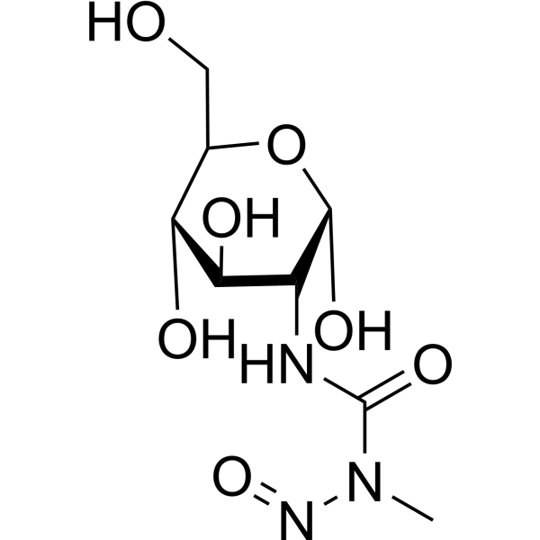
HY-13753
StreptozotocinDiabetes Modeling Agent -
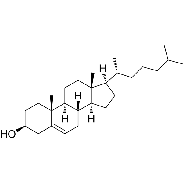
HY-N0322
Cholesterol (from animal)ERRα Agonist -
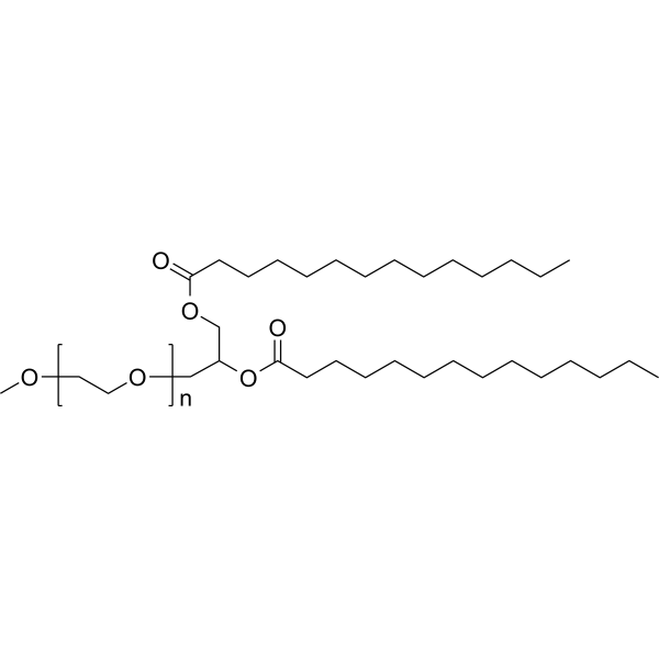
HY-112764
mPEG2000-DMGLiposome -
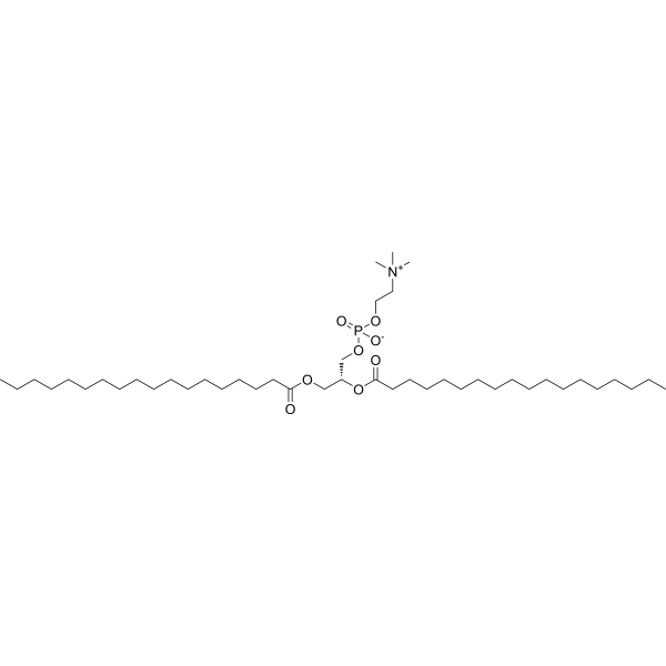
HY-W040193
DSPCLiposome Compound -

HY-B0015
PaclitaxelAntitumor Agent -

HY-15371
ForskolinAdenylate Cyclase Activator -
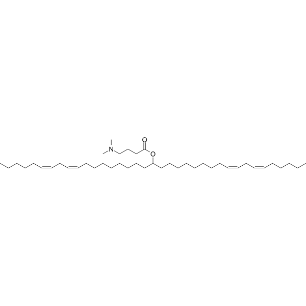
HY-112251
D-Lin-MC3-DMAsiRNA Delivery Vehicle -
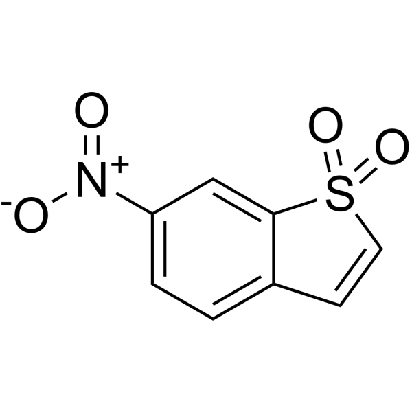
HY-13818
StatticSTAT3 Inhibitor -
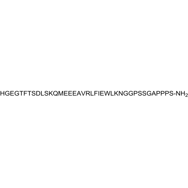
HY-13443
Exendin-4GLP-1 Receptor Agonist -

HY-114118
SemaglutideGLP-1R Agonist -
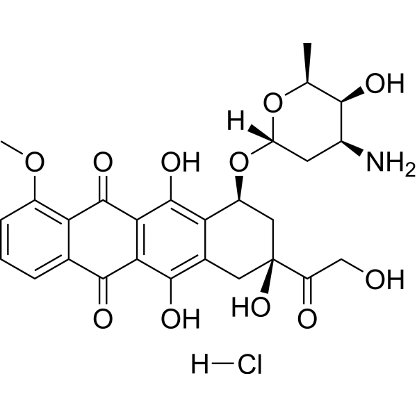
HY-15142
Doxorubicin hydrochlorideTopoisomerase Inhibitor -
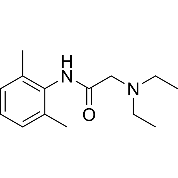
HY-B0185
LidocaineNa+ Channel Blocker
Publications Citing Use of MedChemExpress (MCE) Sodium Citrate Buffer, 0.1M, pH 4.0
More- Bone Res. 2025 Mar 3;13(1):30. [Abstract]
- Autophagy. 2026 Jun 10:1-23. [Abstract]
- Autophagy. 2026 Apr 29. [Abstract]
- ACS Nano. 2025 Aug 25. [Abstract]
- Cancer Lett. 2024 May 28:590:216845. [Abstract]
- Int J Biol Macromol. 2026 Mar:350:150917. [Abstract]
- Life Sci. 2025 Sep 1:376:123746. [Abstract]
- Mol Immunol. 2026 Feb:190:1-10. [Abstract]
- Diabetes Metab Syndr Obes. 2025 Jun 28:18:2041-2055. [Abstract]
- bioRxiv. 2026 Mar 21:2026.03.20.712457. [Abstract]


Biological Activity
Description
In Vitro
Sodium Citrate Buffer, 0.1M, pH 4.0 is applied in heat-induced epitope retrieval (HIER) to reverse antigenicity loss in Formalin-fixed paraffin-embedded tissues by breaking protein cross-links, thereby unmasking antigens and epitopes and ultimately enhancing antibody staining intensity[2][3].
Sodium Citrate Buffer, 0.1M, pH 4.0 can be used as a stabilizer to stabilize proteins and enzymes, hydrate liposomes, and reverse antigen cross-linking[2][3].
Sodium Citrate Buffer, 0.1M, pH 4.0 is widely used in buffer preparation[2].
Sodium Citrate Buffer, 0.1M, pH 4.0 can be used as an RT-PCR enhancement reagent[3].
MedChemExpress (MCE) has not independently confirmed the accuracy of these methods. They are for reference only. Further protocols information, click here.


Chemical Information
-
Appearance Liquid
-
Color Colorless to light yellow
-
SMILES
[Sodium Citrate Buffer, 0.1M, pH 4.0]
-
Shipping
Room temperature in continental US; may vary elsewhere.
-
Storage
Please store the product under the recommended conditions in the Certificate of Analysis.


Publications (10)
-
Journal Impact Factor
-
Most Recent
-
Bone Res
Enhanced SIRT3 expression restores mitochondrial quality control mechanism to reverse osteogenic impairment in type 2 diabetes mellitus. [Abstract]2025 Mar 3;13(1):30. PMID: 40025004 -
Autophagy
MEN1/menin deficiency suppresses hepatocellular carcinogenesis via disrupting mitophagy-mediated mitochondrial homeostasis. [Abstract]2026 Jun 10:1-23. PMID: 42210578 -
Autophagy
Cardiac fibroblast-derived CCN1 aggravates diabetic cardiomyopathy through ITGAV-ITGB1/integrin αvβ1-mediated autophagy inhibition. [Abstract]2026 Apr 29. PMID: 42056922 -
ACS Nano
Dual-Targeting Mn@CeO2 Nanozyme-Modified Probiotic Hydrogel Microspheres Reshape Gut Homeostasis in Inflammatory Bowel Disease. [Abstract]2025 Aug 25. PMID: 40853091 -
Cancer Lett
CAPN2-responsive mesoporous silica nanoparticles: A promising nanocarrier for targeted therapy of pancreatic cancer. [Abstract]2024 May 28:590:216845. PMID: 38589004 -
Int J Biol Macromol
ETS transcription factor 1-insulin-like growth factor 2 mRNA-binding protein 2-interferon-induced transmembrane protein 3 pathway promotes ferroptosis-driven osteoblast senescence in diabetic osteoporosis. [Abstract]2026 Mar:350:150917. PMID: 41692190 -
Life Sci
KAT2A-mediated H3K79 succinylation promotes ferroptosis in diabetic nephropathy by regulating SAT2. [Abstract]2025 Sep 1:376:123746. PMID: 40409584 -
Mol Immunol
Formononetin in Jiawei Qihuangyin inhibits podocyte epithelial-mesenchymal transition and ameliorates diabetic nephropathy via SIRT1/NF-κB axis. [Abstract]2026 Feb:190:1-10. PMID: 41483656 -
Diabetes Metab Syndr Obes
Yiqi Yangyin Tongluo Recipe Alleviates Diabetic Kidney Disease Through AGE-RAGE Signalling Axis. [Abstract]2025 Jun 28:18:2041-2055. PMID: 40606302 -
bioRxiv
Structure-Activity Mapping of Intraperitoneal mRNA-LNPs: Decoupling Tumor and Liver Biodistribution in Pancreatic Cancer. [Abstract]2026 Mar 21:2026.03.20.712457. PMID: 41889911

Protocols
-
Polymer-Based Two-Step IHC Detection
Polymer-based two-step IHC detects tissue antigens by first binding an unlabeled primary antibody to the antigen and then applying an HRP-polymer secondary reagent that carries multiple secondary antibodies and HRP molecules on a polymer backbone; the localized HRP converts chromogens such as DAB or AEC into visible deposits for light-microscopic interpretation. The method is \"two-step\" because the primary antibody step is followed directly by the polymer-enzyme secondary reagent, rather than by separate secondary-antibody and avidin-biotin complex steps; published comparisons reported similar or higher sensitivity than several multistep systems and avoidance of endogenous-biotin interference.
-
Immunohistochemistry-Frozen
Immunohistochemistry-Frozen (IHC-F) of frozen samples is a widely used technique for detecting and locating specific antigens within preserved cellular structures. Unlike formalin-fixed paraffin-embedded samples, frozen tissues retain their natural antigenicity, making them particularly suitable for targets sensitive to chemical fixation, and the procedure is relatively simple and rapid.
-
Avidin-Biotin/Streptavidin-Biotin IHC
Avidin-biotin or streptavidin-biotin immunohistochemistry detects tissue antigens by binding a primary antibody to the target antigen, then detecting that antibody with a biotinylated antibody and an avidin-biotin-enzyme or streptavidin-enzyme detection complex; the enzyme reaction produces a visible chromogenic deposit at the antigen site for light-microscopic localization. The classic ABC method uses the high-affinity avidin-biotin interaction to bridge biotinylated secondary antibody and biotinylated peroxidase, and early comparative studies reported stronger immunoperoxidase staining than PAP-based methods in formalin-fixed tissue sections.
-
ROS/oxidative-stress fluorescent staining
ROS/oxidative-stress fluorescent staining uses cell-permeant fluorogenic probes that become fluorescent after oxidation inside cells or tissues; commonly used examples include DCFH-DA/DCFDA for broad cellular oxidant detection, DHE for superoxide-related signal detection, MitoSOX for mitochondrial superoxide-related signal detection, and CellROX probes for oxidative-stress-associated fluorescence readouts. The assay detects probe oxidation rather than a single ROS species unless the probe and analysis method have been chemically validated for that species. DCFH-DA enters cells, is deacetylated by intracellular esterases to DCFH, and produces fluorescent DCF after oxidation, so the readout is used as an operational measure of total cellular oxidative stress rather than a species-specific ROS measurement. DHE and MitoSOX can report superoxide-related oxidation, but red fluorescence alone can include non-specific ethidium-like oxidation products; HPLC or optimized spectral approaches are
-
Double or multiplex chromogenic IHC
Double or multiplex chromogenic IHC detects two or more protein targets in the same FFPE tissue section by repeated antigen-antibody binding, enzyme-linked detection, chromogen deposition, image capture, and, for higher-plex workflows, removal or destaining before the next staining cycle. Chromogenic readouts are generated as colored precipitates at antigen sites, enabling evaluation of marker expression, cell phenotype, and spatial relationships in preserved tissue architecture. Classic examples include MICSSS, which performs iterative chromogenic IHC staining, scanning, and destaining on a single slide, and p16/Ki-67 dual staining, which uses chromogenic co-detection to identify cervical cells with combined cell-cycle deregulation and proliferation signals.


Purity & Documentation
-
Data Sheet (267 KB)
-
SDS (394 KB)
- English - EN (394 KB)
- Français - FR (394 KB)
- Deutsch - DE (394 KB)
- Norwegian - NO (394 KB)
- Español - ES (394 KB)
- Swedish - SV (394 KB)
- Italian - IT (394 KB)
- Korean - KR (394 KB)
- Portuguese - PT (394 KB)
-
Handling Instructions (2659 KB)
References
[1]. L M Sammel, et al. Investigation of mechanisms by which sodium citrate reduces the pink color defect in cooked ground turkey. Meat Sci. 2006, 72, 4. [Content Brief]
[2]. Huang C, e al., Liver-Specific Ionizable Lipid Nanoparticles Mediated Efficient RNA Interference to Clear "Bad Cholesterol". Int J Nanomedicine. 2023 Dec 19;18:7785-7801. [Content Brief]
[3]. Liu Y, et al. Immobilization of laccase on magnetic bimodal mesoporous carbon and the application in the removal of phenolic compounds. Bioresour Technol. 2012 Jul;115:21-6. [Content Brief]

Calculators
Concentration (start) × Volume (start) = Concentration (final) × Volume (final)


Sodium Citrate Buffer, 0.1M, pH 4.0 Related Classifications
HY-B1610N Related Classifications
 Powered by Bioz