



Sodium sulfite

Based on 1 Customer Validation
Sodium sulfite is an inorganic salt used as an antioxidant and preservative. Sodium sulfite is also used in sulfonation and sulfomethylation reactions. Sodium sulfite can also be used as a bleaching agent, desulfurizer, and dechlorinator. Sodium sulfite inhibits hepatocyte proliferation, promotes hepatocyte apoptosis and necrosis, and impairs mitochondrial integrity. Sodium sulfite induces superoxide anion production, primes neutrophils for enhanced superoxide anion generation, and induces neutrophil gene expression. Sodium sulfite can be used in studies related to pulmonary inflammation and gastric tissue injury.
For research use only. We do not sell to patients.
- Purity : 98.15%
- CAS No.: 7757-83-7
- Formula: Na2SO3
- Molecular Weight:126.04
-
Storage:Powder -20°C, 3 years , 4°C, 2 years ; In solvent -80°C, 6 months , -20°C, 1 month


To place orders, for customer services and technical support, please contact: MedChemExpress USA
Tel: 609-228-6898 E-mail: [email protected] [email protected]


-
 Biological Activity
-
Chemical Information
-
Solvent & Solubility
- Protocols
- Purity & Documentation
- References
-
 Help & FAQs


-

HY-Y1103
Iron(II) sulfate heptahydrate, 99%Iron Salt -
HY-Y0317H
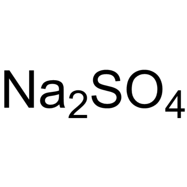
Sodium sulfate anhydrous, for cell cultureDrying Agent -

HY-B1770
Sodium Iodide,99%Iodide Salt -
HY-124011
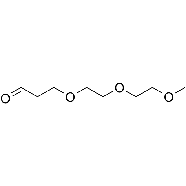
m-PEG3-aldehydePROTAC Linker -

HY-108775A
Sodium thiosulfate (99%, water≤1.0%)Antioxidant -

HY-Y1103A
Iron(II) sulfate heptahydrate, for cell culture, 99%Iron Salt -

HY-Y0849C
Ammonium acetate, for molecular biology, 98%Biochemical Reagent -
HY-B0178A
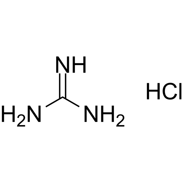
Guanidine hydrochlorideProtein Denaturant -

HY-108775
Sodium thiosulfate (98%)Antioxidant -

HY-23539C
Sodium thiosulfate pentahydrate (GR,≥99.5%)Biochemical Reagent


Biological Activity
Description
In Vitro
Sodium sulfite (10-5-10-2 M; 24-72 h) alters the morphology of human hepatocytes L02 in a time- and concentration-dependent manner, with notable toxic effects occurring at 10-3 M and 10-2 M[1].
Sodium sulfite (10-5-10-2 M; 72 h) inhibits the viability of human hepatocytes L02 in a concentration-dependent manner, with an IC50 of 4.68×10⁻⁴ M after 72 h incubation[1].
Sodium sulfite (10-5-10-2 M; 24-72 h) inhibits the proliferation of human hepatocytes L02 in a time- and concentration-dependent manner, with significant inhibition occurring at concentrations of 5×10⁻⁴ M and higher[1].
Sodium sulfite (10-5-10-2 M; 72 h) induces apoptosis in human hepatocytes L02 in a concentration-dependent manner, with notable apoptosis starting at 10-4 M and severe cell death occurring at 10-2 M within 24 h[1].
Sodium sulfite (4.68×10⁻⁴ M; 72 h) alters the expression of 97 genes in human hepatocytes L02, with 13 genes significantly upregulated and 14 genes significantly downregulated, driving pathways that inhibit cell proliferation, induce apoptosis, and cause mitochondrial damage via reactive oxygen species production[1].
Sodium sulfite (0.1-10 mM; 5-30 min) concentration-dependently induces superoxide production in freshly isolated human neutrophils within 5 to 30 min, and (0.1-10 mM; 30 min pre-incubation followed by 5 min fMLP stimulation) concentration-dependently primes these neutrophils to enhance fMLP-induced superoxide production following 30 min pre-incubation[2].
Sodium sulfite (0.1-10 mM; 4 h) concentration-dependently induces RNA synthesis in freshly isolated human neutrophils following 4 h incubation, but does not induce neutrophil shape changes[2].
Sodium sulfite (0.1-10 mM; 20 h) does not modulate apoptosis rates in freshly isolated human neutrophils following 20 h incubation, nor does it reverse the apoptosis-delaying effect of GM-CSF in these cells[2].
Sodium sulfite (0.001-10 mM; 5-24 h) induces dose-dependent necrotic cell death in RGM1 cells via oxidative stress, with an IC50 of 0.17 mM, and this cytotoxicity is attenuated by the free radical scavenger Trolox[3].
Sodium sulfite (0-5 mM; 20 h) induces necrotic cell death in RGM1 cells independent of the apoptotic signaling pathway, as shown by unchanged caspase/PARP cleavage, caspase activity, and DNA fragmentation, alongside dose-dependent LDH release[3].
Sodium sulfite (0-10 mM; 0-120 min, 24 h) induces oxidative modification, aggregation, and inactivation of purified lysozyme via metal ion-catalyzed free radical formation, independent of superoxide or hydrogen peroxide scavenging[3].
MedChemExpress (MCE) has not independently confirmed the accuracy of these methods. They are for reference only. Further protocols information, click here.
-
Cell Line:human hepatocytes L02
-
Concentration:10-5 M, 10-4 M, 2.5×10⁻⁴ M, 5×10⁻⁴ M, 5×10⁻³ M, 10-2 M
-
Incubation Time:72 h
-
Result:Gradually decreased L02 cell viability with increasing sodium sulfite concentration.
Reached an IC50 of 4.68×10⁻⁴ M for L02 cells at 72 h.
-
Cell Line:human hepatocytes L02
-
Concentration:10-5 M, 10-4 M, 5×10⁻⁴ M, 5×10⁻³ M, 10-2 M
-
Incubation Time:24 h, 48 h, 72 h
-
Result:Significantly inhibited L02 cell proliferation at concentrations ≥5×10⁻⁴ M, with proliferation rate decreasing as incubation time increased.
Had no obvious effect on proliferation activity at low concentrations (10-5 M, 10-4 M).
-
Cell Line:human hepatocytes L02
-
Concentration:10-5 M, 10-4 M, 10-3 M, 10-2 M
-
Incubation Time:72 h
-
Result:Caused cell color changes from green to orange/red (indicating apoptosis) at 10-4 M with increased incubation time.
Induced significant apoptosis as early as 24 h at 10-3 M, indicated by dominant orange and red staining.
Led to majority of cells being dead by 24 h at 10-2 M.
-
Cell Line:human hepatocytes L02
-
Concentration:4.68×10⁻⁴ M (IC50)
-
Incubation Time:72 h
-
Result:Identified 97 significantly regulated differentially expressed genes between sodium sulfite-treated and control cells via transcriptomic analysis.
Confirmed the expression changes of 13 significantly upregulated genes (including SOCS3, FOSL1, PLK3, IL1B) and 14 significantly downregulated genes (including CPS1, FBN1, FKBP3) via qPCR.
Linked these genes to pathways including TNF signaling, IL-17 signaling, apoptosis, metabolism, and inflammatory responses.
-
Cell Line:rat gastric mucosal epithelial cells (RGM1)
-
Concentration:0-5 mM (20 h incubation); 0-5 mM (1 h pre-incubation followed by 20 h in fresh medium)
-
Incubation Time:20 h; 1 h (pre-incubation) + 20 h (fresh medium)
-
Result:Did not alter levels of cleaved caspase-9, cleaved caspase-3, or cleaved PARP, nor change caspase-9 or caspase-3 activity.
Showed no apoptotic DNA fragmentation, but dose-dependently increased LDH release into the culture medium.


Chemical Information
-
CAS No. 7757-83-7
-
Appearance Solid
-
Molecular Weight 126.04
-
Formula Na2SO3
-
Color White to off-white
-
SMILES
O=S(O[Na])([Na])=O
-
Shipping
Room temperature in continental US; may vary elsewhere.
-
Storage
Powder -20°C 3 years 4°C 2 years In solvent -80°C 6 months -20°C 1 month


Solvent & Solubility
In Vitro:
H2O : ≥ 200 mg/mL (1586.80 mM)
* "≥" means soluble, but saturation unknown.
Please refer to the solubility information to select the appropriate solvent. Once prepared, please aliquot and store the solution to prevent product inactivation from repeated freeze-thaw cycles.
Storage method and period of stock solution: -80°C, 6 months; -20°C, 1 month. When stored at -80°C, please use it within 6 months. When stored at -20°C, please use it within 1 month.
* Note: If you choose water as the stock solution, please dilute it to the working solution, then filter and sterilize it with a 0.22 μm filter before use.
Please refer to the solubility information to select the appropriate solvent. Once prepared, please aliquot and store the solution to prevent product inactivation from repeated freeze-thaw cycles.
Storage method and period of stock solution: -80°C, 6 months; -20°C, 1 month. When stored at -80°C, please use it within 6 months. When stored at -20°C, please use it within 1 month.
* Note: If you choose water as the stock solution, please dilute it to the working solution, then filter and sterilize it with a 0.22 μm filter before use.
Concentration (start) × Volume (start) = Concentration (final) × Volume (final)

Protocols
-
RT-PCR
Reverse transcription technology uses RNA as a template to synthesize DNA. RT-PCR is simple, specific and sensitive, and can be used to detect gene expression levels and expression differences in cells; detect RNA virus content; clone cDNA sequences of specific genes.
-
RNA extraction experimental
By lysing cells, releasing RNA, and removing impurities such as proteins and DNA, high-purity RNA products are finally obtained. The commonly used traditional method is the guanidine isothiocyanate/phenol/chloroform method (Trizol), which is suitable for a variety of animal materials including animal tissues, microorganisms, cultured cells, etc., and most plant materials.
-
Apoptosis
Apoptosis, also called programmed cell death, is generally characterized by distinct morphological characteristics.
-
TUNEL staining for apoptotic DNA fragmentation
TUNEL staining detects DNA strand breaks by using terminal deoxynucleotidyl transferase to add labeled nucleotides to exposed 3′-OH DNA termini, generating either microscopic staining in fixed cells or tissue sections, or fluorescence/cytometric signal in cell suspensions. TUNEL positivity reflects DNA fragmentation but should not be interpreted alone as definitive apoptosis, because TUNEL can also label necrotic, autolytic, mechanically damaged, or DNA-repair-associated DNA breaks.
-
Annexin V plus membrane-impermeant dye apoptosis staining
Annexin V-based apoptosis assays rely on the detection of phosphatidylserine (PS) externalization from the inner leaflet of the plasma membrane to the outer leaflet, an early biochemical hallmark of apoptosis. Fluorescently labeled Annexin V binds PS in a calcium-dependent manner, enabling identification of early apoptotic cells by flow cytometry or fluorescence microscopy. When combined with a membrane-impermeant DNA-binding dye (e. g. , propidium iodide), this approach allows discrimination between viable (Annexin V−/dye−), early apoptotic (Annexin V+/dye−), and late apoptotic or necrotic (Annexin V+/dye+) cell populations by assessing membrane integrity and PS exposure.
-
Apoptosis Solutions
Apoptosis is a regulated, generally non-lytic cell-death pathway that removes unwanted, damaged, infected, or abnormal cells through coordinated morphological changes, caspase activation, DNA fragmentation, and membrane remodeling. The intrinsic apoptosis pathway is controlled mainly by mitochondrial outer membrane permeabilization, BCL-2 family proteins, cytochrome c release, apoptosome formation, caspase-9 activation, and downstream executioner caspase-3/7 activation. The extrinsic apoptosis pathway is initiated by death receptors such as Fas, TNFR, and TRAIL receptors, which recruit adaptor proteins and activate caspase-8 before engaging executioner caspases or mitochondrial amplification through BID cleavage. Apoptosis is linked to many phenotypes, including cancer cell killing, tissue homeostasis, immune regulation, neurodegeneration, infection response, and treatment-induced cytotoxicity; unresolved questions include how apoptosis interacts with necroptosis, pyroptosis, ferroptos
-
Research Protocol for Inflammation-related Diseases
The NLRP3 inflammasome is a cytosolic innate immune signaling platform that integrates priming signals and danger-signal activation to promote caspase-1 activation, maturation of IL-1β and IL-18, and gasdermin D-mediated pyroptotic cell death. The core experimental logic is to determine whether inflammatory disease phenotypes are driven by increased NLRP3 expression, ASC-containing inflammasome assembly, caspase-1 cleavage, GSDMD cleavage, and extracellular release of IL-1β/IL-18 rather than by nonspecific cell injury alone. The pathway is strongly linked to inflammation-related disease phenotypes because monosodium urate crystals activate NALP3/NLRP3 inflammasome signaling in gout-like crystal inflammation, cholesterol crystals activate NLRP3 inflammasomes in atherogenesis models, and DSS-induced intestinal inflammation has been reported to involve NLRP3 inflammasome activity. However, experimental colitis studies also show context-dependent protective effects of NLRP3 inflammasome co
-
Real Time qPCR (Q-PCR)
Real-time quantitative PCR (qPCR) quantifies an amplifiable nucleic-acid target by monitoring fluorescence during PCR cycling rather than measuring product only after amplification. The increase in fluorescence tracks accumulation of PCR product, and the quantification cycle (Cq; historically also Ct/CP) is related to the initial amount of target: samples containing more starting target generally reach the defined fluorescence threshold in fewer cycles.
-
Cell Viability Determination by MTT Colorimetric Assay
The following protocol uses the MTT colorimetric assay as a classic literature-established method for assessing cell viability/metabolic activity in cultured mammalian cells. MTT[3-(4,5-dimethylthiazol-2-yl)-2,5-diphenyltetrazolium bromide] is reduced by metabolically active cells to a colored formazan product; the amount of formazan is quantified spectrophotometrically and provides an indirect measure of metabolically active viable cells. Importantly, MTT reduction reflects cellular oxidoreductase/metabolic activity rather than an absolute direct count of living cells, so changes in cellular metabolism can alter the signal independently of cell number.


Purity & Documentation
-
Data Sheet (282 KB)
-
SDS (394 KB)
- English - EN (394 KB)
- Français - FR (394 KB)
- Deutsch - DE (394 KB)
- Norwegian - NO (394 KB)
- Español - ES (394 KB)
- Swedish - SV (394 KB)
- Italian - IT (394 KB)
- Korean - KR (394 KB)
- Portuguese - PT (394 KB)
-
Handling Instructions (2659 KB)
References
[1]. Han X, et al. Mechanism analysis of toxicity of sodium sulfite to human hepatocytes L02. Mol Cell Biochem. 2020;473(1-2):25-37. [Content Brief]
[2]. Labbé P, et al. Functional responses of human neutrophils to sodium sulfite (Na2SO3) in vitro. Hum Exp Toxicol. 1998 Nov;17(11):600-5. [Content Brief]
[3]. Oshimo M, et al. Sodium sulfite causes gastric mucosal cell death by inducing oxidative stress. Free Radic Res. 2021;55(6):731-743. [Content Brief]


Complete Stock Solution Preparation Table
Please refer to the solubility information to select the appropriate solvent. Once prepared, please aliquot and store the solution to prevent product inactivation from repeated freeze-thaw cycles.
Storage method and period of stock solution: -80°C, 6 months; -20°C, 1 month. When stored at -80°C, please use it within 6 months. When stored at -20°C, please use it within 1 month.
| Optional Solvent | Concentration Solvent Mass | 1 mg | 5 mg | 10 mg | 25 mg |
|---|---|---|---|---|---|
| H2O | 1 mM | 7.9340 mL | 39.6699 mL | 79.3399 mL | 198.3497 mL |
| 5 mM | 1.5868 mL | 7.9340 mL | 15.8680 mL | 39.6699 mL | |
| 10 mM | 0.7934 mL | 3.9670 mL | 7.9340 mL | 19.8350 mL | |
| 15 mM | 0.5289 mL | 2.6447 mL | 5.2893 mL | 13.2233 mL | |
| 20 mM | 0.3967 mL | 1.9835 mL | 3.9670 mL | 9.9175 mL | |
| 25 mM | 0.3174 mL | 1.5868 mL | 3.1736 mL | 7.9340 mL | |
| 30 mM | 0.2645 mL | 1.3223 mL | 2.6447 mL | 6.6117 mL | |
| 40 mM | 0.1983 mL | 0.9917 mL | 1.9835 mL | 4.9587 mL | |
| 50 mM | 0.1587 mL | 0.7934 mL | 1.5868 mL | 3.9670 mL | |
| 60 mM | 0.1322 mL | 0.6612 mL | 1.3223 mL | 3.3058 mL | |
| 80 mM | 0.0992 mL | 0.4959 mL | 0.9917 mL | 2.4794 mL | |
| 100 mM | 0.0793 mL | 0.3967 mL | 0.7934 mL | 1.9835 mL |
* Note: If you choose water as the stock solution, please dilute it to the working solution, then filter and sterilize it with a 0.22 μm filter before use.


Sodium sulfite Related Classifications
HY-Y0651 Related Classifications
 Powered by Bioz