



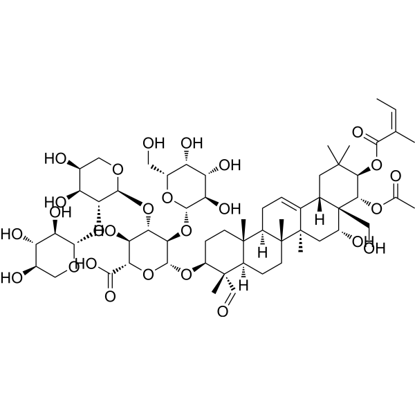
Theasaponin E1

Based on 1 Customer Validation
Theasaponin E1 is an orally effective tea saponin. Theasaponin E1 inhibits the proliferation of cancer cells by activating apoptosis. Theasaponin E1 inhibits angiogenesis in ovarian cancer cells and HUVECs by reducing the expression of VEGF. Theasaponin E1 upregulates the phosphorylation level of ATM protein and the expression level of PTEN protein in cancer cells, decreases the phosphorylation levels of Akt, mTOR, p70S6K and 4E-BP1 proteins, downregulates the expression of HIF-1α and NF-κB, and reduces the protein expression of Notch ligands Dll4 and Jagged1. Theasaponin E1 exerts neuroprotective effects by inhibiting the activity of acetylcholinesterase, activating α-secretase and neprilysin, reducing the concentration of Aβ, and inhibiting the activities of β-secretase and γ-secretase. Theasaponin E1 exhibits toxic effects on cancer cells and quinone reductase-inducing activity, and inhibits tumor growth in vivo. Theasaponin E1 induces ferroptosis in Pomacea canaliculata by synergistically disrupting cholesterol homeostasis and sphingolipid metabolism. Theasaponin E1 possesses anti-biofilm activity against Candida albicans. Theasaponin E1 can be used in the research of ovarian cancer, obesity, Alzheimer's disease and fungal infections.
For research use only. We do not sell to patients.
- Purity : 99.08%
- CAS No.: 220114-28-3
- Formula: C59H90O27
- Molecular Weight:1231.33
-
Storage:
4°C, protect from light
* In solvent : -80°C, 6 months; -20°C, 1 month (protect from light)

To place orders, for customer services and technical support, please contact: MedChemExpress USA
Tel: 609-228-6898 E-mail: [email protected] [email protected]


-
 Biological Activity
-
Chemical Information
- Protocols
- Purity & Documentation
- References
-
 Help & FAQs


All VEGFR Isoforms
More

Biological Activity
Description
IC50 & Target
Quinone reductase (QR)[1]
In Vitro
Theasaponin E1 (1-5 μM; 24 h) potently inhibits the growth of OVCAR-3 (IC50 = 3.5 μM) and A2780/CP70 (IC50 = 2.8 μM) cells in vitro, and exhibits low cytotoxicity against normal ovarian IOSE-364 cells at 24 h (IC50 > 5 μM at 24 h)[1].
Theasaponin E1 (1-4 μM; 24 h) induces apoptosis in OVCAR-3 cells via both intrinsic and extrinsic apoptotic pathways, while upregulating the levels of DNA damage markers[1].
Theasaponin E1 (1-4 μM; 24 h) mildly induces G2/M cell cycle arrest in OVCAR-3 cells by upregulating p-Chk2, p21, p-cdc2 (Tyr15) and Cyclin B1[1].
Theasaponin E1 (1-4 μM; 24 h) inhibits the migration of OVCAR-3 cells and reduces cellular VEGF secretion[1].
Theasaponin E1 (1-4 μM; 24 h) upregulates the phosphorylation level of ATM protein and the expression level of PTEN protein, reduces the phosphorylation levels of Akt, mTOR, p70S6K and 4E-BP1 proteins, and downregulates the protein expression of HIF-1α and VEGF in OVCAR-3 cells[1].
Theasaponin E1 (1-4 μM; 24 h) inhibits the Notch1 signaling pathway in OVCAR-3 cells by reducing the protein expression of NICD, Dll4 and Jagged1[1].
Theasaponin E1 (1-25 μg/mL; 4 h) potently inhibits capillary tube formation in human umbilical vein endothelial cells (HUVECs)[2].
Theasaponin E1 (1-10 μg/mL; 24 h) inhibits the proliferation of HCC-1428, SNU-1005, SNU-432 and SNU-719 in a dose-dependent manner[2].
Theasaponin E1 (5-25 μg/mL; 24 h) inhibits the expression and interaction of VEGF receptor complex components VEGFR-2, PI3K, β-Catenin and VE-Cadherin in HUVECs, and downregulates the mRNA expression of VEGFR-2, PI3K, β-Catenin, VE-Cadherin, Akt and NF-κB in the cells[2].
Theasaponin E1 (1-25 μg/mL; 48 h) inhibits lipid droplet accumulation in differentiating 3T3-L1 adipocytes[2].
Theasaponin E1 (5-20 μg/mL; 10 min) inhibits acetylcholinesterase activity[3].
Theasaponin E1 (5-20 μg/mL; 24 h) activates α-secretase (ADAM10) and neprilysin, inhibits β-secretase (BACE1) and γ-secretase subunits (PS1, NCT), upregulates the mRNA and protein expressions of ADAM10 and neprilysin, and downregulates the mRNA and protein expressions of BACE1, PS1 and NCT in SweAPP N2a cells[3].
Theasaponin E1 (5-20 μg/mL; 24 h) dose-dependently reduces the levels of Aβ peptides and APP in SweAPP N2a cells[3].
Theasaponin E1 (3.55-12 mg/L; 48-96 h) exhibits concentration- and time-dependent molluscicidal activity against adult golden apple snails, with a 96 h LC50 of 5.97 mg/L and a 96 h LC20 of 4.32 mg/L[4].
Theasaponin E1 (4.32 mg/L; 24 h) induces dysregulation of cholesterol homeostasis, membrane integrity pathways, and ferroptosis in the soft tissues of golden apple snails, increases the level of cell death in snail hemocytes, disrupts the cytoskeletal structure and alters the morphology of snail hemocytes, and triggers metabolic reprogramming, including enhanced fatty acyl turnover, oxidative lipid remodeling, and activation of detoxification pathways[4].
Theasaponin E1 (4.32 mg/L; 24 h) upregulates the expression of apoptosis-, sphingolipid metabolism- and ferroptosis-related genes in hemocytes of golden apple snails, while downregulating the expression of CTSL[4].
Theasaponin E1 (4.32 mg/L; 24 h) disrupts sphingolipid homeostasis in hemocytes of golden apple snails, induces plasma membrane rupture, impairs lysosomal function, triggers iron overload and oxidative stress, and causes severe ultrastructural damage to hyalinocytes and agranulocytes of golden apple snails[4].
Theasaponin E1 (0-400 μM; 1.5 h) inhibits the adhesion of Candida albicans ATCC 10231 to polystyrene surfaces, with an IC50 of 33.64 μM[5].
Theasaponin E1 (0-400 μM; 24 h) concentration-dependently inhibits early biofilm formation of Candida albicans ATCC 10231, with an 80% biofilm inhibitory concentration (BIC80) of 71.96 μM, and eradicates mature biofilms of Candida albicans ATCC 10231, with an 80% biofilm eradication concentration (BEC80) of 234.75 μM[5].
Theasaponin E1 (25-100 μM; 1.5 h for adherent cells, 24 h for mature biofilms) reduces the cell surface hydrophobicity of Candida albicans ATCC 10231, and decreases the extracellular phospholipase activity of Candida albicans ATCC 10231 in both adherent and mature biofilm states[5].
Theasaponin E1 (100 μM; 1.5 h) significantly downregulates the expression of multiple virulence-related genes in Candida albicans ATCC 10231, including genes associated with adhesion, hyphal development and signaling pathways[5].
Theasaponin E1 (overnight treatment) exhibits cytotoxic activity against human K562 and HL-60 tumor cell lines, with IC50 values of 17.9 μg/mL and 13.7 μg/mL, respectively[6].
Theasaponin E1 (4-20 μg/mL) exhibits significant quinone reductase-inducing activity in Hepa 1c1c7 wild-type hepatocellular carcinoma cells, with an induction rate of 4.2 (57.0% cell viability) at 4 μg/mL and 4.4 (3.3% cell viability) at 20 μg/mL[6].
Theasaponin E1 (5-40 μM; 24-72 h) dose-dependently inhibits the viability and single-cell sphere-forming capacity of ALDH-positive ovarian cancer stem-like cells derived from the A2780/CP70 and OVCAR-3 cell lines[7].
MedChemExpress (MCE) has not independently confirmed the accuracy of these methods. They are for reference only.
-
Cell Line:OVCAR-3 cells, A2780/CP70 cells, IOSE-364 cells
-
Concentration:0, 1, 2, 3, 4, 5 μM
-
Incubation Time:24 h
-
Result:Inhibited OVCAR-3 and A2780/CP70 cell growth in a concentration-dependent manner, with IC50 values of 3.5 μM (OVCAR-3) and 2.8 μM (A2780/CP70) at 24 h.
Killed approximately 80% of OVCAR-3 cells and 98% of A2780/CP70 cells at 4 μM.
Exhibited an IC50 value of more than 5 μM for normal IOSE-364 cells at 24 h, with no obvious inhibitory effect on IOSE-364 cells at 4 μM.
-
Cell Line:human platinum-resistant ovarian cancer OVCAR-3 cells
-
Concentration:0, 1, 2, 3, 4 μM
-
Incubation Time:24 h
-
Result:Induced apoptotic morphology (fragmented/condensed nuclei) observed via Hoechst 33342 staining.
Increased total early and late apoptotic cells from 10.80% to 74.18% and decreased live cells from 84.92% to 21.48% in a concentration-dependent manner via flow cytometry.
Markedly increased protein levels of cleaved-Caspase-3, cleaved-Caspase-7, cleaved-PARP, phospho-Histone H2A.X (Ser139), proapoptotic Bax, Cytochrome C, pro-Caspase-9, cleaved-Caspase-9, DR4, FADD, and cleaved-Caspase-8.
Reduced antiapoptotic Bcl-xL expression and slightly increased pro-Caspase-8 expression, with no obvious effect on pro-Caspase-3 or pro-Caspase-7.
-
Cell Line:OVCAR-3 cells
-
Concentration:0, 1, 2, 3, 4 μM
-
Incubation Time:24 h
-
Result:Increased the population of cells in G2/M phase from 14.70% to 21.96% via flow cytometry.
Significantly increased protein levels of p-Chk2, p21, and p-cdc2 (Tyr15), and downregulated Cyclin B1 protein expression via Western blot.
-
Cell Line:OVCAR-3 cells
-
Concentration:0-4 μM
-
Incubation Time:24 h
-
Result:Increased the relative space between scratch edges from 57.50% to 100.00% in a concentration-dependent manner at 24 h, indicating inhibited migration.
-
Cell Line:human platinum-resistant ovarian cancer OVCAR-3 cells
-
Concentration:0-4 μM (alone); 2 μM (combined with 100 nM wortmannin)
-
Incubation Time:24 h
-
Result:Significantly increased phosphorylation of ATM (Ser1981) and PTEN protein expression, decreased phosphorylation of Akt (Ser473), mTOR (Ser2448), p70S6K (Thr421/Ser424), and 4E-BP1 (Ser65/Thr70), and downregulated HIF-1α protein expression when used alone.
Enhanced the inhibitory effect on p-Akt, HIF-1α protein expression, and VEGF secretion when combined with 100 nM wortmannin compared to single treatment.
-
Cell Line:human umbilical vein endothelial cells (HUVECs)
-
Concentration:5 μg/mL, 10 μg/mL, 25 μg/mL
-
Incubation Time:24 h
-
Result:Downregulated mRNA expression levels of VEGFR-2, PI3 K, β-Catenin, VE-Cadherin, Akt, and NF-κB in a dose-dependent manner.
Showed the most pronounced reduction of mRNA expression at 25 μg/mL.
-
Cell Line:SweAPP N2a mouse neuroblastoma cells
-
Concentration:5-20 μg/mL
-
Incubation Time:24 h
-
Result:Reduced Aβ peptide levels in a dose-dependent manner, decreasing levels to ~100 pg/mL from a control level of ~180 pg/mL at 20 μg/mL.
Reduced APP levels in a dose-dependent manner, decreasing levels to ~250 pg/mL from a control level of ~380 pg/mL at 20 μg/mL.
-
Cell Line:Candida albicans ATCC 10231
-
Concentration:100 μM
-
Incubation Time:1.5 h
-
Result:Significantly downregulated the expression of adhesion-related genes (ALS1, ALS3, HWP1, EAP1), hyphal development-related genes (UME6, EED1, HGC1, HYR1), signaling pathway genes (RAS1, CPH1, TPK2, EFG1), and secreted hydrolase genes (SAP5, PLB1, PLB2) relative to untreated controls.
In Vivo
Theasaponin E1 (10 μg/g; i.p./intratumoral injection; once daily, once weekly for 4 consecutive weeks) significantly inhibits tumor growth in nude mice bearing human subcutaneous gastric cancer, liver cancer, uterine cancer, and breast cancer, with the largest reduction in tumor volume observed in uterine cancer (decreasing from 200.2 mm to 70.7 mm)[2].
Theasaponin E1 (10 μg/g; p.o.; daily; 40 days) reduces body weight gain in high-fat diet-fed C57Bl/6 J-ob/ob mice and improves their plasma lipid profiles and liver enzyme profiles[2].
MedChemExpress (MCE) has not independently confirmed the accuracy of these methods. They are for reference only.
-
Animal Model:specific-pathogen-free fertile eggs[1]
-
Dosage:4 µM
-
Administration:implanted onto CAM; single administration
-
Result:Reduced CAM blood vessel density to ~20% of the control level.
-
Animal Model:nude mice (6-week-old, female)[2]
-
Dosage:10 μg/g body weight
-
Administration:i.p.; daily; 4 weeks; intratumoral injection; once per week
-
Result:Reduced stomach tumor surface area from 97.5 mm to 56 mm.
Reduced liver tumor surface area from 127.2 mm to 103.6 mm.
Reduced uterus tumor surface area from 200.2 mm to 70.7 mm.
Reduced breast tumor surface area from 156.6 mm to 55.1 mm.
Hardened and shrank tumor surfaces.
-
Animal Model:C57Bl/6 J-ob/ob mice (4-week-old, male)[2]
-
Dosage:10 μg/g body weight
-
Administration:p.o.; daily; 40 days
-
Result:Lowered body weight gain significantly compared to controls over 40 days.
Decreased plasma triglyceride levels by 1.51-fold relative to controls.
Decreased total body cholesterol by 1.34-fold relative to controls.
Increased HDL cholesterol by 1.41-fold relative to controls.
Decreased ALT levels by 1.86-fold relative to controls.
- RNA extraction experimental
- Western Blot
- Human pluripotent stem cell endothelial-cell differentiation
- Vascular/Branching Fractal Analysis
- Annexin V plus membrane-impermeant dye apoptosis staining
- Patient-Derived Orthotopic Xenograft (PDOX)
- Research Protocol for Inflammation-related Diseases
- Research Protocol for Metabolic Diseases
- Cell Viability Determination by MTT Colorimetric Assay
- Protocol For Protein Expression And Purification
- Alzheimer’s Disease Modeling
- Kinase activity and phosphorylation assays
- Detection of Apoptosis
- TUNEL staining for apoptotic DNA fragmentation
- Endothelial Tube Formation Assay
- Patient-Derived Xenograft (PDX)
- Apoptosis Solutions
- Notch Pathway Solutions
- Protocol for Kinase activity and phosphorylation assays
- Ferroptosis Solutions
- Subcutaneous Cell-Line-Derived Xenograft
- Endothelial Cell Migration/Angiogenic Sprouting Assay
- Orthotopic Cell-Line Xenograft


Chemical Information
-
CAS No. 220114-28-3
-
Appearance Solid
-
Molecular Weight 1231.33
-
Formula C59H90O27
-
Color White to off-white
-
SMILES
OC[C@]12[C@](CC(C)([C@H]([C@@H]2OC(C)=O)OC(/C(C)=C\C)=O)C)([H])C3=CC[C@@]([C@@]4([C@@]([C@@](C)([C@H](CC4)O[C@H]5[C@@H]([C@H]([C@@H]([C@H](O5)C(O)=O)O)O[C@H]6[C@@H]([C@H]([C@H](CO6)O)O)O[C@H]7[C@@H]([C@H]([C@@H](CO7)O)O)O)O[C@@H]8O[C@@H]([C@@H]([C@@H]([C@H]8O)O)O)CO)C=O)([H])CC9)C)([H])[C@]9(C)[C@@]3(C[C@H]1O)C
-
Structure Classification
-
Initial Source
-
Shipping
Room temperature in continental US; may vary elsewhere.
-
Storage
4°C, protect from light
* In solvent : -80°C, 6 months; -20°C, 1 month (protect from light)


Purity & Documentation
-
Data Sheet (299 KB)
-
SDS (393 KB)
- English - EN (393 KB)
- Français - FR (393 KB)
- Deutsch - DE (393 KB)
- Norwegian - NO (393 KB)
- Español - ES (393 KB)
- Swedish - SV (393 KB)
- Italian - IT (393 KB)
- Korean - KR (393 KB)
- Portuguese - PT (393 KB)
-
Handling Instructions (2659 KB)
References
[1]. Li B, et al. Theasaponin E1 Inhibits Platinum-Resistant Ovarian Cancer Cells through Activating Apoptosis and Suppressing Angiogenesis. Molecules. 2021;26(6):1681. Published 2021 Mar 17. [Content Brief]
[2]. Kim JD, et al. Theasaponin E₁ as an effective ingredient for anti-angiogenesis and anti-obesity effects. Biosci Biotechnol Biochem. 2014;78(2):279-287. [Content Brief]
[3]. Khan MI, et al. Green Tea Seed Isolated Theasaponin E1 Ameliorates AD Promoting Neurotoxic Pathogenesis by Attenuating Aβ Peptide Levels in SweAPP N2a Cells. Molecules. 2020;25(10):2334. Published 2020 May 16. [Content Brief]
[4]. Ma CJ, Liao GM, Han YW, Chen P, Wang XA, Li JW, Hou Y, Tang B. Theasaponin E1 Induce Ferroptotic Cell Death in Pomacea canaliculata through Coordinated Disruption of Cholesterol Homeostasis and Sphingolipid Metabolism. J Agric Food Chem. 2025 Nov 19;73(46):29581-29592. [Content Brief]
[5]. Chen Y, Gao Y, Li Y, Yin J. Anti-Biofilm Activity of Assamsaponin A, Theasaponin E1, and Theasaponin E2 against Candida albicans. Int J Mol Sci. 2024 Mar 22;25(7):3599. [Content Brief]
[6]. Li N, et al. Phytochemical analysis of the triterpenoids with cytotoxicity and QR inducing properties from the total tea seed saponin of Camellia sinensis. Fitoterapia. 2013;84:321-325. [Content Brief]
[7]. Jia LY, et al. Anti-Proliferation Effect of Theasaponin E₁ on the ALDH-Positive Ovarian Cancer Stem-Like Cells. Molecules. 2018;23(6):1469. Published 2018 Jun 17. [Content Brief]

Calculators
Concentration (start) × Volume (start) = Concentration (final) × Volume (final)


Theasaponin E1 Related Classifications
HY-N11709 Related Classifications
 Powered by Bioz
Keywords
- Theasaponin E1
- 220114-28-3
- Theasaponin E 1
- Theasaponin E-1
- Apoptosis
- VEGFR
- ATM/ATR
- PTEN
- Akt
- mTOR
- HIF/HIF Prolyl-Hydroxylase
- NF-κB
- Notch
- Cholinesterase (ChE)
- Amyloid-β
- γ-secretase
- Ferroptosis
- Fungal
- tea saponin
- OVCAR-3 cells
- A2780/CP70 cells
- Notch1
- HUVECs
- SweAPP N2a cells
- Candida albicans
- ferroptosis
- apoptosis
- golden apple snails
- ovarian cancer
- obesity
- Alzheimer's disease
- fungal infections
- nude mice
- Inhibitor
- inhibitor
- inhibit