



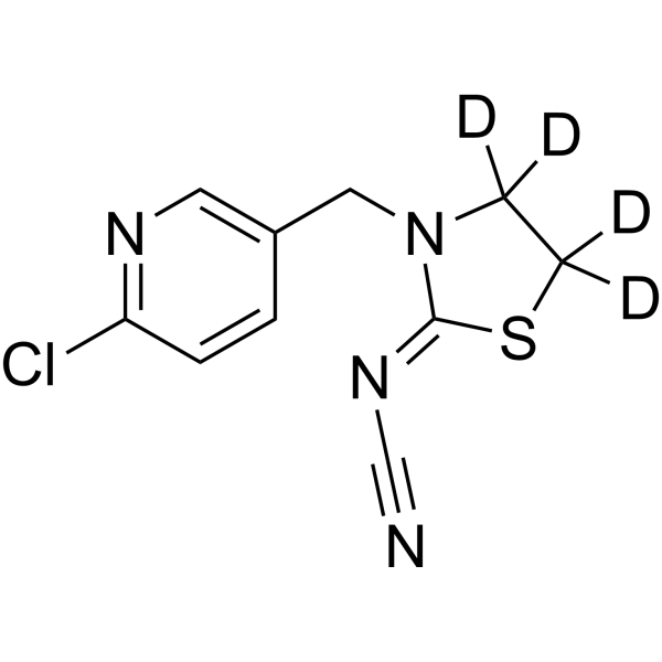
Thiacloprid-d4
Thiacloprid-d4 is the deuterium labeled Thiacloprid. Thiacloprid is an orally active neurotoxic insecticide and also a nAChR agonist. Thiacloprid reduces the viability of healthy cells, depletes reduced glutathione, and increases MDA levels, thereby inducing cytotoxicity and oxidative stress damage. In practical applications, Thiacloprid has lower acute toxicity to honeybees than other compounds of the same class such as Imidacloprid (HY-B0838), but it still significantly impairs the learning and memory function, immune capacity and survival status of honeybees. Thiacloprid induces intestinal microbial dysbiosis and reduces survival rate in middle-aged honeybees, increases the risk of premature collapse in bumblebee colonies, and significantly decreases the final colony weight and reproductive output. Thiacloprid is used in broad-spectrum agricultural pest control, often alone or in combination with Deltamethrin (HY-B1971), and meets the pest management needs of various crops including potatoes, cabbages, various fruits and vegetables, and nuts.
For research use only. We do not sell to patients.
- CAS No.: 1793071-39-2
- Formula: C10H5D4ClN4S
- Molecular Weight:256.75
-
Storage:
Please store the product under the recommended conditions in the Certificate of Analysis.

To place orders, for customer services and technical support, please contact: MedChemExpress USA
Tel: 609-228-6898 E-mail: [email protected] [email protected]


-
 Biological Activity
-
Chemical Information
- Protocols
- Purity & Documentation
- References
-
 Help & FAQs


-
HY-B1198S
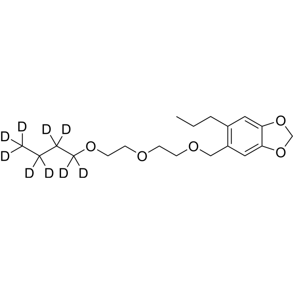
Piperonyl butoxide-d9Stable Isotope -
HY-133167
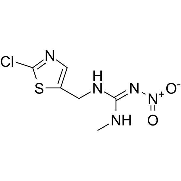
ClothianidinInsecticide -

HY-133167R
Clothianidin (Standard)Insecticide -
HY-12545
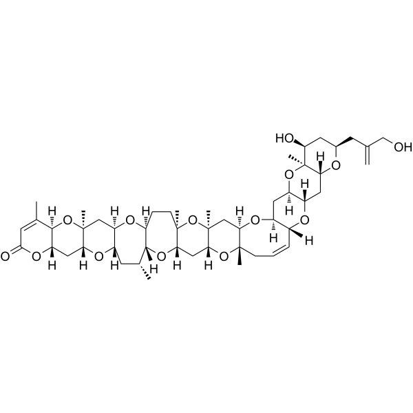
Brevetoxin-3Na+ Channel Activator -

HY-N10653
AltemicidinMonoterpene Alkaloid -
HY-W013170
MGK-264Synergist -

HY-W718787
Fenoxycarb-d3Stable Isotope -
HY-15598S
20-HETE-d6Stable Isotope -

HY-W013170R
MGK-264 (Standard)Synergist


Biological Activity
Description
In Vitro
Stable heavy isotopes of hydrogen, carbon, and other elements have been incorporated into drug molecules, largely as tracers for quantitation during the drug development process. Deuteration has gained attention because of its potential to affect the pharmacokinetic and metabolic profiles of drugs[1].
MedChemExpress (MCE) has not independently confirmed the accuracy of these methods. They are for reference only. Further protocols information, click here.
Application
1. This compound can be used as a tracer
2. This compound can be used as an internal standard for quantitative analysis by NMR, GC-MS, or LC-MS.


Chemical Information
-
CAS No. 1793071-39-2
-
Unlabeled CAS 111988-49-9
-
Molecular Weight 256.75
-
Formula C10H5D4ClN4S
-
SMILES
N#C/N=C(SC([2H])([2H])C1([2H])[2H])/N1CC2=CN=C(Cl)C=C2
-
Shipping
Room temperature in continental US; may vary elsewhere.
-
Storage
Please store the product under the recommended conditions in the Certificate of Analysis.

Protocols
-
Neurotoxicity Study
This protocol assesses in vitro neurotoxicity by combining neuronal viability, mitochondrial/metabolic activity, neurite outgrowth, and optional neuronal network function readouts. Calcein-AM or resazurin/PrestoBlue readouts estimate viable or metabolically active cells; βIII-tubulin immunofluorescence detects neuronal morphology and neurite networks; TMRE detects mitochondrial membrane potential; and MEA recordings detect functional changes in neuronal network activity.
-
Protocol for Shuttle Box Test (TDPA)
The Shuttle Box Test for TDPA, or temporally dissociated passive avoidance, measures hippocampus-dependent associative learning by testing whether a rodent avoids entering a dark compartment that was previously paired with foot shock after a temporal delay between dark-compartment entry and shock delivery. The main behavioral readout is crossover or step-through latency from the light chamber into the dark chamber; increased latency across training or retention trials reflects learned avoidance memory rather than motor performance alone.
-
Cell Cytotoxicity Assay
Cytotoxicity assays are usually based on the assessment of cell membrane damage, which can also be indirectly detected by measuring cell viability. Detection methods include MTT assay, CKK-8 assay, LDH assay and ATP assay, etc.
-
Mammalian live/dead viability and cytotoxicity staining
Live/dead viability and cytotoxicity staining assays are based on the simultaneous detection of intracellular esterase activity in metabolically active (viable) cells and membrane integrity loss in non-viable cells. In commonly used dual-staining approaches, membrane-permeant fluorogenic substrates are converted by intracellular esterases into fluorescent products in live cells, while impermeant DNA-binding dyes selectively enter cells with compromised plasma membranes and label nucleic acids in dead or dying cells, enabling discrimination between viable and non-viable populations by fluorescence microscopy or flow cytometry.
-
ROS/oxidative-stress fluorescent staining
ROS/oxidative-stress fluorescent staining uses cell-permeant fluorogenic probes that become fluorescent after oxidation inside cells or tissues; commonly used examples include DCFH-DA/DCFDA for broad cellular oxidant detection, DHE for superoxide-related signal detection, MitoSOX for mitochondrial superoxide-related signal detection, and CellROX probes for oxidative-stress-associated fluorescence readouts. The assay detects probe oxidation rather than a single ROS species unless the probe and analysis method have been chemically validated for that species. DCFH-DA enters cells, is deacetylated by intracellular esterases to DCFH, and produces fluorescent DCF after oxidation, so the readout is used as an operational measure of total cellular oxidative stress rather than a species-specific ROS measurement. DHE and MitoSOX can report superoxide-related oxidation, but red fluorescence alone can include non-specific ethidium-like oxidation products; HPLC or optimized spectral approaches are
-
Protocol for Water Maze
The Morris Water Maze is a rodent spatial learning and memory assay in which a mouse or rat swims in opaque water to find an escape platform; in the hidden-platform version, the animal cannot see the platform and must use distal extra-maze cues to learn its fixed spatial location. The assay primarily measures hippocampus-dependent spatial learning during acquisition trials and spatial reference memory during probe trials after platform removal; readouts include escape latency, swim path length, swim speed, quadrant occupancy, platform-site crossings, and proximity to the former platform location.
-
Acute Systemic Toxicity Study
Acute systemic toxicity studies evaluate adverse effects occurring after a single exposure, or repeated exposure within a short acute window, and the main in vivo readouts are mortality, moribund condition, clinical signs, body-weight change, and gross pathological findings; acute oral toxicity methods were developed to replace classical LD50 testing with reduced-animal designs such as fixed-dose procedure, acute toxic class method, and up-and-down procedure. The fixed-dose procedure classifies acute toxicity by administering predefined dose levels and observing evident toxicity rather than using death as the primary endpoint, whereas the acute toxic class method uses sequential groups of three animals per step and the up-and-down procedure doses animals sequentially to estimate an LD50 with fewer animals than conventional LD50 testing.
-
Cell Viability Determination by MTT Colorimetric Assay
The following protocol uses the MTT colorimetric assay as a classic literature-established method for assessing cell viability/metabolic activity in cultured mammalian cells. MTT[3-(4,5-dimethylthiazol-2-yl)-2,5-diphenyltetrazolium bromide] is reduced by metabolically active cells to a colored formazan product; the amount of formazan is quantified spectrophotometrically and provides an indirect measure of metabolically active viable cells. Importantly, MTT reduction reflects cellular oxidoreductase/metabolic activity rather than an absolute direct count of living cells, so changes in cellular metabolism can alter the signal independently of cell number.


Purity & Documentation
References
[1]. Russak EM, et al. Impact of Deuterium Substitution on the Pharmacokinetics of Pharmaceuticals. Ann Pharmacother. 2019 Feb;53(2):211-216. [Content Brief]
[3]. Liu YJ, et al. Thiacloprid exposure perturbs the gut microbiota and reduces the survival status in honeybees. J Hazard Mater. 2020;389:121818. [Content Brief]
[4]. Ellis C, et al. The Neonicotinoid Insecticide Thiacloprid Impacts upon Bumblebee Colony Development under Field Conditions. Environ Sci Technol. 2017;51(3):1727-1732. [Content Brief]

Calculators
Concentration (start) × Volume (start) = Concentration (final) × Volume (final)


Thiacloprid-d4 Related Classifications
HY-B1953S Related Classifications
 Powered by Bioz