



Transglutaminase, Streptoverticillium mobaraense

Based on 1 Customer Validation
Transglutaminase, Streptoverticillium mobaraense (TG) is an enzyme that forms cross-links between protein molecules. Transglutaminase, Streptoverticillium mobaraense attaches proteins and peptides to small molecules, polymers, surfaces, DNA and other proteins. Transglutaminase, Streptoverticillium mobaraense is widely used in food applications in the meat, fish, dairy and baking industries.
연구목적의 판매만을 진행합니다. 환자를 대상으로 한 판매는 하지 않습니다.
- CAS No.: 80146-85-6
-
보관:
Please store the product under the recommended conditions in the Certificate of Analysis.


To place orders, for customer services and technical support, please contact: MedChemExpress USA
Tel: 609-228-6898 E-mail: [email protected] [email protected]


-
 Biological Activity
-
Chemical Information
- Protocol
- 순도&문서
- References
-
 Help & FAQs


-

HY-140214
Azido-PEG5-aminePROTAC Linkers -

HY-10182
LaduviglusibGSK-3 Inhibitor -

HY-10162
OlaparibPARP Inhibitor -

HY-10431
SB-431542TGF-β Receptor Kinase Inhibitor -

HY-D0940
H2DCFDAROS-Sensitive Probe -
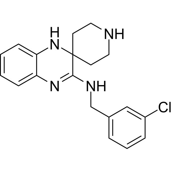
HY-12726
Liproxstatin-1Ferroptosis Inhibitor -

HY-17389
GenipinCrosslinking Reagent -
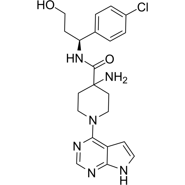
HY-15431
CapivasertibAkt Inhibitor -

HY-B0215
AcetylcysteineMucolytic Agent -

HY-B0015
PaclitaxelAntitumor Agent -

HY-15371
ForskolinAdenylate Cyclase Activator -
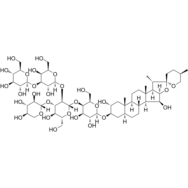
HY-N4000
DigitoninDetergent


Biological Activity
제품 설명
In Vitro
Tissue Transglutaminase is used to detect T cell proliferation experiments[4]
(1) Culture PBMC cells in a 96-well plate using complete RPMI and heat-inactivated 10% human AB serum, with 1×10⁵ cells per well. Add 12 μg/mL of peptide, 50 μg/mL of trypsin-digested gliadin, or phosphate-buffered saline, along with Tissue Transglutaminase. The total volume per well is 200 μL, and incubate at 37°C, 5% CO2, and humid air for 5 days.
(2) Add 1 microcurie of 3H-thymidine to each well. After continuing the incubation for another 18 hours, measure the incorporation of 3H in DNA using a cell harvester and a radioactive counter. The results are expressed as counts per minute (c.p.m.) for the test samples.
Tissue Transglutaminase is used in HLA class II restriction experiments[4]
(1) Gliadin (100 mg/ml) is digested with α-chymotrypsin (weight ratio 200:1) at room temperature in 0.1 M ammonium bicarbonate and 2 M urea.
(2) After the digestion process lasts for 24 hours, terminate the reaction by heating the samples to 98°C for 10 minutes.
(3) After centrifuging the samples at 13,000 g for 10 minutes, collect the supernatant and sterile filter it using a 0.2-micron filter membrane.
(4) Verify the digestion of gliadin by SDS-PAGE and determine the protein concentration.
(5) Treat gliadin (640 μg/mL) and synthetic gliadin peptide segments (15 amino acid segments: 160 μg/mL, other segments: 0.1 mM) with the products of α-chymotrypsin digestion at 37°C using 50 μg/mL of Tissue Transglutaminase in PBS buffer containing 1 mM CaCl2 for 2 hours.
(6) The treated peptide segments and peptide mixtures are aliquoted into sterile 96-well plates and stored frozen at -20°C.
(7) Wash the anti-CD4 or CD8 antibody beads four times with RPMI medium, and incubate them on ice with 5×10⁶ PBMC cells for 30 minutes.
(8) Remove the magnetic beads and perform a cell count.
(9) Pre-incubate 5×10⁶ PBMC cells with monoclonal antibodies against HLA-DR (L243), HLA-DQ (L2), and HLA-DP (B7.21) (10 μg/mL each) at room temperature for 1 hour.
(10) Then add the peptide segments and use the ELISPOT method to detect the secretion of IFN-γ, assessing T cell response to the peptide segments.
MedChemExpress (MCE) has not independently confirmed the accuracy of these methods. They are for reference only. Further protocols information, click here.
Clinical Trial
| NCT Number | Sponsor | Condition | Start Date |
Phase

|
|---|---|---|---|---|
| NCT01329991 | Plexxikon| | 2011-05 | PHASE1 |
EC Number
2.3.2.13
Specific Activity
≥10 U/mg protein
Unit Definition
One unit is defined as the amount of enzyme that form 1 μmol L-Glutamic acid γ-monohydroxamate in per min at 30°C and pH 6.0.


Chemical Information
-
CAS No. 80146-85-6
-
Appearance Solid
-
Color White to yellow
-
SMILES
[Transglutaminase, Streptoverticillium mobaraense]
-
Synonyms
TG
-
선적
Room temperature in continental US; may vary elsewhere.
-
보관
Please store the product under the recommended conditions in the Certificate of Analysis.

Protocol
-
Protocol for Fluorescence In Situ Hybridization (FISH)
Fluorescence in situ hybridization detects specific DNA or RNA sequences inside fixed cells or tissue sections by hybridizing fluorescently labeled nucleic-acid probes to complementary target sequences, allowing the target’s copy number, chromosomal position, spatial distribution, or transcript abundance to be visualized microscopically. DNA-FISH detects genomic loci, chromosomal gains/losses, amplifications, deletions, and rearrangements, while RNA-FISH detects RNA molecules or transcript localization; in cancer cells, mouse tumors, neurons, organoids, macrophages, or drug-screening samples, the readout is fluorescent puncta, fusion/split signals, or localized RNA signal interpreted relative to validated controls.


순도&문서
-
Data Sheet (269 KB)
-
SDS (418 KB)
- English - EN (418 KB)
- Français - FR (418 KB)
- Deutsch - DE (418 KB)
- Norwegian - NO (418 KB)
- Español - ES (418 KB)
- Swedish - SV (418 KB)
- Italian - IT (418 KB)
- Korean - KR (418 KB)
- Portuguese - PT (418 KB)
-
Handling Instructions (2659 KB)
References
[1]. Kieliszek M, Misiewicz A. Microbial transglutaminase and its application in the food industry. A review. Folia Microbiol (Praha). 2014 May;59(3):241-50. [Content Brief]
[2]. Pavel Strop, et al. Versatility of microbial transglutaminase. Bioconjug Chem. 2014 May 21;25(5):855-62. [Content Brief]
[4]. R P Anderson, et al. In vivo antigen challenge in celiac disease identifies a single transglutaminase-modified peptide as the dominant A-gliadin T-cell epitope. Nat Med. 2000 Mar;6(3):337-42. [Content Brief]

Calculators
Concentration (start) × Volume (start) = Concentration (final) × Volume (final)


Transglutaminase, Streptoverticillium mobaraense Related Classifications
HY-P2962 Related Classifications
 Powered by Bioz