



TXA6101
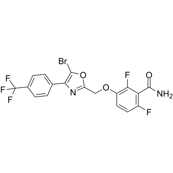
TXA6101 is a bacterial protein FtsZ (filamentous temperature-sensitive protein Z) inhibitor that inhibits bacterial division. TXA6101 has antimicrobial activity against MRSA isolates expressing either the G193D or G196S mutant FtsZ with the MIC value of 1 μg/mL, retains significant activity against the TXA707-resistant FtsZ mutant. TXA6101 can be used as a potential method against Gram-negative bacterial infections.
For research use only. We do not sell to patients.
- CAS No.: 1459695-66-9
- Formula: C18H10BrF5N2O3
- Molecular Weight:477.18
-
Storage:
Please store the product under the recommended conditions in the Certificate of Analysis.

To place orders, for customer services and technical support, please contact: MedChemExpress USA
Tel: 609-228-6898 E-mail: [email protected] [email protected]


-
 Biological Activity
-
Chemical Information
- Protocols
- Purity & Documentation
- References
-
 Help & FAQs




Biological Activity
Description
In Vitro
TXA6101 has antibacterial activity against MRSA with the MIC value of 0.125 μg/mL while TXA707 with the MIC value of 1 μg/mL[2].
TXY6129 inhibits the polymerization of Escherichia coli FtsZ in a concentration-dependent manner and induces morphological changes in Escherichia coli and Klebsiella pneumoniae[3].
MedChemExpress (MCE) has not independently confirmed the accuracy of these methods. They are for reference only.


Chemical Information
-
CAS No. 1459695-66-9
-
Molecular Weight 477.18
-
Formula C18H10BrF5N2O3
-
SMILES
O=C(N)C1=C(F)C=CC(OCC2=NC(C3=CC=C(C(F)(F)F)C=C3)=C(Br)O2)=C1F
-
Shipping
Room temperature in continental US; may vary elsewhere.
-
Storage
Please store the product under the recommended conditions in the Certificate of Analysis.


Purity & Documentation
References
[1]. Andrea Casiraghi, et al. Targeting Bacterial Cell Division: A Binding Site-Centered Approach to the Most Promising Inhibitors of the Essential Protein FtsZ. Antibiotics (Basel). 2020 Feb 7;9(2):69. [Content Brief]
[2]. Junso Fujita, et al. Structural Flexibility of an Inhibitor Overcomes Drug Resistance Mutations in Staphylococcus aureus FtsZ. ACS Chem Biol. 2017 Jul 21;12(7):1947-1955. [Content Brief]
[3]. Jesus D Rosado-Lugo, et al. Evaluation of 2,6-difluoro-3-(oxazol-2-ylmethoxy)benzamide chemotypes as Gram-negative FtsZ inhibitors. J Antibiot (Tokyo). 2022 Jul;75(7):385-395. [Content Brief]

Calculators
Concentration (start) × Volume (start) = Concentration (final) × Volume (final)


TXA6101 Related Classifications
HY-150699 Related Classifications
 Powered by Bioz