



Vari Fluor 647-Streptavidin

Based on 1 Customer Validation
Vari Fluor 647-Streptavidin is a dye marker of Vari Fluor-streptavidin consisting of labeling streptavidin with a Vari Fluor series of fluorescent probes. Streptavidin is a high-affinity tetramer protein, each tetramer consisting of four identical streptavidin subunits. Streptavidin binds to biotin specifically via a reversible non-covalent effect. Streptavidin can achieve rapid and efficient detection of biotin markers, and is often used in immunofluorescence (IF), enzyme-linked immunosorbent assay (ELISA), immunohistochemical staining (IFH), in situ hybridization (ISH) and other experiments. Ex/Em=650 nm/665 nm.
Product Concentration: 0.8 mg/mL
For research use only. We do not sell to patients.
-
Storage:
-20°C, protect from light
* In solvent : -80°C, 6 months; -20°C, 1 month (protect from light)


To place orders, for customer services and technical support, please contact: MedChemExpress USA
Tel: 609-228-6898 E-mail: [email protected] [email protected]


-
 Biological Activity
-
Chemical Information
- Protocols
- Purity & Documentation
- References
-
 Help & FAQs


-
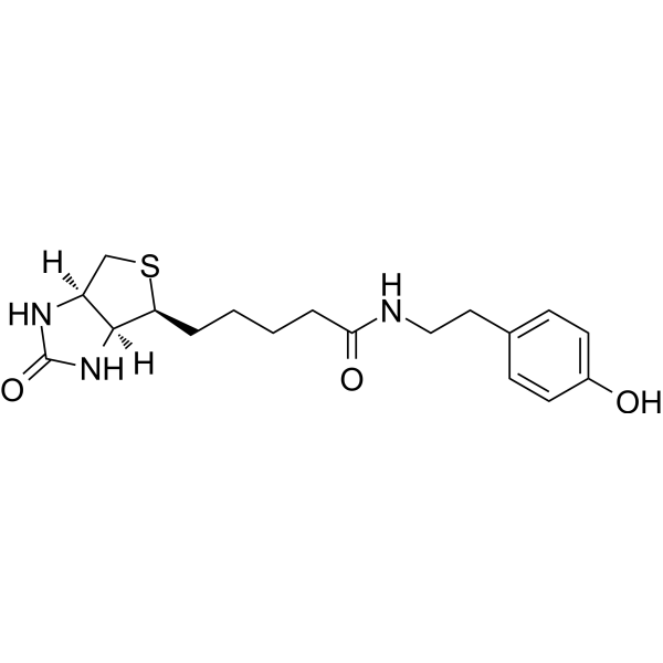
HY-125658
Biotinyl tyramideBiotin Derivative/Reagent -
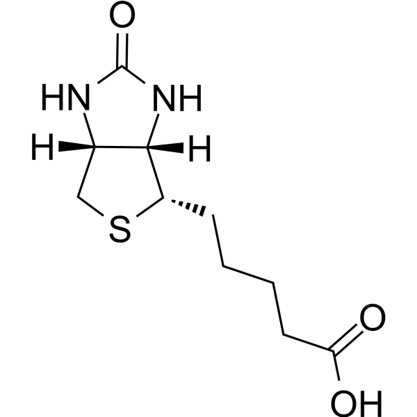
HY-B0511
BiotinVitamin B7 -
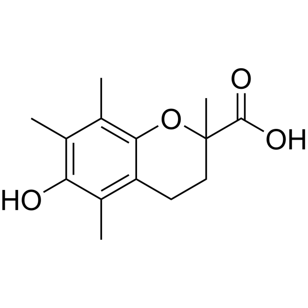
HY-101445
TroloxAntioxidant -

HY-D1807
Vari Fluor 555-StreptavidinFluorescent Dye -

HY-100579
Ferrostatin-1Ferroptosis Inhibitor -

HY-P3152
StreptavidinBiotin-Binding Protein -

HY-D1808
Vari Fluor 488-StreptavidinFluorescent Dye -

HY-D1806
Vari Fluor 594-StreptavidinFluorescent Dye -
HY-K0208
Streptavidin Magnetic Beads -

HY-D1809
Vari Fluor 405-StreptavidinFluorescent Dye


Biological Activity
Description
Product Concentration: 0.8 mg/mL
In Vitro
General Protocol
1.Protein treatment
Before use, centrifuge at 5000× g for 3 min, only the supernatant is used for experiments to eliminate protein aggregates and reduce non-specific background staining.
2.Labeling
Dilute Vari Fluor-Streptavidin at 1:500-1:1000, which can be adjusted according to the specific conditions of the protein or antibody.
Storage
-20°C,
Protect from light
Precautions
1. The actual content of the dye is small, dissolve it directly in the tube after receiving it for experiments.
2. Before use, please centrifuge the product to the bottom of the tube instantaneously before subsequent experiments.
3. This product is intended for scientific research only by professionals and is not to be used for clinical diagnosis or treatment, food or medicine.
4. For your safety and health, please wear lab coat and disposable gloves.
MedChemExpress (MCE) has not independently confirmed the accuracy of these methods. They are for reference only. Further protocols information, click here.
Emission (Em)
665
Excitation (Ex)
650


Chemical Information
-
Appearance Liquid
-
Color Light blue to blue
-
SMILES
[Vari Fluor 647-Streptavidin]
-
Shipping
Room temperature in continental US; may vary elsewhere.
-
Storage
-20°C, protect from light
* In solvent : -80°C, 6 months; -20°C, 1 month (protect from light)

Protocols
-
Enzyme-Linked Immunosorbent Assay (ELISA)
Enzyme-linked immunosorbent assay (ELISA) is a biochemical technology widely used to detect and quantify proteins, antibodies, hormones and other biomolecules. This method uses enzyme-labeled antibodies or antigens to specifically bind to the test substance, and generates a measurable signal through an enzyme-catalyzed reaction. ELISA has the characteristics of high sensitivity, specificity and high-throughput, and is suitable for medical diagnosis, biomedical research and drug development and other fields.
-
Antibody-based immunofluorescence/immunocytochemistry staining
Antibody-based immunofluorescence/immunocytochemistry detects the cellular or subcellular localization of a target antigen by binding a primary antibody to the target and detecting that antibody directly with a fluorophore-conjugated primary antibody or indirectly with a fluorophore-conjugated secondary antibody. Indirect immunofluorescence can amplify signal because multiple secondary antibodies can bind one primary antibody. The assay readout is fluorescence intensity and localization measured by fluorescence or confocal microscopy, and the result reflects antigen distribution only when the antibody has been validated for the target, sample type, fixation condition, and imaging workflow. Antibody specificity must not be assumed from catalog information alone, and appropriate validation or control experiments are required for serious interpretation.
- Immunocytochemistry/Immunofluorescence
-
In situ hybridization
The labeled nucleic acid probe is hybridized with the DNA or RNA on tissues and cells by using the complementary base sequence between the single strands of nucleic acid molecules. The detected DNA or RNA molecules in situ in cells can be displayed by autoradiography, fluorescence detection or enzyme color development.


Purity & Documentation
-
Data Sheet (259 KB)
-
SDS (251 KB)
- English - EN (251 KB)
- Français - FR (251 KB)
- Deutsch - DE (251 KB)
- Norwegian - NO (251 KB)
- Español - ES (251 KB)
- Swedish - SV (251 KB)
- Italian - IT (251 KB)
- Korean - KR (251 KB)
- Portuguese - PT (251 KB)
-
Handling Instructions (2659 KB)
References

Calculators
Concentration (start) × Volume (start) = Concentration (final) × Volume (final)


Vari Fluor 647-Streptavidin Related Classifications
HY-D1805 Related Classifications
 Powered by Bioz