



ZIKV-IN-1
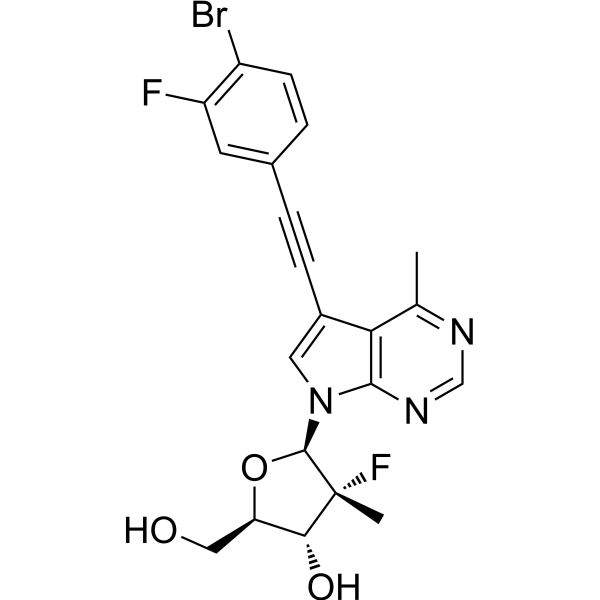
ZIKV-IN-1 is a potent zika virus inhibitor with an EC50 of 2.8 μM and EC90 of 6.8 μM. ZIKV-IN-1 shows anti-ZIKV activity with low cytotoxicity. ZIKV-IN-1 shows a strong affinity to ZIKV RdRp domain. ZIKV-IN-1 is a click chemistry reagent, it contains an Alkyne group and can undergo copper-catalyzed azide-alkyne cycloaddition (CuAAc) with molecules containing Azide groups.
For research use only. We do not sell to patients.
- CAS No.: 2762166-06-1
- Formula: C21H18BrF2N3O3
- Molecular Weight:478.29
-
Storage:
Please store the product under the recommended conditions in the Certificate of Analysis.

To place orders, for customer services and technical support, please contact: MedChemExpress USA
Tel: 609-228-6898 E-mail: [email protected] [email protected]


-
 Biological Activity
-
Chemical Information
- Protocols
- Purity & Documentation
- References
-
 Help & FAQs




Biological Activity
Description
IC50 & Target
EC50: 2.8 µM (ZIKV)[1]
Cellular Effect
|
Cell Line

|
Type | Value | Description | References |
|---|---|---|---|---|
| A549 | CC50 |
>50 μM
Compound: 38
|
Cytotoxicity against human A549 cells assessed as cell growth inhibition measured after 24 hrs by MTT assay
Cytotoxicity against human A549 cells assessed as cell growth inhibition measured after 24 hrs by MTT assay
|
[PMID: 35306290] |
| Huh-7 | CC50 |
43.8 μM
Compound: 38
|
Cytotoxicity against human Huh-7 cells assessed as cell growth inhibition measured after 24 hrs by MTT assay
Cytotoxicity against human Huh-7 cells assessed as cell growth inhibition measured after 24 hrs by MTT assay
|
[PMID: 35306290] |
| SNB-19 | CC50 |
49.4 μM
Compound: 38
|
Cytotoxicity against human SNB-19 cells assessed as cell growth inhibition measured after 24 hrs by MTT assay
Cytotoxicity against human SNB-19 cells assessed as cell growth inhibition measured after 24 hrs by MTT assay
|
[PMID: 35306290] |
| Vero | CC50 |
46.8 μM
Compound: 38
|
Cytotoxicity against African green monkey Vero cells assessed as cell growth inhibition measured after 24 hrs by MTT assay
Cytotoxicity against African green monkey Vero cells assessed as cell growth inhibition measured after 24 hrs by MTT assay
|
[PMID: 35306290] |
In Vitro
ZIKV-IN-1 (compound 38) (0-80 µM) shows anti-ZIKV activity in a dose-dependent manner in A549 cells[1].
ZIKV-IN-1 (5, 10, 25 µM; 48 h) decreases the expression of ZIKV E and cleaved caspase 3 protein[1].
ZIKV-IN-1 (1.25, 2.5, 5, 10, 20 µM; 0-250 s) shows strong affinity to ZIKV RdRp domain (Kd=1.87 µM) in A549 cells[1].
MedChemExpress (MCE) has not independently confirmed the accuracy of these methods. They are for reference only.
-
Cell Line:A549 cells
-
Concentration:0-50 µM
-
Incubation Time:
-
Result:Showed anti-ZIKV activity with an EC50 of 2.8 µM, EC90 of 6.8 µM, and CC50 of >50 µM.
-
Cell Line:A549, Vero cells
-
Concentration:5, 10, 25 µM
-
Incubation Time:48 h
-
Result:Decreased the expression of ZIKV E and cleaved caspase 3 protein.
-
Cell Line:Vero, SNB19, Huh7, A549 cells
-
Concentration:0-80 µM
-
Incubation Time:
-
Result:Showed low cytotoxicity with CC50s of 46.8, 49.4, 43.8, 54.1 µM for ZG01 strain of Vero, SNB19, Huh7, A549 cells, and CC50s of 470.5, 485.2, 512.8, 160.8 µM for MR766 strain of Vero, SNB19, Huh7, A549 cells, respectively.


Chemical Information
-
CAS No. 2762166-06-1
-
Molecular Weight 478.29
-
Formula C21H18BrF2N3O3
-
SMILES
CC1=NC=NC2=C1C(C#CC3=CC(F)=C(C=C3)Br)=CN2[C@@H]4O[C@@H]([C@H]([C@]4(F)C)O)CO
-
Shipping
Room temperature in continental US; may vary elsewhere.
-
Storage
Please store the product under the recommended conditions in the Certificate of Analysis.


Purity & Documentation
References

Calculators
Concentration (start) × Volume (start) = Concentration (final) × Volume (final)


ZIKV-IN-1 Related Classifications
HY-146957 Related Classifications
 Powered by Bioz