



Zilovertamab vedotin
Based on 1 publication(s) in Google Scholar
Zilovertamab vedotin (VLS-101) is a novel antibody-drug conjugate comprising the humanized monoclonal antibody zilovertamab and and the anti-microtubule cytotoxin monomethyl vedotin. Zilovertamab vedotin binding to tumor cell ROR1 results in rapid internalization, trafficking to lysosomes, antibody–agent conjugate cleavage, and monomethyl vedotin release. Zilovertamab vedotin induces apoptosis. Zilovertamab vedotin can be used in research of cancer.
For research use only. We do not sell to patients.
- Purity : 99.77%
- Molecular Weight:151000 (average)
-
Storage:
Please store the product under the recommended conditions in the Certificate of Analysis.

To place orders, for customer services and technical support, please contact: MedChemExpress USA
Tel: 609-228-6898 E-mail: [email protected] [email protected]


-
 Biological Activity
-
Chemical Information
- Protocols
- Purity & Documentation
- References
-
 Help & FAQs


-

HY-132254A
Sacituzumab govitecan (solution)Trop-2 ADC -

HY-P3371
Ifinatamab deruxtecanDNA Topoisomerase I Inhibitor -

HY-P99675
IvonescimabPD-1/VEGF Bispecific Antibody -

HY-P9921
Trastuzumab emtansineAntibody-drug Conjugate -

HY-132258A
Mirvetuximab soravtansine (solution)ADC -
HY-P99349
Loncastuximab tesirineAntibody-Drug Conjugate -
HY-70002
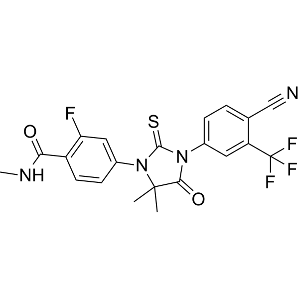
EnzalutamideAndrogen Receptor Antagonist -

HY-138298A
Trastuzumab deruxtecanHER2 ADC -

HY-141598
Datopotamab deruxtecan (solution)TROP2-Targeted ADC -

HY-P99201
ZilovertamabROR1 Inhibitor -
HY-15870
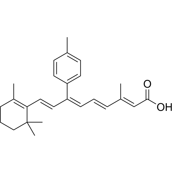
SR 11302AP-1 Inhibitor -

HY-P99001
Human IgG1 kappa, Isotype ControlIgG1κ Isotype Control
Publications Citing Use of MedChemExpress (MCE) Zilovertamab vedotin
More

Biological Activity
Description
In Vitro
Zilovertamab vedotin (VLS-101; 0-100 μg/mL) induces cytotoxicity in ROR1+ primary MCL cells in a dose-dependent manner[1].
Zilovertamab vedotin (24h; ibrutinib-sensitive (JeKo-1) and resistant (JeKo BTK KD_2) cell lines) induces cell apoptosis and cell cycle arrest at G2/M[1].
MedChemExpress (MCE) has not independently confirmed the accuracy of these methods. They are for reference only.
In Vivo
MedChemExpress (MCE) has not independently confirmed the accuracy of these methods. They are for reference only.
-
Animal Model:NSG PDX mice models with dual resistance to ibrutinib and CAR T. A PDX model[1]
-
Dosage:2.5 mg/kg
-
Administration:intravenous injection; weekly, for 3 weeks
-
Result:Eliminated subcutaneous tumor growth of BA-resistant PDX model (PDX-1).
Prolonged tumor bearing mouse survival.
Clinical Trial
| NCT Number | Sponsor | Condition | Start Date |
Phase

|
|---|---|---|---|---|
| NCT01329991 | 2011-05 | PHASE1 |


Chemical Information
-
Appearance Liquid
-
Molecular Weight 151000 (average)
-
Color Colorless to light yellow
-
SMILES
[Zilovertamab vedotin]
-
Synonyms
VLS-101; MK-2140
-
Shipping
Shipping with dry ice.
-
Storage
Please store the product under the recommended conditions in the Certificate of Analysis.


Publications (1)
-
Journal Impact Factor
-
Most Recent


Purity & Documentation
-
Data Sheet (259 KB)
-
SDS (251 KB)
- English - EN (251 KB)
- Français - FR (251 KB)
- Deutsch - DE (251 KB)
- Norwegian - NO (251 KB)
- Español - ES (251 KB)
- Swedish - SV (251 KB)
- Italian - IT (251 KB)
- Korean - KR (251 KB)
- Portuguese - PT (251 KB)
-
Handling Instructions (2659 KB)
References

Calculators
Concentration (start) × Volume (start) = Concentration (final) × Volume (final)


Zilovertamab vedotin Related Classifications
HY-P99956 Related Classifications
 Powered by Bioz