



Δ14-Triamcinolone acetonide

Based on 1 Customer Validation
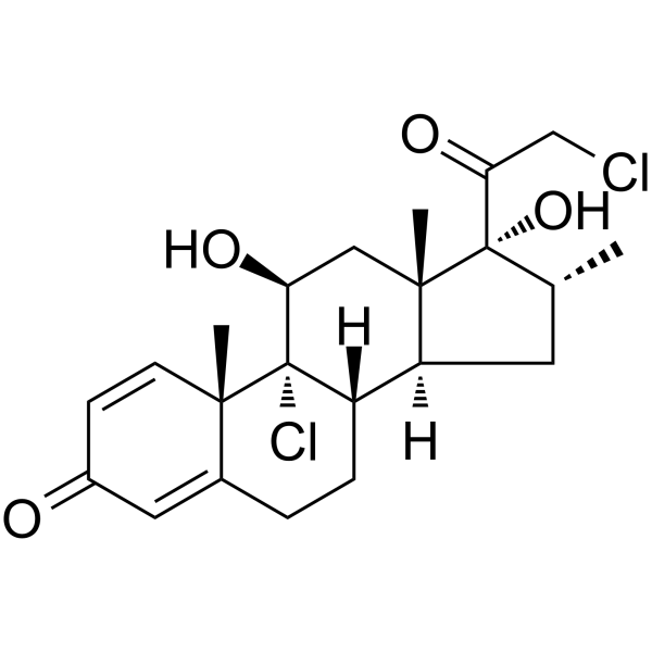
Δ14-Triamcinolone acetonide is a potential impurity. Triamcinolone acetonide inhibits basic fibroblast growth factor (bFGF) induced proliferation of retinal endothelial cells. Triamcinolone acetonide reduces chondrocyte viability and leads to cartilage destruction. Triamcinolone acetonide can be used in the study of diseases such as atopic dermatitis.
For research use only. We do not sell to patients.
- CAS No.: 1260149-96-9
- Formula: C24H29FO6
- Molecular Weight:432.48
-
Storage:
Please store the product under the recommended conditions in the Certificate of Analysis.


To place orders, for customer services and technical support, please contact: MedChemExpress USA
Tel: 609-228-6898 E-mail: [email protected] [email protected]


-
 Biological Activity
-
Chemical Information
- Protocols
- Purity & Documentation
- References
-
 Help & FAQs


-
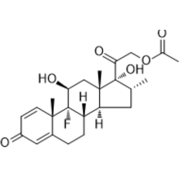
HY-14648A
Dexamethasone acetateCorticosteroid Ester -
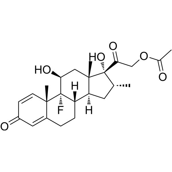
HY-14648AR
Dexamethasone acetate (Standard)Analytical Standard -
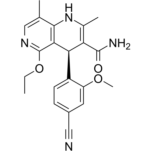
HY-111372
FinerenoneMineralocorticoid Receptor Antagonist -
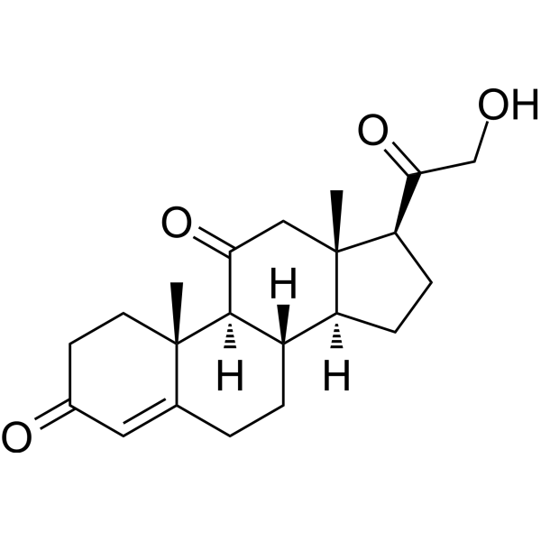
HY-113447
11-DehydrocorticosteroneEndogenous Corticosteroid -

HY-111372R
Finerenone (Standard)Mineralocorticoid Receptor Antagonist -
HY-B0629
MometasoneInhaled Glucocorticoid -

HY-B0629R
Mometasone (Standard)Inhaled Glucocorticoid -

HY-W749404
Clobetasone 17-Propionate


Biological Activity
Description


Chemical Information
-
CAS No. 1260149-96-9
-
Molecular Weight 432.48
-
Formula C24H29FO6
-
SMILES
OCC([C@]12[C@@]3(C([C@@]4([H])[C@]([C@H](C3)O)([C@@]5(C(CC4)=CC(C=C5)=O)C)F)=C[C@@]1([H])OC(C)(O2)C)C)=O
-
Shipping
Room temperature in continental US; may vary elsewhere.
-
Storage
Please store the product under the recommended conditions in the Certificate of Analysis.

Protocols
-
TPA/Croton Oil Ear Edema and Dermatitis
The TPA (12-O-tetradecanoylphorbol-13-acetate) and croton oil-induced mouse ear edema model is a well-established acute cutaneous inflammation system used to evaluate topical anti-inflammatory activity by measuring edema formation, neutrophil infiltration, vascular permeability, and cytokine-mediated skin responses in vivo. The inflammatory response is triggered by topical application of phorbol esters (TPA) or croton oil constituents, leading to rapid activation of protein kinase C signaling, leukocyte recruitment, and increased vascular permeability, which can be quantified by ear thickness, weight, dye extravasation, and biochemical markers such as myeloperoxidase (MPO) activity and pro-inflammatory mediators in ear tissue homogenates. This model is widely used for screening anti-inflammatory agents, where reductions in edema and inflammatory biomarkers reflect suppression of acute dermal inflammation and immune cell infiltration. Histological evaluation typically confirms epidermal
-
Endothelial Tube Formation Assay
Endothelial tube formation assay evaluates the ability of endothelial cells to attach, migrate, align, and organize into capillary-like networks when cultured on gelled basement membrane extract or Matrigel; the readout is the morphology and quantity of tube-like networks, which reflects an in vitro endothelial morphogenesis step related to angiogenesis. Basement membrane extract/Matrigel provides laminin-rich extracellular matrix cues that support endothelial differentiation into capillary-like structures, but it can contain biologically active growth factors, so growth-factor-reduced matrix is preferred when testing defined angiogenic stimulators or inhibitors.
-
Cell Viability Determination by MTT Colorimetric Assay
The following protocol uses the MTT colorimetric assay as a classic literature-established method for assessing cell viability/metabolic activity in cultured mammalian cells. MTT[3-(4,5-dimethylthiazol-2-yl)-2,5-diphenyltetrazolium bromide] is reduced by metabolically active cells to a colored formazan product; the amount of formazan is quantified spectrophotometrically and provides an indirect measure of metabolically active viable cells. Importantly, MTT reduction reflects cellular oxidoreductase/metabolic activity rather than an absolute direct count of living cells, so changes in cellular metabolism can alter the signal independently of cell number.


Purity & Documentation
-
Data Sheet (273 KB)
-
SDS (252 KB)
- Français - FR (252 KB)
- Deutsch - DE (252 KB)
- Norwegian - NO (252 KB)
- Español - ES (252 KB)
- Swedish - SV (252 KB)
- Italian - IT (252 KB)
- Korean - KR (252 KB)
- Portuguese - PT (252 KB)
-
Handling Instructions (2659 KB)
References

Calculators
Concentration (start) × Volume (start) = Concentration (final) × Volume (final)


Δ14-Triamcinolone acetonide Related Classifications
HY-Z8648 Related Classifications
 Powered by Bioz