



3-Nitro-L-tyrosine

Based on 1 Customer Validation
3-Nitro-L-tyrosine serves as a biomarker of oxidative stress. 3-Nitro-L-tyrosine attenuates the pressor and vasoconstrictive effects of angiotensin II by inhibiting the α1-adrenergic receptor-mediated signaling pathway, and participates in hemodynamic regulation under pathological conditions such as inflammation and ischemia. 3-Nitro-L-tyrosine can be used in studies related to atherosclerosis, ischemia-reperfusion and sepsis.
For research use only. We do not sell to patients.
- Purity : 99.84%
- CAS No.: 621-44-3
- Formula: C9H10N2O5
- Molecular Weight:226.19
-
Storage:Powder -20°C, 3 years , 4°C, 2 years ; In solvent -80°C, 6 months , -20°C, 1 month

To place orders, for customer services and technical support, please contact: MedChemExpress USA
Tel: 609-228-6898 E-mail: [email protected] [email protected]


-
 Biological Activity
-
Chemical Information
-
Solvent & Solubility
- Protocols
- Purity & Documentation
- References
-
 Help & FAQs


-
HY-Y0017
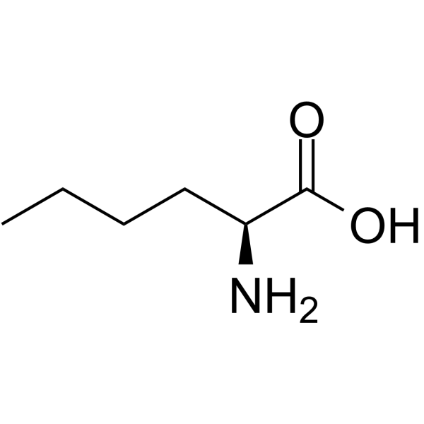
L-Norleucine -

HY-113248R
3-Nitro-L-tyrosine (Standard)Standard -

HY-Y0017R
L-Norleucine (Standard) -
HY-N0667
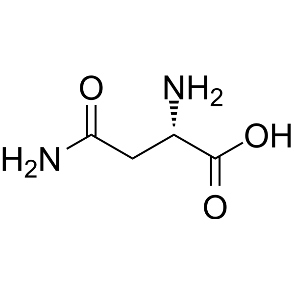
L-AsparagineEndogenous Metabolite -
HY-107848
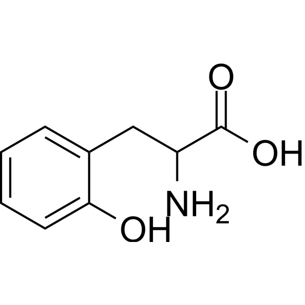
DL-O-TyrosineAmino Acid Derivative -
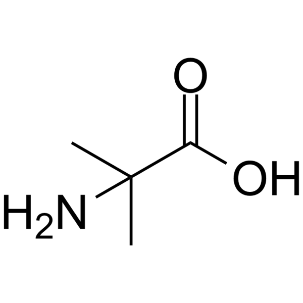
HY-Y0124
NSC 16590 -
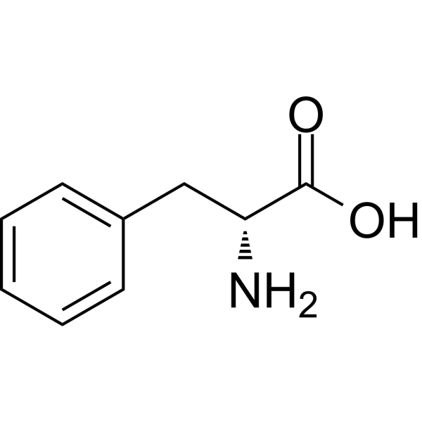
HY-Y0079
D-PhenylalanineBacterial Biofilm Formation Inhibibitor -

HY-N0667R
L-Asparagine (Standard)Endogenous Metabolite -

HY-Y0079R
D-Phenylalanine (Standard) -
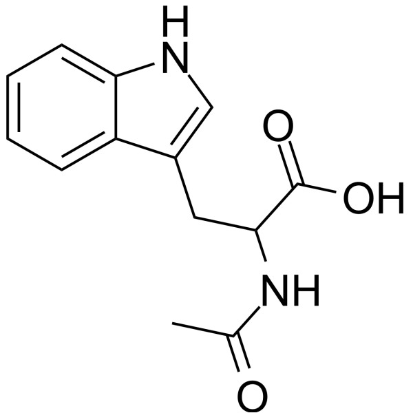
HY-W011982
Ac-DL-Trp-OH -

HY-Y0124R
NSC 16590 (Standard) -

HY-W011982R
Ac-DL-Trp-OH (Standard)


Biological Activity
Description
IC50 & Target
|
Human Endogenous Metabolite |
In Vitro
3-Nitro-L-tyrosine can be found in the brain as a product of superoxide and nitric oxide, and serves as a marker for oxidative stress induced by reactive oxygen species and nitric oxide[1].
MedChemExpress (MCE) has not independently confirmed the accuracy of these methods. They are for reference only. Further protocols information, click here.
In Vivo
MedChemExpress (MCE) has not independently confirmed the accuracy of these methods. They are for reference only.
-
Animal Model:Sprague-Dawley (male, 250-350 g, pentobarbital-anesthetized, surgically implanted with hemodynamic monitoring catheters and Doppler flow probes)[2]
-
Dosage:2.5 μmol/kg
-
Administration:i.v.; single bolus
-
Result:Produced a significant 36% increase in baseline hindquarter vascular resistance at 120-180 min post-treatment.
Diminished the dose-dependent pressor responses to angiotensin II at 120-180 min post-treatment.
Significantly reduced the dose-dependent vasoconstrictor effects of angiotensin II in renal and mesenteric beds at 30-60 min post-treatment, with further attenuation evident at 120-180 min post-treatment.
Did not alter the minor effects of angiotensin II on hindquarter vascular resistance.


Chemical Information
-
CAS No. 621-44-3
-
Appearance Solid
-
Molecular Weight 226.19
-
Formula C9H10N2O5
-
Color Light yellow to yellow
-
SMILES
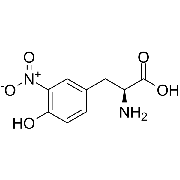
N[C@@H](CC1=CC([N+]([O-])=O)=C(C=C1)O)C(O)=O
-
Structure Classification
-
Shipping
Room temperature in continental US; may vary elsewhere.
-
Storage
Powder -20°C 3 years 4°C 2 years In solvent -80°C 6 months -20°C 1 month


Solvent & Solubility
In Vitro:
H2O : 4.76 mg/mL (21.04 mM; ultrasonic and adjust pH to 2 with 1 M HCL)
Please refer to the solubility information to select the appropriate solvent. Once prepared, please aliquot and store the solution to prevent product inactivation from repeated freeze-thaw cycles.
Storage method and period of stock solution: -80°C, 6 months; -20°C, 1 month. When stored at -80°C, please use it within 6 months. When stored at -20°C, please use it within 1 month.
* Note: If you choose water as the stock solution, please dilute it to the working solution, then filter and sterilize it with a 0.22 μm filter before use.
Please refer to the solubility information to select the appropriate solvent. Once prepared, please aliquot and store the solution to prevent product inactivation from repeated freeze-thaw cycles.
Storage method and period of stock solution: -80°C, 6 months; -20°C, 1 month. When stored at -80°C, please use it within 6 months. When stored at -20°C, please use it within 1 month.
* Note: If you choose water as the stock solution, please dilute it to the working solution, then filter and sterilize it with a 0.22 μm filter before use.
Concentration (start) × Volume (start) = Concentration (final) × Volume (final)

Protocols
-
LPS-Induced Endotoxemia/Systemic Inflammation
Lipopolysaccharide (LPS)-induced endotoxemia is a widely used in vivo model of acute systemic inflammation in which LPS, a Gram-negative bacterial endotoxin, activates innate immune signaling primarily through TLR4, leading to rapid and transient induction of pro-inflammatory cytokines such as TNF-α, IL-6, and IL-1β in circulation and tissues. This cytokine surge is commonly used as a measurable readout of systemic inflammatory activation and immune dysregulation, and is typically assessed within hours after intraperitoneal LPS administration in mouse models of endotoxemia. The model captures key features of systemic inflammatory response syndrome, including cytokine release, immune cell activation, and downstream tissue responses, and has been used to evaluate anti-inflammatory interventions such as cytokine modulation, lipid mediators, and immune cell-targeting therapies.
-
ROS/oxidative-stress fluorescent staining
ROS/oxidative-stress fluorescent staining uses cell-permeant fluorogenic probes that become fluorescent after oxidation inside cells or tissues; commonly used examples include DCFH-DA/DCFDA for broad cellular oxidant detection, DHE for superoxide-related signal detection, MitoSOX for mitochondrial superoxide-related signal detection, and CellROX probes for oxidative-stress-associated fluorescence readouts. The assay detects probe oxidation rather than a single ROS species unless the probe and analysis method have been chemically validated for that species. DCFH-DA enters cells, is deacetylated by intracellular esterases to DCFH, and produces fluorescent DCF after oxidation, so the readout is used as an operational measure of total cellular oxidative stress rather than a species-specific ROS measurement. DHE and MitoSOX can report superoxide-related oxidation, but red fluorescence alone can include non-specific ethidium-like oxidation products; HPLC or optimized spectral approaches are
-
Research Protocol for Cardiovascular Diseases
Cardiovascular disease can be modeled as maladaptive cardiac remodeling, where ischemic injury or pressure overload activates inflammatory signaling, fibroblast activation, extracellular-matrix deposition, cardiomyocyte hypertrophy, vascular remodeling, and progressive ventricular dysfunction. The TGF-β/SMAD axis is a central profibrotic pathway after myocardial injury and pressure overload, while innate immune and cytokine pathways regulate leukocyte recruitment, scar formation, and adverse remodeling. Key unresolved questions include which inflammatory signals are reparative versus harmful, when fibrosis is protective versus maladaptive, and whether pathway inhibition improves function without weakening necessary infarct healing or compensatory remodeling.
-
Research Protocol for Inflammation-related Diseases
The NLRP3 inflammasome is a cytosolic innate immune signaling platform that integrates priming signals and danger-signal activation to promote caspase-1 activation, maturation of IL-1β and IL-18, and gasdermin D-mediated pyroptotic cell death. The core experimental logic is to determine whether inflammatory disease phenotypes are driven by increased NLRP3 expression, ASC-containing inflammasome assembly, caspase-1 cleavage, GSDMD cleavage, and extracellular release of IL-1β/IL-18 rather than by nonspecific cell injury alone. The pathway is strongly linked to inflammation-related disease phenotypes because monosodium urate crystals activate NALP3/NLRP3 inflammasome signaling in gout-like crystal inflammation, cholesterol crystals activate NLRP3 inflammasomes in atherogenesis models, and DSS-induced intestinal inflammation has been reported to involve NLRP3 inflammasome activity. However, experimental colitis studies also show context-dependent protective effects of NLRP3 inflammasome co


Purity & Documentation
-
Data Sheet (282 KB)
-
SDS (393 KB)
- English - EN (393 KB)
- Français - FR (393 KB)
- Deutsch - DE (393 KB)
- Norwegian - NO (393 KB)
- Español - ES (393 KB)
- Swedish - SV (393 KB)
- Italian - IT (393 KB)
- Korean - KR (393 KB)
- Portuguese - PT (393 KB)
-
Handling Instructions (2659 KB)
References
[1]. Maruyama W, et al. Identification of 3-nitro-L-tyrosine, a product of nitric oxide and superoxide, as an indicator of oxidative stress in the human brain. J Chromatogr B Biomed Appl. 1996 Feb 9;676(1):153-8. [Content Brief]
[2]. Kooy NW, et al. The peroxynitrite product 3-nitro-L-tyrosine attenuates the hemodynamic responses to angiotensin II in vivo. Eur J Pharmacol. 1996 Nov 14;315(2):165-70. [Content Brief]


Complete Stock Solution Preparation Table
Please refer to the solubility information to select the appropriate solvent. Once prepared, please aliquot and store the solution to prevent product inactivation from repeated freeze-thaw cycles.
Storage method and period of stock solution: -80°C, 6 months; -20°C, 1 month. When stored at -80°C, please use it within 6 months. When stored at -20°C, please use it within 1 month.
| Optional Solvent | Concentration Solvent Mass | 1 mg | 5 mg | 10 mg | 25 mg |
|---|---|---|---|---|---|
| H2O | 1 mM | 4.4211 mL | 22.1053 mL | 44.2106 mL | 110.5265 mL |
| 5 mM | 0.8842 mL | 4.4211 mL | 8.8421 mL | 22.1053 mL | |
| 10 mM | 0.4421 mL | 2.2105 mL | 4.4211 mL | 11.0527 mL | |
| 15 mM | 0.2947 mL | 1.4737 mL | 2.9474 mL | 7.3684 mL | |
| 20 mM | 0.2211 mL | 1.1053 mL | 2.2105 mL | 5.5263 mL |
* Note: If you choose water as the stock solution, please dilute it to the working solution, then filter and sterilize it with a 0.22 μm filter before use.


3-Nitro-L-tyrosine Related Classifications
HY-113248 Related Classifications
 Powered by Bioz