



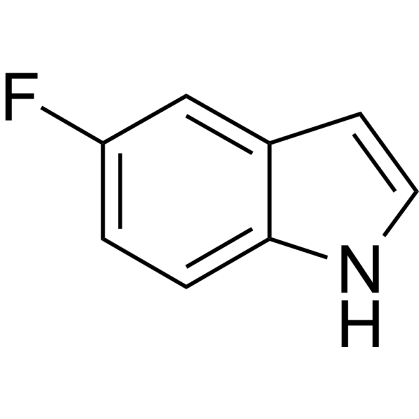
5-Fluoroindole

Based on 1 Customer Validation
5-Fluoroindole is an orally active fluorinated indole derivative and antibacterial agent. 5-Fluoroindole induces ROS accumulation and triggers Apoptosis. 5-Fluoroindole inhibits the growth of pan-susceptible Mycobacterium tuberculosis (Mtb) H37Rv strains. 5-Fluoroindole has significant bactericidal activity against Pseudomonas syringae pv. actinidiae (Psa) with an EC50 of 15.34 μg/mL. 5-Fluoroindole introduces fluorine labeling for protein research. 5-Fluoroindole can be used in the study of tuberculosis and kiwifruit bacterial canker.
For research use only. We do not sell to patients.
- Purity : 99.88%
- CAS No.: 399-52-0
- Formula: C8H6FN
- Molecular Weight:135.14
-
Storage:
4°C, protect from light, stored under nitrogen
* In solvent : -80°C, 2 years; -20°C, 1 year (protect from light, stored under nitrogen)

To place orders, for customer services and technical support, please contact: MedChemExpress USA
Tel: 609-228-6898 E-mail: [email protected] [email protected]


-
 Biological Activity
-
Chemical Information
-
Solvent & Solubility
- Protocols
- Purity & Documentation
- References
-
 Help & FAQs


-
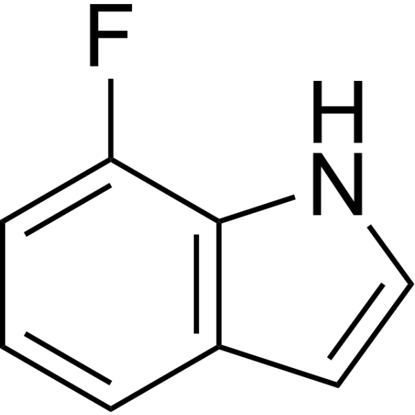
HY-W007613
7-Fluoro-1H-indoleBiochemical Reagent -

HY-21194
Perfluorohexanoic acid -
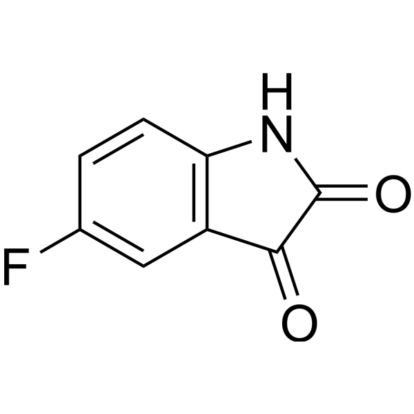
HY-W007549
5-FluoroisatinBiochemical Reagent -

HY-21190
Perfluorobutylsulfonyl fluorideIntermediate -
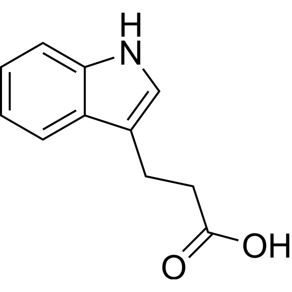
HY-W015229
3-Indolepropionic acid -
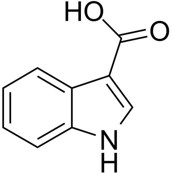
HY-40161
Indole-3-carboxylic acidEndogenous Metabolite -
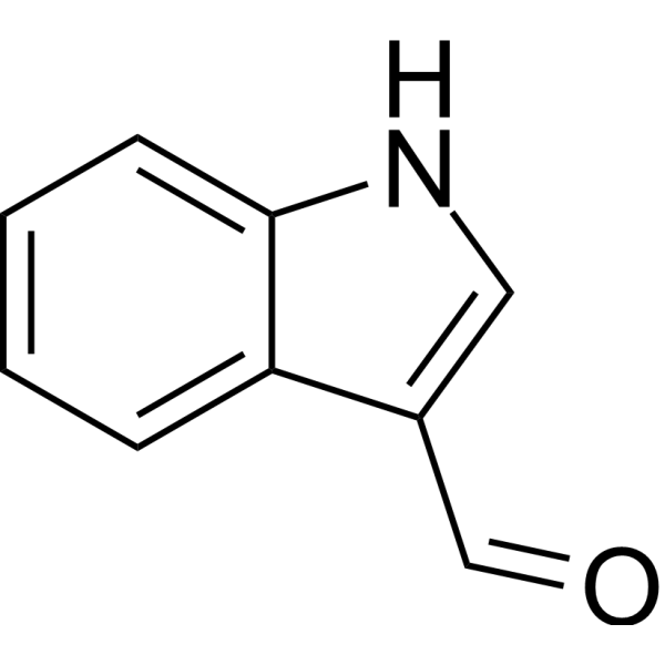
HY-W007376
Indole-3-carboxaldehydeEndogenous Metabolite -
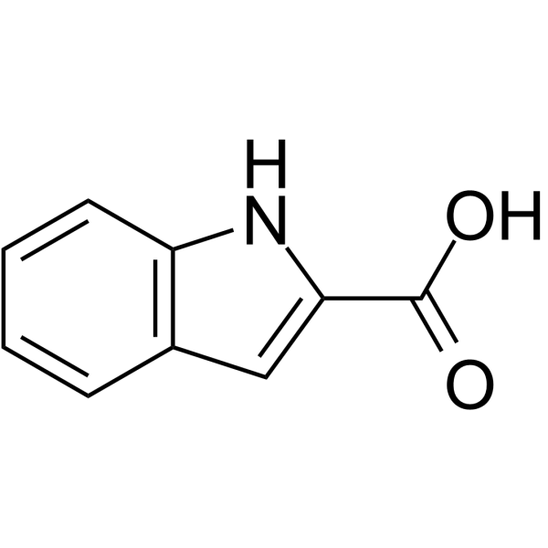
HY-I0096
Indole-2-carboxylic acidLipid Peroxidation Inhibitor -

HY-W007376R
Indole-3-carboxaldehyde (Standard)Endogenous Metabolite -

HY-W015229R
3-Indolepropionic acid (Standard) -

HY-40161R
Indole-3-carboxylic acid (Standard)Endogenous Metabolite -
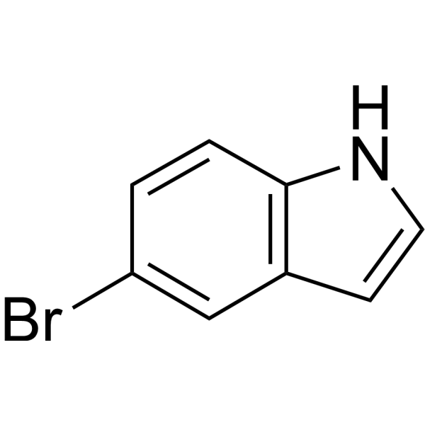
HY-30236
5-BromoindoleAntibacterial Agent


Biological Activity
Description
In Vitro
5-Fluoroindole (4.7-29.1 μM) inhibits the growth of pansensitive Mycobacterium tuberculosis (Mtb) H37Rv strain and drug-resistant clinical isolates[1].
5-Fluoroindole has high metabolic stability (Clint 9.0 mL/min/kg) in rat liver microsomes[1].
5-Fluoroindole exhibits significant bactericidal activity against Pseudomonas syringae pv. actinidiae (Psa), with an EC50 of 15.34 μg/mL[2].
MedChemExpress (MCE) has not independently confirmed the accuracy of these methods. They are for reference only. Further protocols information, click here.
In Vivo


Chemical Information
-
CAS No. 399-52-0
-
Appearance Solid
-
Molecular Weight 135.14
-
Formula C8H6FN
-
Color White to light brown
-
SMILES
FC1=CC=C2NC=CC2=C1
-
Shipping
Room temperature in continental US; may vary elsewhere.
-
Storage
4°C, protect from light, stored under nitrogen
* In solvent : -80°C, 2 years; -20°C, 1 year (protect from light, stored under nitrogen)


Solvent & Solubility
In Vitro:
DMSO : 100 mg/mL (739.97 mM; Need ultrasonic; Hygroscopic DMSO has a significant impact on the solubility of product, please use newly opened DMSO)
Please refer to the solubility information to select the appropriate solvent. Once prepared, please aliquot and store the solution to prevent product inactivation from repeated freeze-thaw cycles.
Storage method and period of stock solution: -80°C, 2 years; -20°C, 1 year (protect from light, stored under nitrogen). When stored at -80°C, please use it within 2 years. When stored at -20°C, please use it within 1 year.
Please refer to the solubility information to select the appropriate solvent. Once prepared, please aliquot and store the solution to prevent product inactivation from repeated freeze-thaw cycles.
Storage method and period of stock solution: -80°C, 2 years; -20°C, 1 year (protect from light, stored under nitrogen). When stored at -80°C, please use it within 2 years. When stored at -20°C, please use it within 1 year.
Concentration (start) × Volume (start) = Concentration (final) × Volume (final)
In Vivo:
Select the appropriate dissolution method based on your experimental animal and administration route.
- For the following dissolution methods, please ensure to first prepare a clear stock solution using an In Vitro approach and then sequentially add co-solvents:
- To ensure reliable experimental results, the clarified stock solution can be appropriately stored based on storage conditions. As for the working solution for In Vivo experiments, it is recommended to prepare freshly and use it on the same day.
- The percentages shown for the solvents indicate their volumetric ratio in the final prepared solution. If precipitation or phase separation occurs during preparation, heat and/or sonication can be used to aid dissolution.
Add each solvent one by one: 10% DMSO 40% PEG300 5% Tween-80 45% Saline
Solubility: ≥ 2.5 mg/mL (18.50 mM); Clear solution
This protocol yields a clear solution of ≥ 2.5 mg/mL (saturation unknown).
Taking 1 mL working solution as an example, add 100 μL DMSO stock solution (25.0 mg/mL) to 400 μL PEG300, and mix evenly; then add 50 μL Tween-80 and mix evenly; then add 450 μL Saline to adjust the volume to 1 mL.
Preparation of Saline: Dissolve 0.9 g sodium chloride in ddH₂O and dilute to 100 mL to obtain a clear Saline solution.
Add each solvent one by one: 10% DMSO 90% (20% SBE-β-CD in Saline)
Solubility: ≥ 2.5 mg/mL (18.50 mM); Clear solution
This protocol yields a clear solution of ≥ 2.5 mg/mL (saturation unknown).
Taking 1 mL working solution as an example, add 100 μL DMSO stock solution (25.0 mg/mL) to 900 μL 20% SBE-β-CD in Saline, and mix evenly.
Preparation of 20% SBE-β-CD in Saline (4°C, storage for one week): 2 g SBE-β-CD powder is dissolved in 10 mL Saline, completely dissolve until clear.
In Vivo Dissolution Calculator
Please enter the basic information of animal experiments:
-
-
-
-
Recommended: Prepare an additional quantity of animals to account for potential losses during experiments.
Please enter your animal formula composition:
-
%DMSO +
Recommended: Keep the proportion of DMSO in working solution below 2% if your animal is weak.
-
%+
-
+%Tween-80 + +
-
%Saline +
The co-solvents required include: DMSO, . All of co-solvents are available by MedChemExpress (MCE). , Tween 80. All of co-solvents are available by MedChemExpress (MCE).
Working solution concentration: 0.22 mg/mL
Method for preparing stock solution: mg drug dissolved in μL DMSO. Stock solution concentration: mg/mL. * In solvent : -80°C, 2 years; -20°C, 1 year (protect from light, stored under nitrogen)
1. Take μL DMSO stock solution;
2. Add μL .
μL , mix evenly;
3. Then add μL Tween 80, mix evenly;
4. Then add μL
Please ensure that the stock solution in the first step is dissolved to a clear state, and add co-solvents in sequence. You can use ultrasonic heating (ultrasonic cleaner, recommended frequency 20-40 kHz), vortexing, etc. to assist dissolution.

Protocols
-
Research Protocol for Infectious Diseases
Infectious-disease experiments test how pathogens interact with host barriers, innate immune receptors, inflammatory signaling, pathogen replication, and tissue injury; pattern-recognition receptors such as TLRs, RIG-I-like receptors, NOD-like receptors, and inflammasomes detect microbial molecules and activate NF-κB, interferon, and cytokine responses. The central hypothesis is that infection severity reflects the balance between pathogen burden and host response: protective inflammation restricts pathogen growth, whereas excessive or mislocalized inflammation contributes to tissue damage and disease phenotype. Unresolved questions include which host pathways are protective versus pathogenic, why some infection models fail to translate to human disease, and which combined readouts best predict clinically relevant infection outcomes.
-
Apoptosis
Apoptosis, also called programmed cell death, is generally characterized by distinct morphological characteristics.
-
TUNEL staining for apoptotic DNA fragmentation
TUNEL staining detects DNA strand breaks by using terminal deoxynucleotidyl transferase to add labeled nucleotides to exposed 3′-OH DNA termini, generating either microscopic staining in fixed cells or tissue sections, or fluorescence/cytometric signal in cell suspensions. TUNEL positivity reflects DNA fragmentation but should not be interpreted alone as definitive apoptosis, because TUNEL can also label necrotic, autolytic, mechanically damaged, or DNA-repair-associated DNA breaks.
-
Annexin V plus membrane-impermeant dye apoptosis staining
Annexin V-based apoptosis assays rely on the detection of phosphatidylserine (PS) externalization from the inner leaflet of the plasma membrane to the outer leaflet, an early biochemical hallmark of apoptosis. Fluorescently labeled Annexin V binds PS in a calcium-dependent manner, enabling identification of early apoptotic cells by flow cytometry or fluorescence microscopy. When combined with a membrane-impermeant DNA-binding dye (e. g. , propidium iodide), this approach allows discrimination between viable (Annexin V−/dye−), early apoptotic (Annexin V+/dye−), and late apoptotic or necrotic (Annexin V+/dye+) cell populations by assessing membrane integrity and PS exposure.
-
ROS/oxidative-stress fluorescent staining
ROS/oxidative-stress fluorescent staining uses cell-permeant fluorogenic probes that become fluorescent after oxidation inside cells or tissues; commonly used examples include DCFH-DA/DCFDA for broad cellular oxidant detection, DHE for superoxide-related signal detection, MitoSOX for mitochondrial superoxide-related signal detection, and CellROX probes for oxidative-stress-associated fluorescence readouts. The assay detects probe oxidation rather than a single ROS species unless the probe and analysis method have been chemically validated for that species. DCFH-DA enters cells, is deacetylated by intracellular esterases to DCFH, and produces fluorescent DCF after oxidation, so the readout is used as an operational measure of total cellular oxidative stress rather than a species-specific ROS measurement. DHE and MitoSOX can report superoxide-related oxidation, but red fluorescence alone can include non-specific ethidium-like oxidation products; HPLC or optimized spectral approaches are
-
Apoptosis Solutions
Apoptosis is a regulated, generally non-lytic cell-death pathway that removes unwanted, damaged, infected, or abnormal cells through coordinated morphological changes, caspase activation, DNA fragmentation, and membrane remodeling. The intrinsic apoptosis pathway is controlled mainly by mitochondrial outer membrane permeabilization, BCL-2 family proteins, cytochrome c release, apoptosome formation, caspase-9 activation, and downstream executioner caspase-3/7 activation. The extrinsic apoptosis pathway is initiated by death receptors such as Fas, TNFR, and TRAIL receptors, which recruit adaptor proteins and activate caspase-8 before engaging executioner caspases or mitochondrial amplification through BID cleavage. Apoptosis is linked to many phenotypes, including cancer cell killing, tissue homeostasis, immune regulation, neurodegeneration, infection response, and treatment-induced cytotoxicity; unresolved questions include how apoptosis interacts with necroptosis, pyroptosis, ferroptos
-
Bacterial live/dead nucleic-acid viability staining
The LIVE/DEAD bacterial viability staining method is based on differential permeability of nucleic-acid-binding fluorescent dyes, most commonly SYTO 9 and propidium iodide (PI), which enables discrimination of bacterial populations with intact versus compromised cytoplasmic membranes. SYTO 9 penetrates both intact and damaged bacterial membranes and binds nucleic acids to produce green fluorescence, whereas propidium iodide penetrates only cells with compromised membranes and fluoresces red while also reducing SYTO 9 signal through competitive binding and fluorescence interactions. The resulting fluorescence pattern is interpreted as a proxy for membrane integrity, which is widely used as an indicator of bacterial viability in microscopy, flow cytometry, and spectroscopic platforms. However, mechanistic studies show that SYTO 9 and PI interactions involve displacement and fluorescence resonance energy transfer effects, which can influence signal interpretation depending on dye ratios a


Purity & Documentation
-
Data Sheet (275 KB)
-
SDS (394 KB)
- English - EN (394 KB)
- Français - FR (394 KB)
- Deutsch - DE (394 KB)
- Norwegian - NO (394 KB)
- Español - ES (394 KB)
- Swedish - SV (394 KB)
- Italian - IT (394 KB)
- Korean - KR (394 KB)
- Portuguese - PT (394 KB)
-
Handling Instructions (2659 KB)
References
[1]. Neves CE, et al., 5-Fluoroindole Reduces the Bacterial Burden in a Murine Model of Mycobacterium tuberculosis Infection. ACS Omega. 2024 Jul 16;9(30):32969-32979. [Content Brief]
[2]. Zhang C, et al. 5-Fluoroindole Inhibits Its Putative Target Methionine Synthase and Pseudomonas syringae pv. actinidiae Virulence. J Agric Food Chem. 2025 Jul 2;73(26):16193-16204. [Content Brief]
[3]. Pan B, et al. Fluorine-19 labeling of the tryptophan residues in the G protein-coupled receptor NK1R using the 5-fluoroindole precursor in Pichia pastoris expression. J Biomol NMR. 2024 Sep;78(3):133-138. [Content Brief]


Complete Stock Solution Preparation Table
Please refer to the solubility information to select the appropriate solvent. Once prepared, please aliquot and store the solution to prevent product inactivation from repeated freeze-thaw cycles.
Storage method and period of stock solution: -80°C, 2 years; -20°C, 1 year (protect from light, stored under nitrogen). When stored at -80°C, please use it within 2 years. When stored at -20°C, please use it within 1 year.
| Optional Solvent | Concentration Solvent Mass | 1 mg | 5 mg | 10 mg | 25 mg |
|---|---|---|---|---|---|
| DMSO | 1 mM | 7.3997 mL | 36.9987 mL | 73.9973 mL | 184.9933 mL |
| 5 mM | 1.4799 mL | 7.3997 mL | 14.7995 mL | 36.9987 mL | |
| 10 mM | 0.7400 mL | 3.6999 mL | 7.3997 mL | 18.4993 mL | |
| 15 mM | 0.4933 mL | 2.4666 mL | 4.9332 mL | 12.3329 mL | |
| 20 mM | 0.3700 mL | 1.8499 mL | 3.6999 mL | 9.2497 mL | |
| 25 mM | 0.2960 mL | 1.4799 mL | 2.9599 mL | 7.3997 mL | |
| 30 mM | 0.2467 mL | 1.2333 mL | 2.4666 mL | 6.1664 mL | |
| 40 mM | 0.1850 mL | 0.9250 mL | 1.8499 mL | 4.6248 mL | |
| 50 mM | 0.1480 mL | 0.7400 mL | 1.4799 mL | 3.6999 mL | |
| 60 mM | 0.1233 mL | 0.6166 mL | 1.2333 mL | 3.0832 mL | |
| 80 mM | 0.0925 mL | 0.4625 mL | 0.9250 mL | 2.3124 mL | |
| 100 mM | 0.0740 mL | 0.3700 mL | 0.7400 mL | 1.8499 mL |


5-Fluoroindole Related Classifications
HY-40156 Related Classifications
 Powered by Bioz