



Adrenic acid
Based on 1 publication(s) in Google Scholar
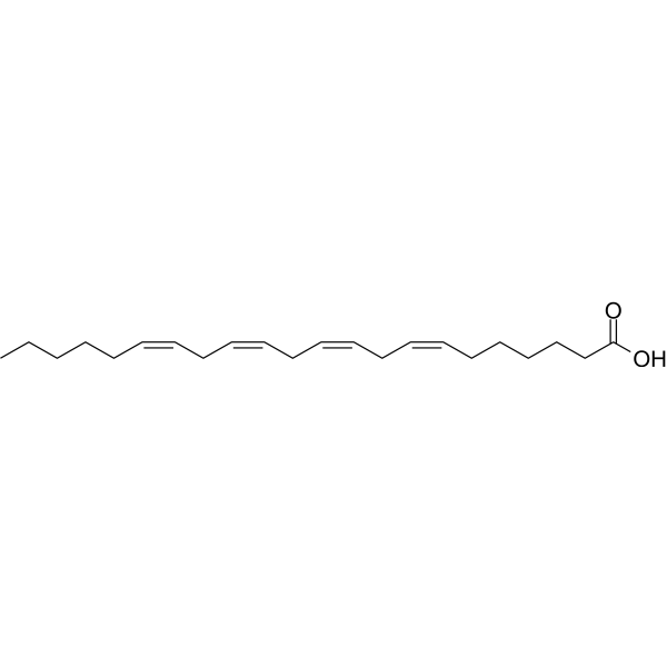
Adrenic Acid (cis-7,10,13,16-Docosatetraenoic acid) is a naturally polyunsaturated fatty acid in the adrenal gland, brain, kidney, and vasculature. Adrenic Acid can regulate the vascular tone in arteries of the adrenal cortex. Adrenic Acid also is an inflammation enhancer in non-alcoholic fatty liver disease.
For research use only. We do not sell to patients.
- Purity : 99.94%
- CAS No.: 28874-58-0
- Formula: C22H36O2
- Molecular Weight:332.52
-
Storage:
Solution, -20°C, 2 years

To place orders, for customer services and technical support, please contact: MedChemExpress USA
Tel: 609-228-6898 E-mail: [email protected] [email protected]


-
 Biological Activity
-
Chemical Information
- Protocols
- Purity & Documentation
- References
-
 Help & FAQs


-
HY-109590
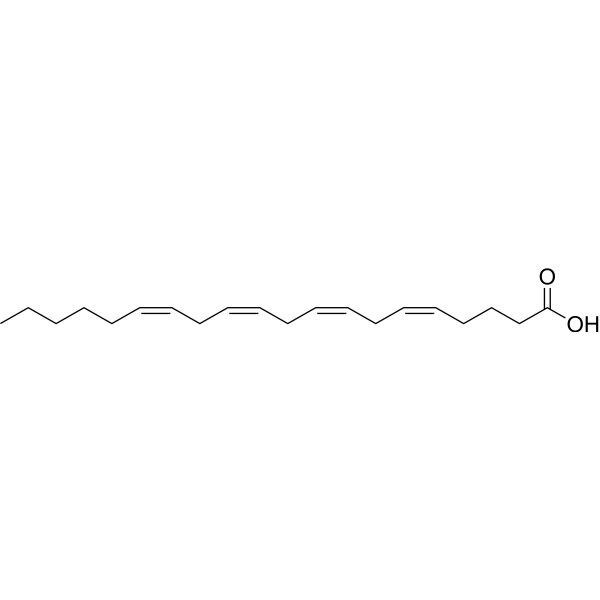
Arachidonic acidPolyunsaturated Fatty Acid -
HY-A0143
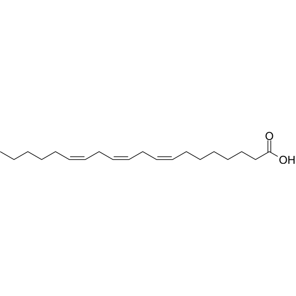
Dihomo-γ-linolenic acidEndogenous Metabolite -
HY-N7864
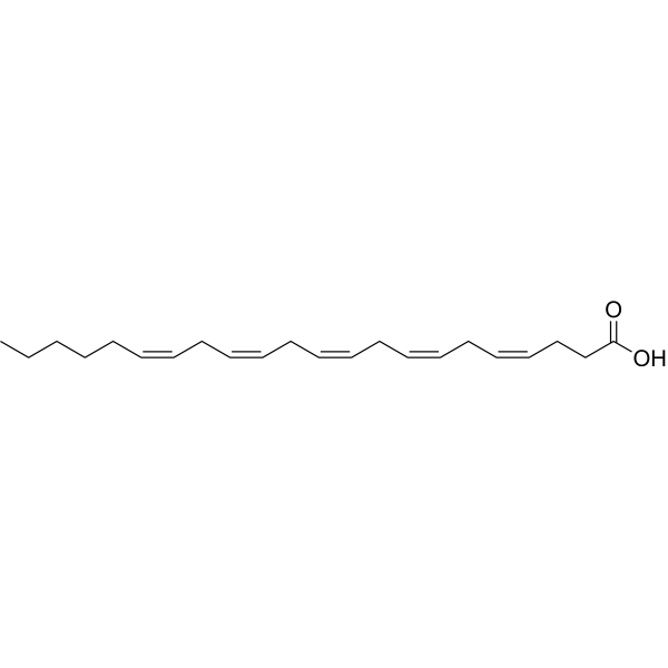
Osbond acidFatty acid -
HY-N7140
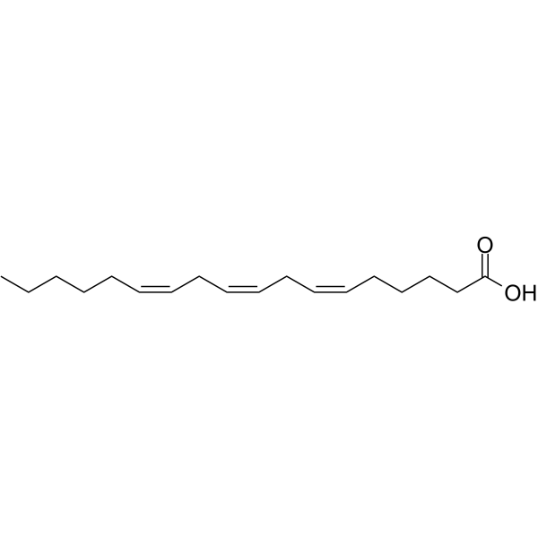
Gamma-Linolenic acidEndogenous Metabolite -
HY-113159
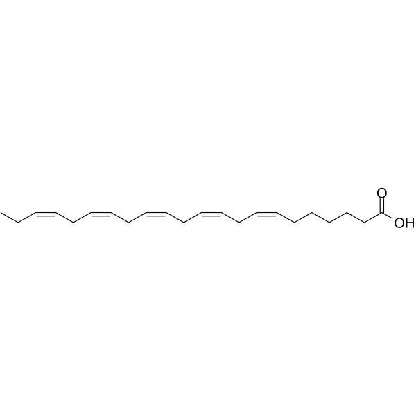
Docosapentaenoic acid (22n-3)Component Of Phospholipids -
HY-B0660
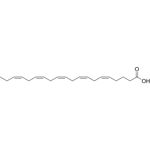
Eicosapentaenoic AcidLC-PUFA -
HY-B2167
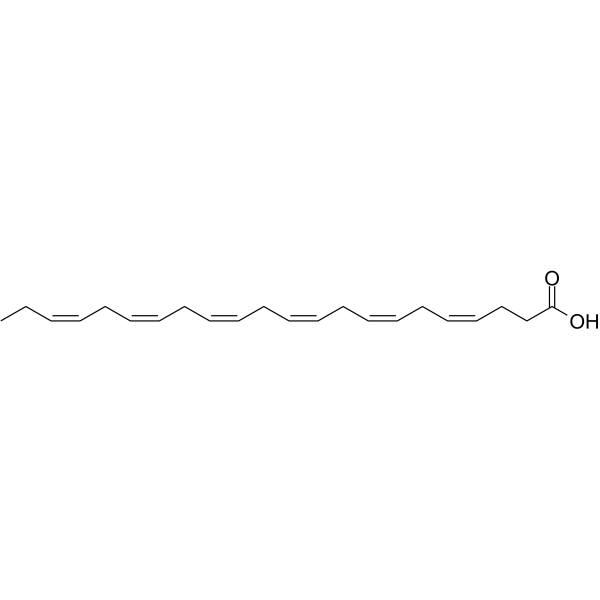
Docosahexaenoic acidomega-3 Fatty Acid -
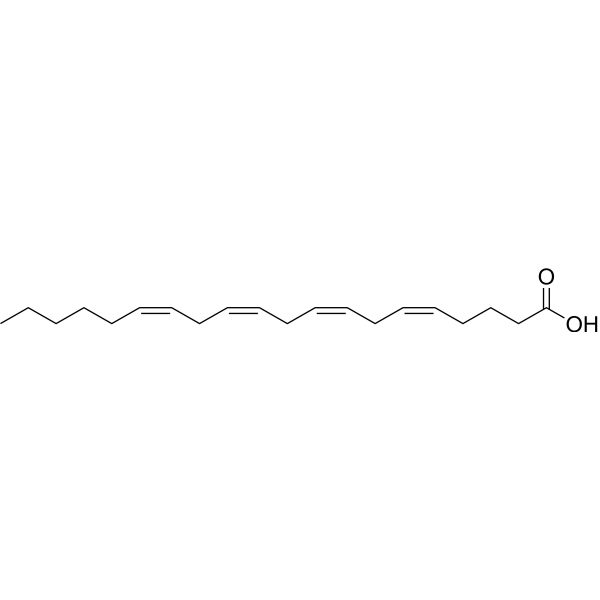
HY-109590R
Arachidonic acid (Standard)Analytical Standard of Arachidonic acid -
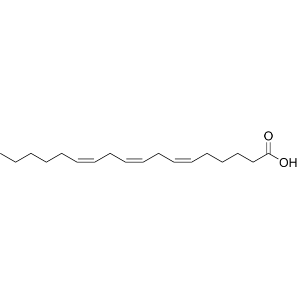
HY-N7140R
Gamma-Linolenic acid (Standard) -
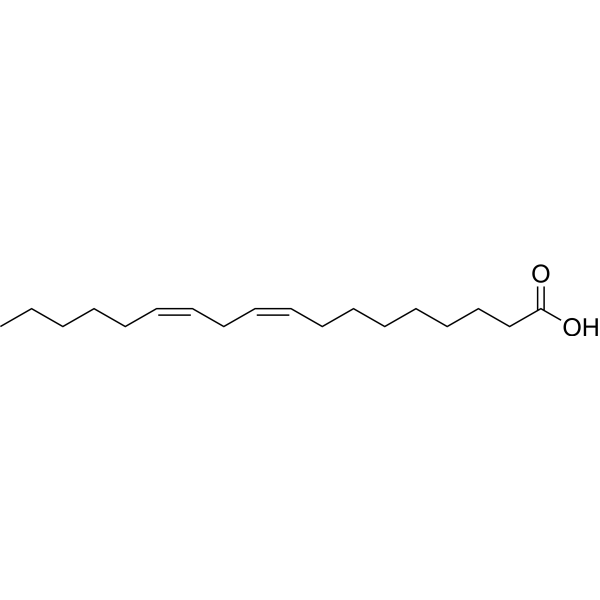
HY-N0729
Linoleic acidHuman Endogenous Metabolite -
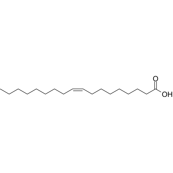
HY-N1446
Oleic acidMonounsaturated Fatty Acid -
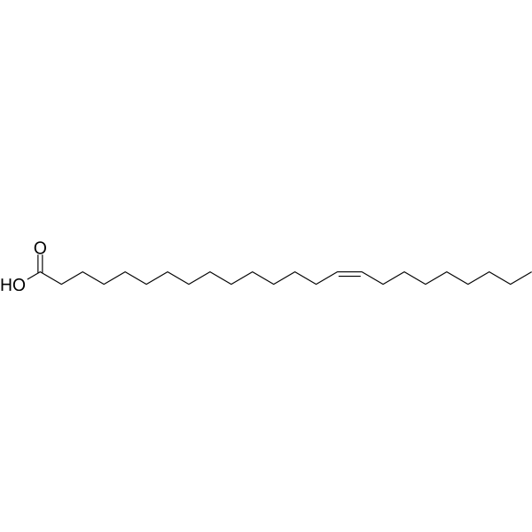
HY-N2526
Nervonic acidEndogenous Metabolite
Publications Citing Use of MedChemExpress (MCE) Adrenic acid
MoreAll Endogenous Metabolite Isoforms
More

Biological Activity
Description
IC50 & Target
|
Human Endogenous Metabolite |
In Vitro
Adrenic acid is produced via the elongation of arachidonic acid by ELOVL2 and 5. Interestingly, the hepatic level of free Adrenic Acid (22:4n6) is markedly higher in the CDAHFD-fed db/db mice than in the SD-fed db/db mice. In plasma, the difference in the adrenic acid level reaches statistical significance (p<0.001). It is also found that the concentrations of phospholipid species containing Adrenic Acid, such as PC or PE (40:4) (containing side chain 18:0/22:4), and LPC 22:4 (sn-1/sn-2), are also significantly increased. (sn-1/sn-2). The hepatic and plasma levels of Adrenic Acid, which is an omega 6 polyunsaturated fatty acid (PUFA), exhibit greater differences between the CDAHFD-fed db/db mice and the SD-fed db/db mice (8.6-fold higher vs. 1.6-fold higher in liver tissue, 5.3-fold higher vs. 2.2-fold higher in plasma) than those of well-known proinflammatory PUFA, such as Arachidonic Acid[1].
MedChemExpress (MCE) has not independently confirmed the accuracy of these methods. They are for reference only. Further protocols information, click here.


Chemical Information
-
CAS No. 28874-58-0
-
Appearance Liquid
-
Molecular Weight 332.52
-
Formula C22H36O2
-
Color Colorless to light yellow
-
SMILES
CCCCC/C=C\C/C=C\C/C=C\C/C=C\CCCCCC(O)=O
-
Synonyms
cis-7,10,13,16-Docosatetraenoic acid
-
Structure Classification
-
Initial Source
-
Shipping
Room temperature in continental US; may vary elsewhere.
-
Storage
Solution, -20°C, 2 years


Publications (1)
-
Journal Impact Factor
-
Most Recent
-
Adv Sci (Weinh)
CypD Dependent mPTP Opening Is Crucial for Oxidized Mitochondrial DNA Release in Ferroptosis. [Abstract]2026 Apr;13(20):e02239. PMID: 41700459

Protocols
-
Research Protocol for Inflammation-related Diseases
The NLRP3 inflammasome is a cytosolic innate immune signaling platform that integrates priming signals and danger-signal activation to promote caspase-1 activation, maturation of IL-1β and IL-18, and gasdermin D-mediated pyroptotic cell death. The core experimental logic is to determine whether inflammatory disease phenotypes are driven by increased NLRP3 expression, ASC-containing inflammasome assembly, caspase-1 cleavage, GSDMD cleavage, and extracellular release of IL-1β/IL-18 rather than by nonspecific cell injury alone. The pathway is strongly linked to inflammation-related disease phenotypes because monosodium urate crystals activate NALP3/NLRP3 inflammasome signaling in gout-like crystal inflammation, cholesterol crystals activate NLRP3 inflammasomes in atherogenesis models, and DSS-induced intestinal inflammation has been reported to involve NLRP3 inflammasome activity. However, experimental colitis studies also show context-dependent protective effects of NLRP3 inflammasome co
-
Research Protocol for Metabolic Diseases
AMP-activated protein kinase, AMPK, is a conserved cellular energy sensor that responds to reduced cellular energy status and coordinates metabolism by increasing ATP-generating catabolic pathways while suppressing ATP-consuming anabolic processes. In metabolic disease research, the AMPK pathway is experimentally relevant because it regulates hepatic lipid synthesis, fatty acid oxidation, glucose production, skeletal-muscle glucose disposal, mTORC1-linked biosynthesis, autophagy, mitochondrial homeostasis, and whole-body energy balance. The central pathway logic is that energy stress, metformin, exercise-like stimulation, or direct AMPK activators increase AMPKα Thr172 phosphorylation and downstream substrate phosphorylation, including ACC and RAPTOR. Phosphorylation of ACC suppresses lipogenesis and supports fatty acid oxidation, whereas phosphorylation of RAPTOR suppresses mTORC1 signaling and links cellular energy status to growth and protein synthesis control. The pathway is linked


Purity & Documentation
-
Data Sheet (264 KB)
-
SDS (396 KB)
- English - EN (396 KB)
- Français - FR (396 KB)
- Deutsch - DE (396 KB)
- Norwegian - NO (396 KB)
- Español - ES (396 KB)
- Swedish - SV (396 KB)
- Italian - IT (396 KB)
- Korean - KR (396 KB)
- Portuguese - PT (396 KB)
-
Handling Instructions (2659 KB)
References
[1]. SautHoras H Nababan, et al. Adrenic acid as an inflammation enhancer in non-alcoholic fatty liver disease. Arch Biochem Biophys. 2017 Jun 1;623-624:64-75. [Content Brief]
[2]. Kopf PG, et, al. Adrenic acid metabolites as endogenous endothelium-derived and zona glomerulosa-derived hyperpolarizing factors. Hypertension. 2010 Feb;55(2):547-54. [Content Brief]

Calculators
Concentration (start) × Volume (start) = Concentration (final) × Volume (final)


Adrenic acid Related Classifications
HY-W013215 Related Classifications
 Powered by Bioz