



Talfirastide
Based on 18 publication(s) in Google Scholar
Angiotensin 1-7 (Ang-(1-7)) is an endogenous heptapeptide from the renin-angiotensin system (RAS) with a cardioprotective role due to its anti-inflammatory and anti-fibrotic activities in cardiac cells. Angiotensin 1-7 inhibits purified canine ACE activity (IC50=0.65 μM). Angiotensin 1-7 acts as a local synergistic modulator of kinin-induced vasodilation by inhibiting ACE and releasing nitric oxide. Angiotensin 1-7 blocks Ang II-induced smooth muscle cell proliferation and hypertrophy and shows antiangiogenic and growth-inhibitory effects on the endothelium. Angiotensin 1-7 shows anti-inflammatory activity .
For research use only. We do not sell to patients.
- Purity : 99.90%
- CAS No.: 51833-78-4
- Formula: C41H62N12O11
- Molecular Weight:899.00
-
Storage:
Sealed storage, away from moisture.
Powder -80°C, 2 years , -20°C, 1 year* In solvent : -80°C, 6 months; -20°C, 1 month (sealed storage, away from moisture)

To place orders, for customer services and technical support, please contact: MedChemExpress USA
Tel: 609-228-6898 E-mail: [email protected] [email protected]


-
 Biological Activity
-
Chemical Information
-
Solvent & Solubility
- Protocols
- Purity & Documentation
- References
-
 Help & FAQs


-
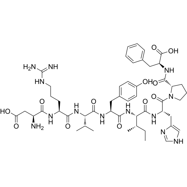
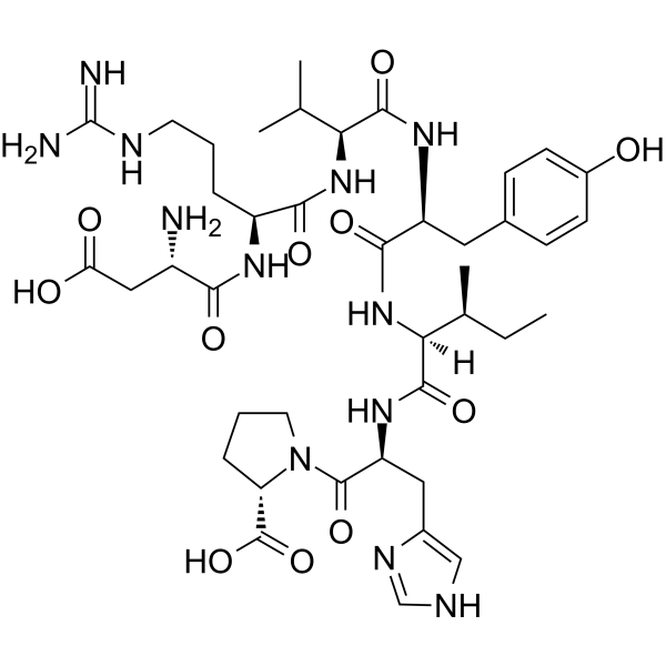
HY-13948
Angiotensin II humanVasoconstrictor -
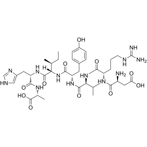
HY-P0216
A 779Mas Receptor Antagonist -
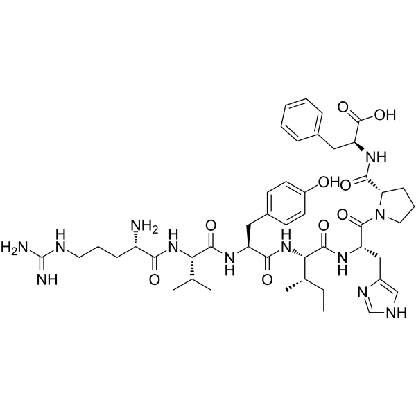
HY-P1540
Angiotensin III, human, mouseAT2R Agonist -
HY-17512
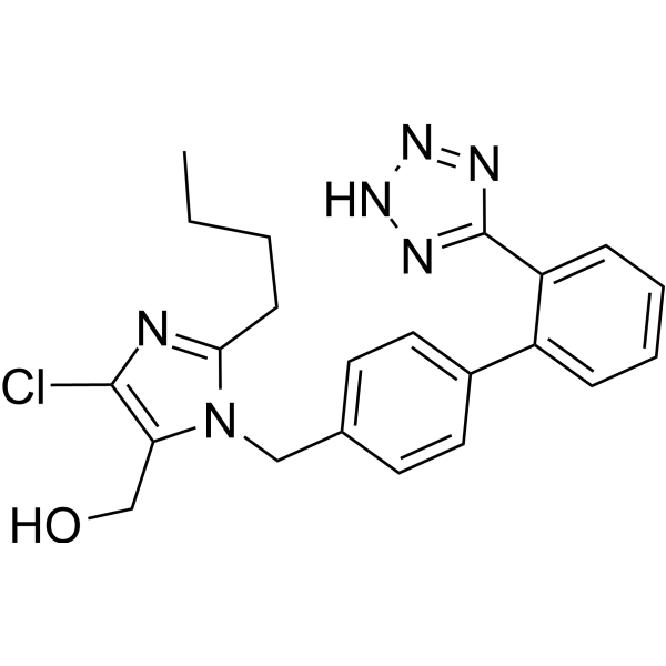
LosartanAngiotensin II Receptor Antagonist -
HY-P1032
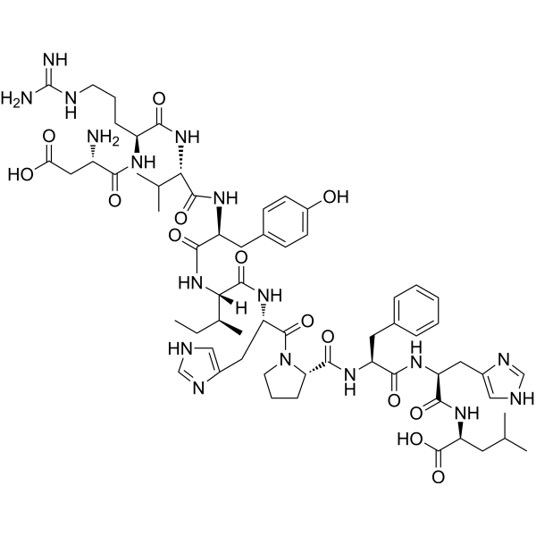
Angiotensin I (human, mouse, rat)Decapeptide Hormone -
HY-P3108
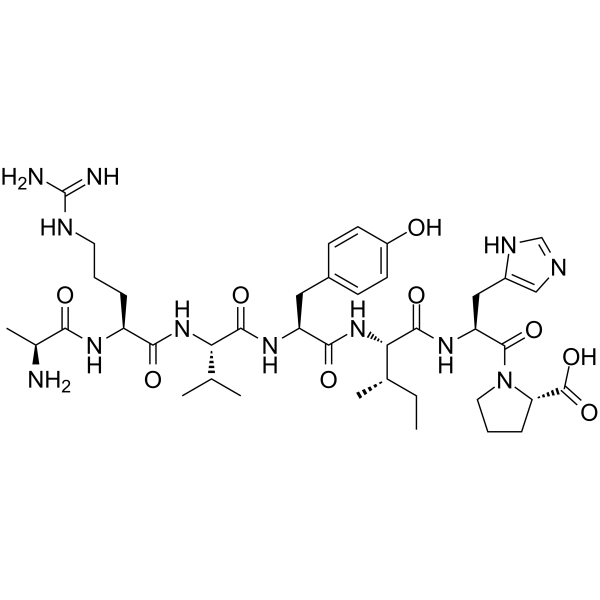
AlamandineMrgD Ligand -
HY-15778
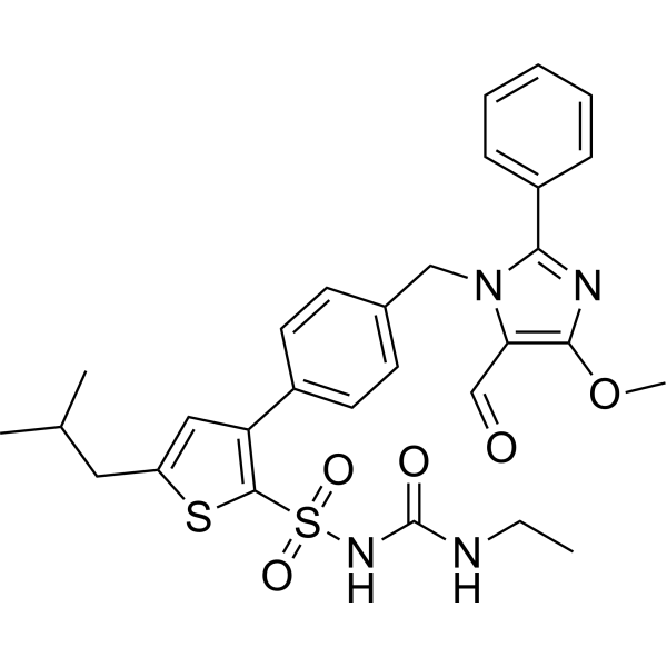
AVE 0991Ang-(1-7) Receptor Agonist -
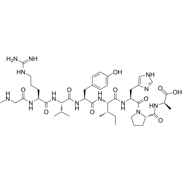
HY-P2141
TRV-120027AT1R Antagonist -
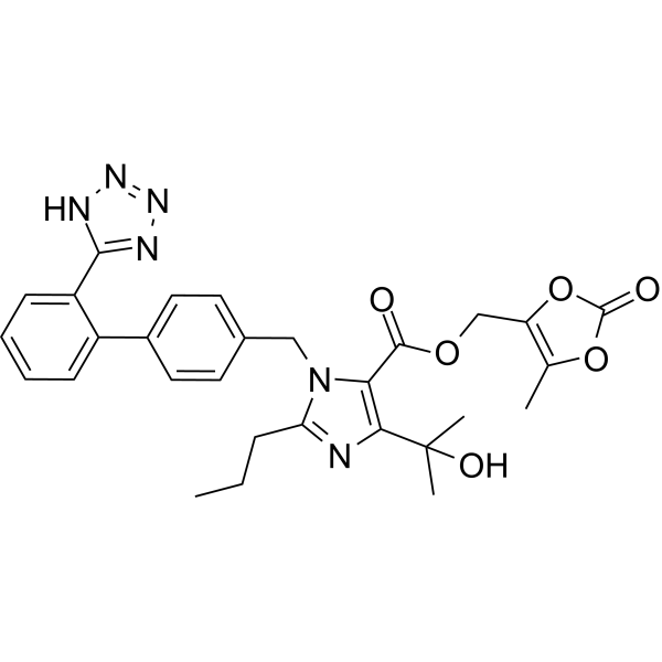
HY-17005
Olmesartan medoxomil -
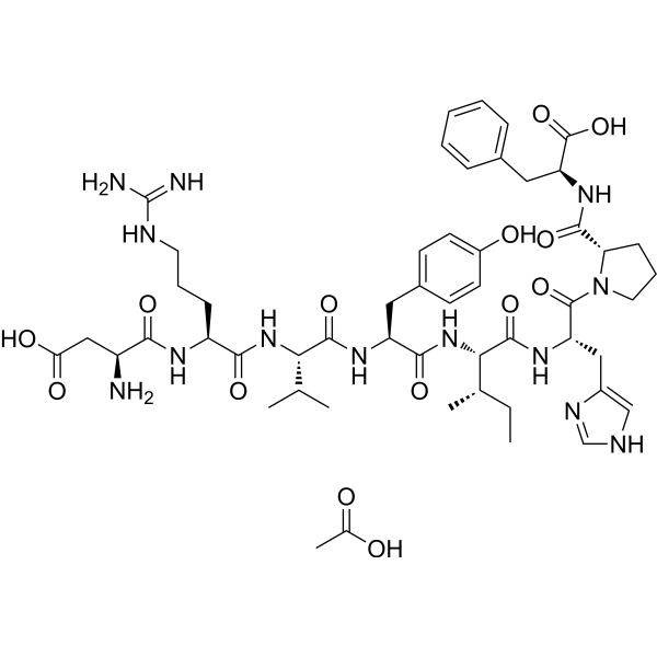
HY-13948A
Angiotensin II human acetateVasoconstrictor -

HY-P9970
InfliximabTNF-α Inhibitor -

HY-12403R
Talfirastide (Standard)Vasodilator Agent
Publications Citing Use of MedChemExpress (MCE) Talfirastide
More- Phytomedicine. 2025 Nov 25:148:157371. [Abstract]
- J Renin Angiotensin Aldosterone Syst. 2026 Apr 7.
- Proc Natl Acad Sci U S A. 2025 Dec 9;122(49):e2515641122. [Abstract]
- Chin Chem Lett. 2023 Mar;34(3):107522. [Abstract]
- Cell Biosci. 2023 Feb 4;13(1):23. [Abstract]
- Acta Physiol. 2026 Apr;242(4):e70200. [Abstract]
- Front Cell Dev Biol. 2021 Jun 11:9:659809. [Abstract]
- Chem Biol Interact. 2025 Sep 5:418:111596. [Abstract]
- Sci Rep. 2025 Nov 25. [Abstract]
- Biol Proced Online. 2022 Oct 25;24(1):15. [Abstract]
- J Inflamm Res. 2024 Jan 24:17:469-485. [Abstract]
- Front Biosci (Landmark Ed). 2024 Sep 24;29(9):334. [Abstract]
- BMC Pulm Med. 2023 Nov 2;23(1):422. [Abstract]
- Endokrynol Pol. 2023;74(5):544-552. [Abstract]
- Int J Clin Exp Med. 2019;12(5):4773-4780.
- bioRxiv. 2026 Mar 25.
- SSRN. 2025 Feb 28.
- Research Square Preprint. 2024 Feb 19.
 Customer Validation & Images

-
Cell Imaging/Staining
-
WB
-
Flow Cytometry
-
Flow Cytometry
-
RT-PCR
All Angiotensin Receptor Isoforms
More

Biological Activity
Description
IC50 & Target
|
AT1 Receptor |
In Vitro
Angiotensin 1-7 (Ang-(1-7)) inhibits cultured vascular smooth muscle cell growth, whereas equal molar concentration of Ang II stimulates cell growth[2].
Angiotensin 1-7 (Ang 1-7) abrogates the methylglyoxal-modified albumin (MGA)-stimulated myofibroblast phenotype by inhibiting the chronic stimulation of the TGF-β-ERK pathway in NRK-52E cells[4].
Angiotensin 1-7 signals through the Mas receptor ( MasR) in opposition to Ang II/angiotensin II type 1 receptor (AT1R), promoting anti-inflammatory,vasodilatory, and neuroprotective effects[5].
MedChemExpress (MCE) has not independently confirmed the accuracy of these methods. They are for reference only. Further protocols information, click here.
In Vivo
MedChemExpress (MCE) has not independently confirmed the accuracy of these methods. They are for reference only.
Clinical Trial
| NCT Number | Sponsor | Condition | Start Date |
Phase

|
|---|---|---|---|---|
| NCT01329991 | Plexxikon| | 2011-05 | PHASE1 |


Chemical Information
-
CAS No. 51833-78-4
-
Appearance Solid
-
Molecular Weight 899.00
-
Formula C41H62N12O11
-
Color White to off-white
-
Synonyms
TXA127; Angiotensin (1-7); Ang-(1-7)
-
Sequence
Asp-Arg-Val-Tyr-Ile-His-Pro
-
Sequence Shortening
DRVYIHP
-
Initial Source
-
Shipping
Room temperature in continental US; may vary elsewhere.
-
Storage
Sealed storage, away from moisture
Powder -80°C 2 years -20°C 1 year * In solvent : -80°C, 6 months; -20°C, 1 month (sealed storage, away from moisture)


Publications (18)
-
Journal Impact Factor
-
Most Recent
-
Phytomedicine
Baicalin and ginsenoside Rb1 suppress the activation of cardiac fibroblasts via regulating the GRK2/AT1R/MasR network. [Abstract]2025 Nov 25:148:157371. PMID: 41110355 -
-
Proc Natl Acad Sci U S A
Ace2 safeguards embryonic hematopoietic stem and progenitor cell production by restraining Nlrp3-mediated pyroptosis. [Abstract]2025 Dec 9;122(49):e2515641122. PMID: 41348733 -
Chin Chem Lett
Nanokit coupled electrospray ionization mass spectrometry for analysis of angiotensin converting enzyme 2 activity in single living cell. [Abstract]2023 Mar;34(3):107522. PMID: 35602918 -
Cell Biosci
Ang-(1-7)/MasR axis promotes functional recovery after spinal cord injury by regulating microglia/macrophage polarization. [Abstract]2023 Feb 4;13(1):23. PMID: 36739421
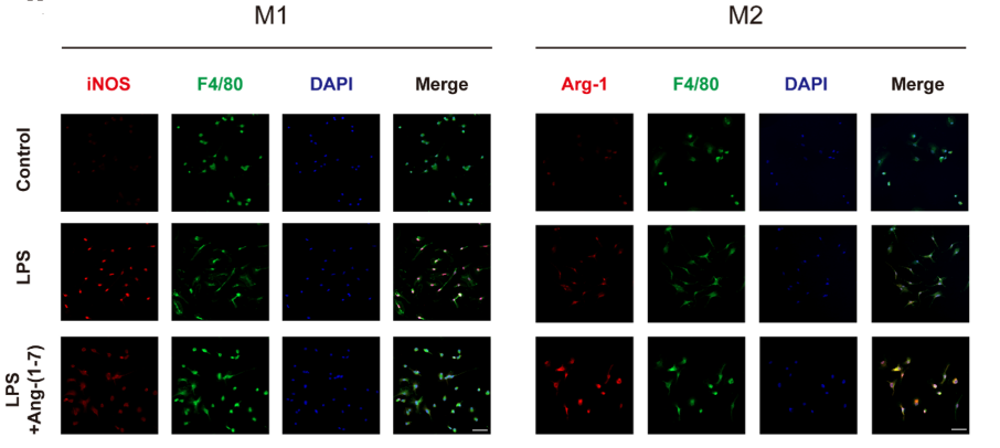
Talfirastide purchased from MedChemExpress. Usage Cited in: Cell Biosci. 2023 Feb 4;13(1):23. [Abstract]
Ang-(1–7) (Talfirastide, 10−6 mol/L, 24 h) regulates the polarization of macrophages towards the M2 phenotype. Double-staining for F4/80 (green)/iNOS (red) or F4/80 (green)/Arg-1 (red) in each group of macrophages (scale bar: 50 μm, n = 5).
Talfirastide purchased from MedChemExpress. Usage Cited in: Cell Biosci. 2023 Feb 4;13(1):23. [Abstract]
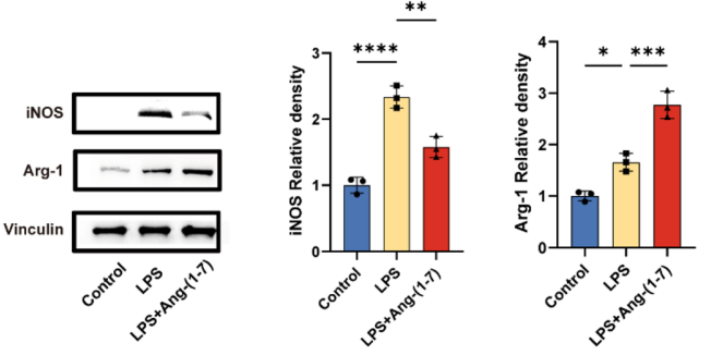
(Talfirastide, 10−6 mol/L). Representative immunoblots and quantification of macrophages in each group showing the expression of iNOS and Arg-1 after treatment for 24 h (n = 3).
Talfirastide purchased from MedChemExpress. Usage Cited in: Cell Biosci. 2023 Feb 4;13(1):23. [Abstract]
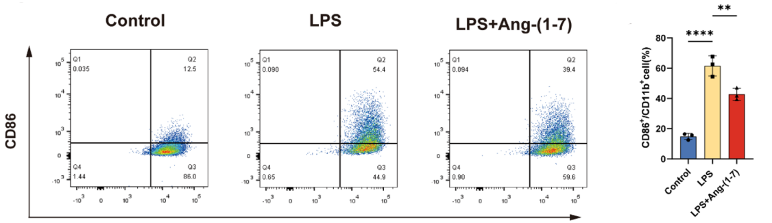
Talfirastide (10−6 mol/L, 24h). The ratio of M1 macrophage phenotype-related surface markers, CD86/CD11b, were determined by flow cytometry for each group (n = 3).
Talfirastide purchased from MedChemExpress. Usage Cited in: Cell Biosci. 2023 Feb 4;13(1):23. [Abstract]
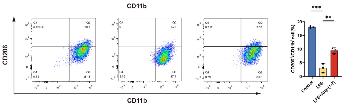
Talfirastide (10−6 mol/L, 24h). The ratio of M2 macrophage phenotype-related surface markers, CD206/ CD11b, were determined by flow cytometry for each group (n = 3).
Talfirastide purchased from MedChemExpress. Usage Cited in: Cell Biosci. 2023 Feb 4;13(1):23. [Abstract]
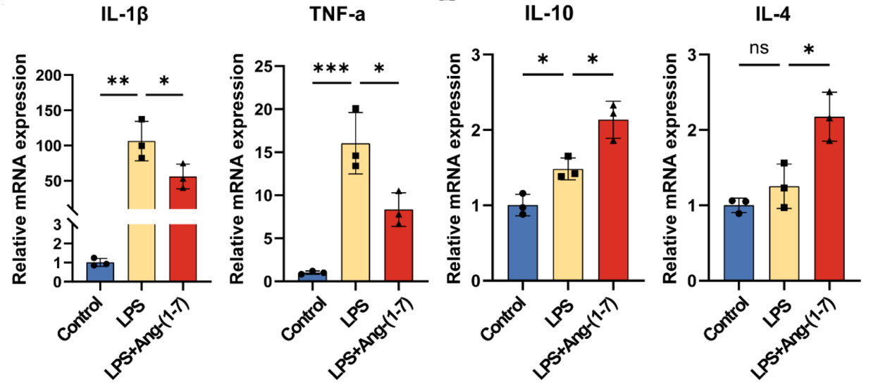
Ang-(1–7) (Talfirastide, 10−6 mol/L, 24h) inhibits inflammation response by modulating the expression of inflammatory cytokines in BMDMs. Quantitative RT-qPCR analysis of macrophages showing mRNA expression of IL-1β, TNF-α, IL-10, IL-4 after treatment for 24 h. GAPDH: loading control. Data are expressed as fold change compared to the control group (n = 3).
Talfirastide purchased from MedChemExpress. Usage Cited in: Cell Biosci. 2023 Feb 4;13(1):23. [Abstract]
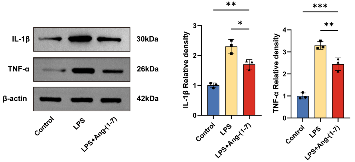
Ang-(1–7)(Talfirastide, 10−6 mol/L, 24h) inhibits inflammation response by modulating the expression of inflammatory cytokines. Representative immunoblots and quantification showing protein expression of IL-1β and TNF-α in the macrophages after treatment for 24 h.
Talfirastide purchased from MedChemExpress. Usage Cited in: Cell Biosci. 2023 Feb 4;13(1):23. [Abstract]
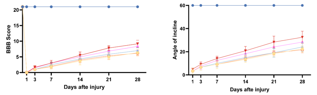
Ang-(1–7) (Talfirastide; administered immediately by intrathecal injection; 10 μL; 500 ng) improved the motor function of rats after SCI. a Basso, Beattie and Bresnahan (BBB) scores (n = 6) and Inclined plane test scores (n = 6).
Talfirastide purchased from MedChemExpress. Usage Cited in: Cell Biosci. 2023 Feb 4;13(1):23. [Abstract]
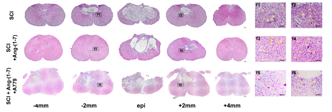
Continuous sections of the spinal cord by HE staining were obtained on the 28th day post-surgery, and the remaining neurons in the SCI group, SCI + Ang-(1–7) groups, and SCI + Ang-(1–7) + A779 groups at the same distance from the epicenter were displayed. (scale bar: 100 μm and 40 μm, n = 3).
-
Acta Physiol
Angiotensin-(1-7) Alleviates Isoproterenol-Induced Cardiac Hypertrophy by Suppressing Autophagy and Apoptosis Through the Synergistic Action of Mas Receptor and Angiotensin II Type 2 Receptor. [Abstract]2026 Apr;242(4):e70200. PMID: 41886752 -
Front Cell Dev Biol
2021 Jun 11:9:659809. PMID: 34178985 -
Chem Biol Interact
Mechanism of ferroptosis in hypoxia-induced pulmonary vascular remodeling in hypoxia pulmonary hypertension: a study based on the ACE2-Ang-(1-7)-Mas axis. [Abstract]2025 Sep 5:418:111596. PMID: 40466901 -
Sci Rep
Ginsenoside Rg1 alleviates HG-induced autophagy of retinal microvascular endothelial cells by activating ACE2/Ang1-7/Mas in vitro. [Abstract]2025 Nov 25. PMID: 41290961 -
Biol Proced Online
Angiotensin-converting enzyme 2 identifies immuno-hot tumors suggesting angiotensin-(1-7) as a sensitizer for chemotherapy and immunotherapy in breast cancer. [Abstract]2022 Oct 25;24(1):15. PMID: 36284262 -
J Inflamm Res
Angiotensin-(1-7) Modulates the Warburg Effect to Alleviate Inflammation in LPS-Induced Macrophages and Septic Mice. [Abstract]2024 Jan 24:17:469-485. PMID: 38282712 -
Front Biosci (Landmark Ed)
ACE2 Alleviates Endoplasmic Reticulum Stress and Protects against Pyroptosis by Regulating Ang1-7/Mas in Ventilator-Induced Lung Injury. [Abstract]2024 Sep 24;29(9):334. PMID: 39344337 -
BMC Pulm Med
Angiotensin-(1-7) suppresses airway inflammation and airway remodeling via inhibiting ATG5 in allergic asthma. [Abstract]2023 Nov 2;23(1):422. PMID: 37919667 -
Endokrynol Pol
Angiotensin (1-7) reverses glucose-induced islet β cell dedifferentiation by Wnt/β-catenin/FoxO1 signalling pathway. [Abstract]2023;74(5):544-552. PMID: 37779373 -
-
-
-


Solvent & Solubility
In Vitro:
H2O : 25 mg/mL (27.81 mM; ultrasonic and warming and heat to 60°C)
Please refer to the solubility information to select the appropriate solvent. Once prepared, please aliquot and store the solution to prevent product inactivation from repeated freeze-thaw cycles.
Storage method and period of stock solution: -80°C, 6 months; -20°C, 1 month (sealed storage, away from moisture). When stored at -80°C, please use it within 6 months. When stored at -20°C, please use it within 1 month.
* Note: If you choose water as the stock solution, please dilute it to the working solution, then filter and sterilize it with a 0.22 μm filter before use.
Please refer to the solubility information to select the appropriate solvent. Once prepared, please aliquot and store the solution to prevent product inactivation from repeated freeze-thaw cycles.
Storage method and period of stock solution: -80°C, 6 months; -20°C, 1 month (sealed storage, away from moisture). When stored at -80°C, please use it within 6 months. When stored at -20°C, please use it within 1 month.
* Note: If you choose water as the stock solution, please dilute it to the working solution, then filter and sterilize it with a 0.22 μm filter before use.
Concentration (start) × Volume (start) = Concentration (final) × Volume (final)
In Vivo:
For the following dissolution methods, please prepare the working solution directly:
It is recommended to prepare fresh solutions and use them promptly within a short period of time.
The percentages shown for the solvents indicate their volumetric ratio in the final prepared solution. If precipitation or phase separation occurs during preparation, heat and/or sonication can be used to aid dissolution.
Add each solvent one by one: PBS
Solubility: 100 mg/mL (111.23 mM); Clear solution; Need ultrasonic

Protocols
-
BrdU Incorporation Assay
Bromodeoxyuridine (BrdU) incorporation assay is based on the principle that BrdU, a thymidine analog, is incorporated into newly synthesized DNA during the S phase of the cell cycle, thereby serving as a marker of DNA replication and cellular proliferation. Incorporated BrdU can be detected using anti-BrdU antibodies following DNA denaturation, enabling visualization or quantification of proliferating cells through immunochemical detection methods such as immunofluorescence or immunohistochemistry.
-
Somatic Cell Culture
A method of simulating the in vivo environment in vitro to maintain the cell growth, differentation and main functions.
-
CFSE Dye Dilution Proliferation Assay
The CFSE (carboxyfluorescein diacetate succinimidyl ester) dye dilution proliferation assay is based on the covalent labeling of intracellular proteins by a cell-permeant fluorescent dye that becomes fluorescent upon intracellular ester cleavage and then is stably retained within cells. As labeled cells divide, the dye is partitioned equally between daughter cells, resulting in a stepwise halving of fluorescence intensity that can be quantified by flow cytometry to determine the number of cell divisions undergone by each cell population. This fluorescence dilution approach enables quantitative tracking of lymphocyte proliferation at the single-cell level over multiple rounds of division. CFSE-based proliferation analysis has been widely applied to measure antigen-driven lymphocyte expansion in vitro, where discrete fluorescence peaks correspond to successive cell divisions and allow reconstruction of proliferative history within heterogeneous populations.
-
Dye-dilution cell tracking and proliferation staining
Dye-dilution cell tracking assays quantify cell proliferation by covalently labeling intracellular proteins with a stable fluorescent dye that is equally partitioned between daughter cells during mitosis, resulting in stepwise halving of fluorescence intensity with each cell division as measured by flow cytometry histograms. Carboxyfluorescein diacetate succinimidyl ester (CFSE) is a prototypical dye that diffuses into cells, is enzymatically converted into a fluorescent compound, and then covalently binds intracellular amine groups, producing long-lived fluorescence suitable for tracking multiple rounds of division in vitro and in vivo. Successive generations of dividing cells form discrete peaks of decreasing fluorescence intensity, enabling estimation of proliferation history, precursor frequency, and division index within heterogeneous populations. Alternative dyes such as CellTrace Violet (CTV) and far-red membrane dyes (e. g. , PKH26) follow the same dilution principle but differ
-
CCK-8/WST-8 Cell Proliferation Assay
The CCK-8/WST-8 assay is based on the reduction of the water-soluble tetrazolium salt WST-8 to a water-soluble formazan product by cellular dehydrogenases in metabolically active cells, where the generated formazan amount is proportional to the number of living cells and is quantified by measuring absorbance in the visible range, providing a colorimetric readout for cell viability and proliferation assessment. This class of tetrazolium-based assays improves upon earlier MTT-based systems by producing a water-soluble formazan, eliminating the need for organic solubilization steps and enabling direct spectrophotometric measurement in culture medium.
-
Protocol for Cell Cycle
Cell-cycle analysis by flow cytometry measures DNA content in single cells to estimate the fraction of cells in G0/G1, S, and G2/M phases. Propidium iodide intercalates into DNA, and after RNA removal with RNase, fluorescence intensity reflects cellular DNA content: 2N cells are assigned to G0/G1, cells between 2N and 4N to S phase, and 4N cells to G2/M. DNA-content analysis alone cannot reliably separate G0 from G1 or G2 from M. Ki-67 can distinguish quiescent G0 cells from cycling cells, EdU or BrdU incorporation marks active DNA synthesis in S phase, and phospho-histone H3 staining identifies mitotic cells within the 4N population.
-
Research Protocol for Inflammation-related Diseases
The NLRP3 inflammasome is a cytosolic innate immune signaling platform that integrates priming signals and danger-signal activation to promote caspase-1 activation, maturation of IL-1β and IL-18, and gasdermin D-mediated pyroptotic cell death. The core experimental logic is to determine whether inflammatory disease phenotypes are driven by increased NLRP3 expression, ASC-containing inflammasome assembly, caspase-1 cleavage, GSDMD cleavage, and extracellular release of IL-1β/IL-18 rather than by nonspecific cell injury alone. The pathway is strongly linked to inflammation-related disease phenotypes because monosodium urate crystals activate NALP3/NLRP3 inflammasome signaling in gout-like crystal inflammation, cholesterol crystals activate NLRP3 inflammasomes in atherogenesis models, and DSS-induced intestinal inflammation has been reported to involve NLRP3 inflammasome activity. However, experimental colitis studies also show context-dependent protective effects of NLRP3 inflammasome co
-
Cell Counting-Based Growth Curve Assay
Cell counting-based growth curve assays quantify cell proliferation by directly measuring changes in viable cell number over time using manual or automated counting methods such as hemocytometer-based counting or instrument-assisted cell enumeration, enabling construction of growth curves that reflect population expansion dynamics in response to culture conditions. A widely used approach is trypan blue exclusion with hemocytometer counting, where membrane-compromised (non-viable) cells take up the dye, allowing discrimination between viable and non-viable cells while simultaneously enabling total cell number quantification. Repeated sampling across time points allows estimation of proliferation rate, growth phases, and comparative growth kinetics between experimental conditions.
-
Colony Formation (Clonogenic) Assay
The clonogenic (colony formation) assay measures the ability of a single cell to retain reproductive viability and form a macroscopic colony, typically defined as a cluster derived from one progenitor cell after a defined growth period. This assay is widely used to evaluate cell survival after exposure to ionizing radiation or cytotoxic treatments and is considered a standard method in radiation biology for generating dose-response relationships of reproductive cell death. Colony formation reflects long-term proliferative capacity rather than short-term metabolic activity, and survival is quantified by comparing treated versus untreated conditions based on colony number and derived survival fractions.
-
Cell Viability Determination by MTT Colorimetric Assay
The following protocol uses the MTT colorimetric assay as a classic literature-established method for assessing cell viability/metabolic activity in cultured mammalian cells. MTT[3-(4,5-dimethylthiazol-2-yl)-2,5-diphenyltetrazolium bromide] is reduced by metabolically active cells to a colored formazan product; the amount of formazan is quantified spectrophotometrically and provides an indirect measure of metabolically active viable cells. Importantly, MTT reduction reflects cellular oxidoreductase/metabolic activity rather than an absolute direct count of living cells, so changes in cellular metabolism can alter the signal independently of cell number.
-
EdU Incorporation Assay (Click Chemistry-Based DNA Synthesis Measurement)
The EdU incorporation assay measures DNA synthesis by adding the thymidine analog 5-ethynyl-2′-deoxyuridine to cells or tissues, where it is incorporated into newly synthesized DNA during S phase. Incorporated EdU is detected by copper-catalyzed azide-alkyne cycloaddition, in which a fluorescent azide covalently reacts with the ethynyl group on EdU, allowing S-phase cells to be detected by fluorescence microscopy, flow cytometry, or high-content imaging. EdU detection does not require DNA denaturation or anti-BrdU antibody access, which preserves sample structure and improves compatibility with immunostaining and multiparameter cytometry compared with BrdU-based detection. EdU can be cytotoxic in a cell-type- and exposure-dependent manner, so pulse duration, concentration, and continuous-labeling designs should be validated for each cell type.
-
Ki-67 Immunostaining Proliferation Assay
Ki-67 immunostaining measures the growth fraction of a cell population by detecting Ki-67, a nuclear antigen present in proliferating cells and absent in quiescent G0 cells. The readout is the percentage of Ki-67-positive nuclei among total counted cells, commonly called the Ki-67 labeling index or proliferation index.
-
Cell Cytotoxicity Assay
Cytotoxicity assays are usually based on the assessment of cell membrane damage, which can also be indirectly detected by measuring cell viability. Detection methods include MTT assay, CKK-8 assay, LDH assay and ATP assay, etc.
-
PCNA Immunodetection Proliferation Assay
PCNA immunodetection measures proliferative activity by detecting proliferating cell nuclear antigen, a nuclear protein associated with DNA polymerase δ function and DNA replication. The assay readout is the proportion of PCNA-positive nuclei among total counted cells, but PCNA labeling is not identical to BrdU labeling because PCNA can mark late G1/early S-associated replication competence and may persist beyond active DNA synthesis depending on fixation and extraction conditions.
-
Protocol for Cell Counting and Cell Density Analysis
Cell counting and cell-density analysis estimate the number of cells in a known volume or field area. Manual hemocytometer counting uses a chamber of defined geometry to convert counted cells into cells/mL, while automated counters and image-analysis workflows detect cell objects from optical, brightfield, fluorescence, impedance, or digital-image features. Trypan blue viability counting is based on dye exclusion: viable cells with intact membranes exclude dye, while non-viable cells with compromised membranes stain blue. The readout is total cell density, viable-cell density, dead-cell density, and percent viability. Cell density can also be estimated from microscopy images by counting objects per image area, from flow cytometry using calibrated volume or reference particles, or from in situ microscopy in bioreactors after calibration against reference methods such as hemocytometer or flow cytometry.
-
Cotton Pellet Granuloma
Cotton pellet granuloma is a classical in vivo chronic inflammation model used to evaluate the anti-inflammatory potential of test substances by measuring their ability to inhibit granuloma tissue formation around an implanted foreign body (cotton pellet) in rodents. The method is based on the biological response to a sterile implanted material, which induces proliferative phase inflammation characterized by fibroblast proliferation and collagen-rich granuloma formation, and the final readout reflects the extent of chronic inflammatory tissue growth surrounding the pellet. In multiple preclinical pharmacological evaluations, inhibition of cotton pellet-induced granuloma formation has been used as an indicator of anti-inflammatory activity in both synthetic and natural product screening contexts.
-
Carrageenan-Induced Paw Edema
Carrageenan-induced paw edema is an acute inflammation model in which intraplantar injection of carrageenan induces localized inflammatory swelling characterized by vascular permeability, leukocyte infiltration, and production of inflammatory mediators such as prostaglandins and cytokines, making it widely used to evaluate anti-inflammatory agents in vivo. The resulting paw volume or thickness increase is quantified over time as a direct readout of inflammatory intensity and drug efficacy, typically reflecting cyclooxygenase-mediated prostaglandin-driven edema formation and immune cell recruitment in peripheral tissue[20].
-
MTT Cell Proliferation Assay
The MTT assay is a colorimetric endpoint assay for estimating viable cell number, cell growth, cytotoxicity, or cell activation in cultured mammalian cells. Living cells reduce the yellow tetrazolium salt MTT into purple/blue formazan, while dead cells do not generate the same signal; the resulting color can be quantified with a multiwell spectrophotometer. MTT reduction is commonly interpreted as a readout of metabolic activity that often correlates with viable cell number, but it should not be treated as a direct cell-counting method unless the assay is optimized for the cell type and experimental condition. Studies show that MTT reduction can involve mitochondrial and non-mitochondrial reducing systems, and formazan may accumulate in intracellular lipid droplets rather than simply marking mitochondria.


Purity & Documentation
-
Data Sheet (294 KB)
-
SDS (394 KB)
- English - EN (394 KB)
- Français - FR (394 KB)
- Deutsch - DE (394 KB)
- Norwegian - NO (394 KB)
- Español - ES (394 KB)
- Swedish - SV (394 KB)
- Italian - IT (394 KB)
- Korean - KR (394 KB)
- Portuguese - PT (394 KB)
-
Handling Instructions (2659 KB)
References
[1]. Gómez-Mendoza DP, et al. Angiotensin-(1-7) oral treatment after experimental myocardial infarction leads to downregulation of CXCR4. J Proteomics. 2019;208:103486. [Content Brief]
[2]. Li P, et al. Angiotensin-(1-7) augments bradykinin-induced vasodilation by competing with ACE and releasing nitric oxide. Hypertension. 1997 Jan;29(1 Pt 2):394-400. [Content Brief]
[3]. Khajah MA, et al. Anti-Inflammatory Action of Angiotensin 1-7 in Experimental Colitis. PLoS One. 2016 Mar 10;11(3):e0150861. [Content Brief]
[4]. Alzayadneh EM, et al. Angiotensin-(1-7) abolishes AGE-induced cellular hypertrophy and myofibroblast transformation via inhibition of ERK1/2. Cell Signal. 2014 Sep 19. pii: S0898-6568(14)00314-3. [Content Brief]
[5]. Janatpour ZC, et al. Subcutaneous Administration of Angiotensin-(1-7) Improves Recovery after Traumatic Brain Injury in Mice. J Neurotrauma. 2019;36(22):3115-3131. [Content Brief]


Complete Stock Solution Preparation Table
Please refer to the solubility information to select the appropriate solvent. Once prepared, please aliquot and store the solution to prevent product inactivation from repeated freeze-thaw cycles.
Storage method and period of stock solution: -80°C, 6 months; -20°C, 1 month (sealed storage, away from moisture). When stored at -80°C, please use it within 6 months. When stored at -20°C, please use it within 1 month.
| Optional Solvent | Concentration Solvent Mass | 1 mg | 5 mg | 10 mg | 25 mg |
|---|---|---|---|---|---|
| H2O | 1 mM | 1.1123 mL | 5.5617 mL | 11.1235 mL | 27.8087 mL |
| 5 mM | 0.2225 mL | 1.1123 mL | 2.2247 mL | 5.5617 mL | |
| 10 mM | 0.1112 mL | 0.5562 mL | 1.1123 mL | 2.7809 mL | |
| 15 mM | 0.0742 mL | 0.3708 mL | 0.7416 mL | 1.8539 mL | |
| 20 mM | 0.0556 mL | 0.2781 mL | 0.5562 mL | 1.3904 mL | |
| 25 mM | 0.0445 mL | 0.2225 mL | 0.4449 mL | 1.1123 mL |
* Note: If you choose water as the stock solution, please dilute it to the working solution, then filter and sterilize it with a 0.22 μm filter before use.


Talfirastide Related Classifications
HY-12403 Related Classifications
 Powered by Bioz