



COTI-2
Based on 4 publication(s) in Google Scholar
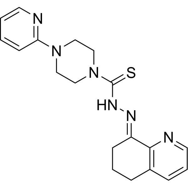
COTI-2, an anti-cancer agent with low toxicity, is an orally available third generation activator of p53 mutant forms. COTI-2 acts both by reactivating mutant p53 and inhibiting the PI3K/AKT/mTOR pathway. COTI-2 induces apoptosis in multiple human tumor cell lines. COTI-2 exhibits antitumor activity in HNSCC through p53-dependent and -independent mechanisms. COTI-2 converts mutant p53 to wild-type conformation.
For research use only. We do not sell to patients.
- Purity : 99.02%
- CAS No.: 1039455-84-9
- Formula: C19H22N6S
- Molecular Weight:366.48
-
Storage:Powder -20°C, 3 years , 4°C, 2 years ; In solvent -80°C, 2 years , -20°C, 1 year

To place orders, for customer services and technical support, please contact: MedChemExpress USA
Tel: 609-228-6898 E-mail: [email protected] [email protected]


-
 Biological Activity
-
Chemical Information
-
Solvent & Solubility
- Protocols
- Purity & Documentation
- References
-
 Help & FAQs


-
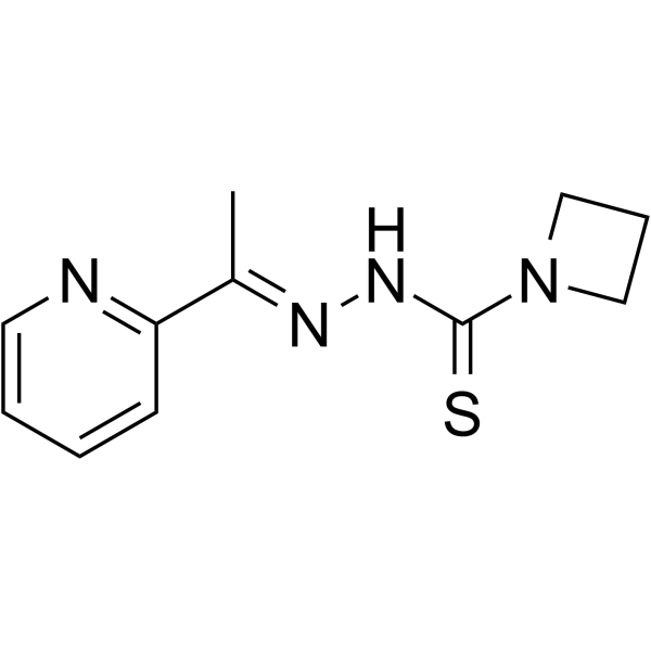
HY-18634
NSC319726MDM-2/p53 p53 Activator -
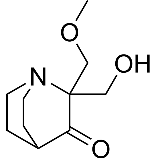
HY-19980
Eprenetapoptp53 Activator -
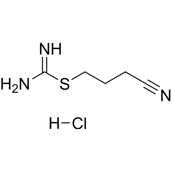
HY-16271
Kevetrin hydrochloridep53 Activator -
HY-19726
NSC59984MDM-2/p53 p53 Activator -
HY-19980A
PRIMA-1Mutant p53 Reactivator -
HY-110088
SCH529074P53 Activator -
HY-13424
RITAp53-HDM-2 Interaction Inhibitor -
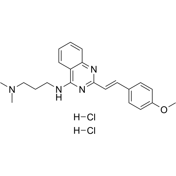
HY-18343A
CP-31398 dihydrochloridep53 Stabilizer -
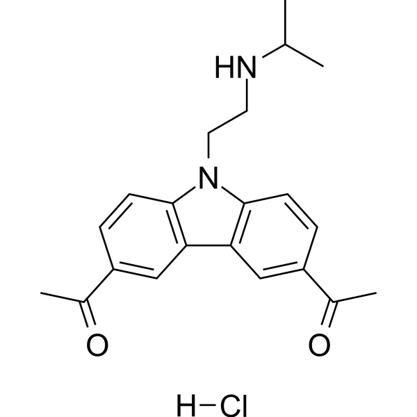
HY-18935A
CBL0137 hydrochlorideNF-κB Inhibitor -
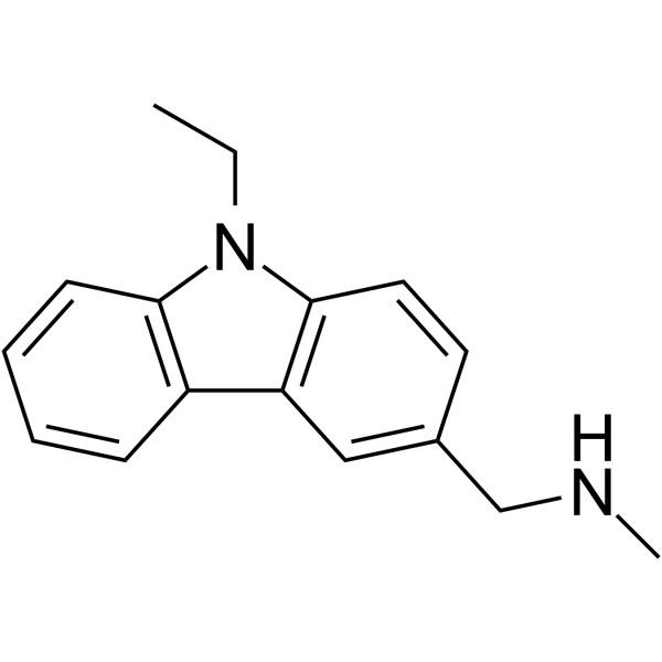
HY-108637
PhiKan 083MDM-2/p53 p53 Activator -
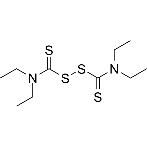
HY-B0240
DisulfiramALDH1 Inhibitor -
HY-12040
ElesclomolCuproptosis Inducer
Publications Citing Use of MedChemExpress (MCE) COTI-2
More

Biological Activity
Description
IC50 & Target
p53[1]
In Vitro
COTI-2 efficiently inhibits the proliferation rate of all the tested cell lines following 72 h of treatment. COTI-2 is significantly effective at inhibiting tumor cell proliferation in all three cell lines (COLO-205, HCT-15, and SW620). Relatively low concentrations of COTI-2 are active against all human glioblastoma cell lines tested (U87-MG, SNB-19, SF-268, and SF-295). COTI-2 treatment of SHP-77 cells with approximate IC50 concentrations results in the induction of early apoptosis among 40 to 47% of total cells[2].
MedChemExpress (MCE) has not independently confirmed the accuracy of these methods. They are for reference only. Further protocols information, click here.
In Vivo
MedChemExpress (MCE) has not independently confirmed the accuracy of these methods. They are for reference only.
Clinical Trial
| NCT Number | Sponsor | Condition | Start Date |
Phase

|
|---|---|---|---|---|
| NCT01329991 | Plexxikon| | 2011-05 | PHASE1 |


Chemical Information
-
CAS No. 1039455-84-9
-
Appearance Solid
-
Molecular Weight 366.48
-
Formula C19H22N6S
-
Color Light yellow to yellow
-
SMILES
S=C(N1CCN(C2=NC=CC=C2)CC1)N/N=C3CCCC4=C\3N=CC=C4
-
Shipping
Room temperature in continental US; may vary elsewhere.
-
Storage
Powder -20°C 3 years 4°C 2 years In solvent -80°C 2 years -20°C 1 year


Publications (4)
-
Journal Impact Factor
-
Most Recent
-
Cell Death Dis
2023 Dec 14;14(12):831. PMID: 38097548 -
Cell Rep
Repurposing antiparasitic antimonials to noncovalently rescue temperature-sensitive p53 mutations. [Abstract]2022 Apr 12;39(2):110622. PMID: 35417717 -
Int J Mol Sci
Improving Reporter Gene Assay Methodology for Evaluating the Ability of Compounds to Restore P53 Activity. [Abstract]2022 Nov 10;23(22):13867. PMID: 36430341 -
Iran J Basic Med Sci
COTI-2 suppresses the malignancy of bladder cancer by inducing apoptosis via the AMPK-mTOR signaling pathway. [Abstract]2025;28(3):240-246. PMID: 39906622


Solvent & Solubility
In Vitro:
DMSO : 5 mg/mL (13.64 mM; Need ultrasonic; Hygroscopic DMSO has a significant impact on the solubility of product, please use newly opened DMSO)
Please refer to the solubility information to select the appropriate solvent. Once prepared, please aliquot and store the solution to prevent product inactivation from repeated freeze-thaw cycles.
Storage method and period of stock solution: -80°C, 2 years; -20°C, 1 year. When stored at -80°C, please use it within 2 years. When stored at -20°C, please use it within 1 year.
Please refer to the solubility information to select the appropriate solvent. Once prepared, please aliquot and store the solution to prevent product inactivation from repeated freeze-thaw cycles.
Storage method and period of stock solution: -80°C, 2 years; -20°C, 1 year. When stored at -80°C, please use it within 2 years. When stored at -20°C, please use it within 1 year.
Concentration (start) × Volume (start) = Concentration (final) × Volume (final)
In Vivo:
Select the appropriate dissolution method based on your experimental animal and administration route.
- For the following dissolution methods, please ensure to first prepare a clear stock solution using an In Vitro approach and then sequentially add co-solvents:
- To ensure reliable experimental results, the clarified stock solution can be appropriately stored based on storage conditions. As for the working solution for In Vivo experiments, it is recommended to prepare freshly and use it on the same day.
- The percentages shown for the solvents indicate their volumetric ratio in the final prepared solution. If precipitation or phase separation occurs during preparation, heat and/or sonication can be used to aid dissolution.
For the following dissolution methods, please prepare the working solution directly:
It is recommended to prepare fresh solutions and use them promptly within a short period of time.
The percentages shown for the solvents indicate their volumetric ratio in the final prepared solution. If precipitation or phase separation occurs during preparation, heat and/or sonication can be used to aid dissolution.
Add each solvent one by one: 50% PEG300 50% Saline
Solubility: 5 mg/mL (13.64 mM); Suspended solution; Need ultrasonic
Add each solvent one by one: 15% Cremophor EL 85% Saline
Solubility: 5 mg/mL (13.64 mM); Suspended solution; Need ultrasonic
In Vivo Dissolution Calculator
Please enter the basic information of animal experiments:
-
-
-
-
Recommended: Prepare an additional quantity of animals to account for potential losses during experiments.
Please enter your animal formula composition:
-
%DMSO +
Recommended: Keep the proportion of DMSO in working solution below 2% if your animal is weak.
-
%+
-
+%Tween-80 + +
-
%Saline +
The co-solvents required include: DMSO, . All of co-solvents are available by MedChemExpress (MCE). , Tween 80. All of co-solvents are available by MedChemExpress (MCE).
Working solution concentration: 0.22 mg/mL
Method for preparing stock solution: mg drug dissolved in μL DMSO. Stock solution concentration: mg/mL.
1. Take μL DMSO stock solution;
2. Add μL .
μL , mix evenly;
3. Then add μL Tween 80, mix evenly;
4. Then add μL
Please ensure that the stock solution in the first step is dissolved to a clear state, and add co-solvents in sequence. You can use ultrasonic heating (ultrasonic cleaner, recommended frequency 20-40 kHz), vortexing, etc. to assist dissolution.

Protocols
-
Apoptosis
Apoptosis, also called programmed cell death, is generally characterized by distinct morphological characteristics.
-
TUNEL staining for apoptotic DNA fragmentation
TUNEL staining detects DNA strand breaks by using terminal deoxynucleotidyl transferase to add labeled nucleotides to exposed 3′-OH DNA termini, generating either microscopic staining in fixed cells or tissue sections, or fluorescence/cytometric signal in cell suspensions. TUNEL positivity reflects DNA fragmentation but should not be interpreted alone as definitive apoptosis, because TUNEL can also label necrotic, autolytic, mechanically damaged, or DNA-repair-associated DNA breaks.
-
Annexin V plus membrane-impermeant dye apoptosis staining
Annexin V-based apoptosis assays rely on the detection of phosphatidylserine (PS) externalization from the inner leaflet of the plasma membrane to the outer leaflet, an early biochemical hallmark of apoptosis. Fluorescently labeled Annexin V binds PS in a calcium-dependent manner, enabling identification of early apoptotic cells by flow cytometry or fluorescence microscopy. When combined with a membrane-impermeant DNA-binding dye (e. g. , propidium iodide), this approach allows discrimination between viable (Annexin V−/dye−), early apoptotic (Annexin V+/dye−), and late apoptotic or necrotic (Annexin V+/dye+) cell populations by assessing membrane integrity and PS exposure.
-
Apoptosis Solutions
Apoptosis is a regulated, generally non-lytic cell-death pathway that removes unwanted, damaged, infected, or abnormal cells through coordinated morphological changes, caspase activation, DNA fragmentation, and membrane remodeling. The intrinsic apoptosis pathway is controlled mainly by mitochondrial outer membrane permeabilization, BCL-2 family proteins, cytochrome c release, apoptosome formation, caspase-9 activation, and downstream executioner caspase-3/7 activation. The extrinsic apoptosis pathway is initiated by death receptors such as Fas, TNFR, and TRAIL receptors, which recruit adaptor proteins and activate caspase-8 before engaging executioner caspases or mitochondrial amplification through BID cleavage. Apoptosis is linked to many phenotypes, including cancer cell killing, tissue homeostasis, immune regulation, neurodegeneration, infection response, and treatment-induced cytotoxicity; unresolved questions include how apoptosis interacts with necroptosis, pyroptosis, ferroptos
-
Subcutaneous Cell-Line-Derived Xenograft
Subcutaneous cell-line-derived xenograft (CDX) models are established by implanting cultured human cancer cell lines into immunodeficient mice, where the injected cells form localized tumors that can be monitored in vivo as a measure of tumorigenic potential, growth kinetics, and treatment response. These models are widely used in oncology research because they allow reproducible tumor formation and enable comparative assessment of tumor growth between different cell lines or genetic manipulations in a controlled in vivo microenvironment. Subcutaneous implantation of cancer cells in immunodeficient mice is a standard approach for evaluating tumor growth behavior and therapeutic response across multiple cancer types, including prostate, esophageal, pancreatic, and colon cancer models.


Purity & Documentation
-
Data Sheet (281 KB)
-
SDS (392 KB)
- English - EN (392 KB)
- Français - FR (392 KB)
- Deutsch - DE (392 KB)
- Norwegian - NO (392 KB)
- Español - ES (392 KB)
- Swedish - SV (392 KB)
- Italian - IT (392 KB)
- Korean - KR (392 KB)
- Portuguese - PT (392 KB)
-
Handling Instructions (2659 KB)
References
[1]. Duffy MJ, et al. Mutant p53 as a target for cancer treatment. Eur J Cancer. 2017 Sep;83:258-265. [Content Brief]
[2]. Salim KY, et al. COTI-2, a novel small molecule that is active against multiple human cancer cell lines in vitro and in vivo. Oncotarget. 2016 Jul 5;7(27):41363-41379. [Content Brief]
[3]. Lindemann A, et al. COTI-2, A Novel Thiosemicarbazone Derivative, Exhibits Antitumor Activity in HNSCC through p53-dependent and -independent Mechanisms. Clin Cancer Res. 2019 Sep 15;25(18):5650-5662. [Content Brief]


Complete Stock Solution Preparation Table
Please refer to the solubility information to select the appropriate solvent. Once prepared, please aliquot and store the solution to prevent product inactivation from repeated freeze-thaw cycles.
Storage method and period of stock solution: -80°C, 2 years; -20°C, 1 year. When stored at -80°C, please use it within 2 years. When stored at -20°C, please use it within 1 year.
| Optional Solvent | Concentration Solvent Mass | 1 mg | 5 mg | 10 mg | 25 mg |
|---|---|---|---|---|---|
| DMSO | 1 mM | 2.7287 mL | 13.6433 mL | 27.2866 mL | 68.2165 mL |
| 5 mM | 0.5457 mL | 2.7287 mL | 5.4573 mL | 13.6433 mL | |
| 10 mM | 0.2729 mL | 1.3643 mL | 2.7287 mL | 6.8217 mL |


COTI-2 Related Classifications
HY-19896 Related Classifications
 Powered by Bioz