



Epertinib
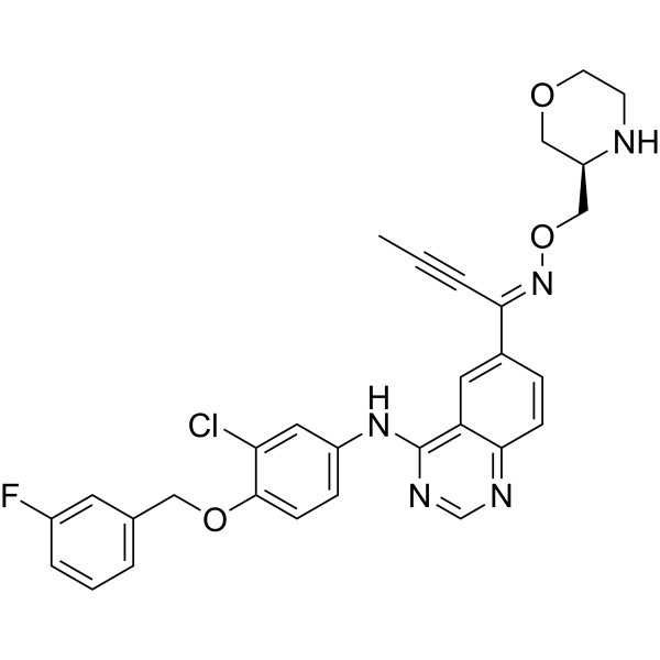
Epertinib (S-22611) is a potent, orally active, reversible, and selective tyrosine kinase inhibitor of EGFR, HER4 and HER2, with IC50s of 1.48 nM, 2.49 nM and 7.15 nM, respectively. Epertinib shows potent antitumor activity. Epertinib is a click chemistry reagent, it contains an Alkyne group and can undergo copper-catalyzed azide-alkyne cycloaddition (CuAAc) with molecules containing Azide groups.
For research use only. We do not sell to patients.
- CAS No.: 908305-13-5
- Formula: C30H27ClFN5O3
- Molecular Weight:560.02
-
Storage:
4°C, protect from light
* In solvent : -80°C, 6 months; -20°C, 1 month (protect from light)

To place orders, for customer services and technical support, please contact: MedChemExpress USA
Tel: 609-228-6898 E-mail: [email protected] [email protected]


-
 Biological Activity
-
Chemical Information
- Protocols
- Purity & Documentation
- References
-
 Help & FAQs


All EGFR Isoforms
More

Biological Activity
Description
IC50 & Target
[1]|
EGFR 1.48 ± 0.0 nM (IC50) |
HER4 2.49 ± 0.1 nM (IC50) |
HER2 7.15 ± 0.5 nM (IC50) |
In Vitro
Epertinib inhibits the phosphorylation of EGFR and HER2 in NCI-N87 cells, with IC50 values of 4.5 and 1.6 nM, respectively[2].
Epertinib shows inhibitory activity against MDA-MB-361 cell, with an IC50 of 26.5 nM[1].
Epertinib (0-10 μM, 72 h) can selectively inhibit the proliferation of a range of cancer cell lines expressing EGFR and/or HER2[2].
MedChemExpress (MCE) has not independently confirmed the accuracy of these methods. They are for reference only. Further protocols information, click here.
-
Cell Line:NCI-N87 (stomach), BT-474 (breast), SK-BR-3 (breast), MDA-MB-453 (breast), MDA-MB-175VII (breast), HT115 (colon), Calu-3 (lung), fR2 (breast), and MRC-5 (lung)
-
Concentration:0-10 μM
-
Incubation Time:72 h
-
Result:Inhibited the growth of NCI-N87, BT-474, SK-BR-3, MDA-MB-453, MDA-MB-175VII, HT115, Calu-3, fR2, and MRC-5, with IC50 values of 8.3 ± 2.6, 9.9 ± 0.8, 14.0 ± 3.6, 48.6 ± 3.1, 21.6 ± 4.3, 53.3 ± 8.6, 241.5 ± 29.2, 5366.7 ± 65.2, and 4964.6 ± 340.3.
In Vivo
Epertinib (50 mg/kg, Orally, once daily for 30 days) significantly reduces the brain tumor volume[1].
Epertinib (0-50 mg/kg, Orally, once daily for 10-28 days) significantly inhibits the tumor growth in a dose-dependent manner[2].
MedChemExpress (MCE) has not independently confirmed the accuracy of these methods. They are for reference only.
-
Animal Model:Nude mice (BALB/cAJcl-nu/nu, implanted intracranially with MDA-MB-361 or BR2 cells)[1]
-
Dosage:12.5, 25, 50, 100 mg/kg
-
Administration:Orally, once daily for 28 days
-
Result:Showed antitumor activity in the mammary fat pad implantation model using both cell lines and the ED50 values were comparable (24.1 mg/kg and 26.5 mg/kg for MDA-MB-361 and BR2 (MDA-MB-361-luc-BR2), respectively).
-
Animal Model:Nude mice (BALB/cAJcl-nu/nu, implanted intracranially with MDA-MB-361 or BR2 cells)[1]
-
Dosage:50 mg/kg
-
Administration:Orally, once daily for 30 days
-
Result:Significantly reduced the brain tumor volume, indicating that epertinib could have potent antitumor activity in brain metastasis even in the presence of an intact BTB (blood-tumor barrier).
-
Animal Model:Nude mice (BALB/cAJcl-nu/nu, prepared by subcutaneous implantation of human gastric cancer cells, NCI-N87 into the back of nude mice)[2]
-
Dosage:0, 6.25, 12.5, 25, and 50 mg/kg
-
Administration:Oral gavage, daily for 10-28 days
-
Result:Significantly inhibited the tumor growth in a dose-dependent manner.


Chemical Information
-
CAS No. 908305-13-5
-
Appearance Solid
-
Molecular Weight 560.02
-
Formula C30H27ClFN5O3
-
Color Light yellow to yellow
-
SMILES
CC#C/C(C1=CC2=C(NC3=CC=C(OCC4=CC=CC(F)=C4)C(Cl)=C3)N=CN=C2C=C1)=N\OC[C@@H]5NCCOC5
-
Synonyms
S-22611
-
Shipping
Room temperature in continental US; may vary elsewhere.
-
Storage
4°C, protect from light
* In solvent : -80°C, 6 months; -20°C, 1 month (protect from light)

Protocols
-
EdU Incorporation Assay (Click Chemistry-Based DNA Synthesis Measurement)
The EdU incorporation assay measures DNA synthesis by adding the thymidine analog 5-ethynyl-2′-deoxyuridine to cells or tissues, where it is incorporated into newly synthesized DNA during S phase. Incorporated EdU is detected by copper-catalyzed azide-alkyne cycloaddition, in which a fluorescent azide covalently reacts with the ethynyl group on EdU, allowing S-phase cells to be detected by fluorescence microscopy, flow cytometry, or high-content imaging. EdU detection does not require DNA denaturation or anti-BrdU antibody access, which preserves sample structure and improves compatibility with immunostaining and multiparameter cytometry compared with BrdU-based detection. EdU can be cytotoxic in a cell-type- and exposure-dependent manner, so pulse duration, concentration, and continuous-labeling designs should be validated for each cell type.
-
Subcutaneous Cell-Line-Derived Xenograft
Subcutaneous cell-line-derived xenograft (CDX) models are established by implanting cultured human cancer cell lines into immunodeficient mice, where the injected cells form localized tumors that can be monitored in vivo as a measure of tumorigenic potential, growth kinetics, and treatment response. These models are widely used in oncology research because they allow reproducible tumor formation and enable comparative assessment of tumor growth between different cell lines or genetic manipulations in a controlled in vivo microenvironment. Subcutaneous implantation of cancer cells in immunodeficient mice is a standard approach for evaluating tumor growth behavior and therapeutic response across multiple cancer types, including prostate, esophageal, pancreatic, and colon cancer models.


Purity & Documentation
-
Data Sheet (274 KB)
-
SDS (252 KB)
- English - EN (252 KB)
- Français - FR (252 KB)
- Deutsch - DE (252 KB)
- Norwegian - NO (252 KB)
- Español - ES (252 KB)
- Swedish - SV (252 KB)
- Italian - IT (252 KB)
- Korean - KR (252 KB)
- Portuguese - PT (252 KB)
-
Handling Instructions (2659 KB)
References
[1]. Tanaka Y, et al. Distribution analysis of epertinib in brain metastasis of HER2-positive breast cancer by imaging mass spectrometry and prospect for antitumor activity. Sci Rep. 2018 Jan 10;8(1):343. [Content Brief]
[2]. Tanaka H, et al. Preclinical antitumor activity of S-222611, an oral reversible tyrosine kinase inhibitor of epidermal growth factor receptor and human epidermal growth factor receptor 2. Cancer Sci. 2014 Aug;105(8):1040-8. [Content Brief]

Calculators
Concentration (start) × Volume (start) = Concentration (final) × Volume (final)


Epertinib Related Classifications
HY-107367 Related Classifications
 Powered by Bioz