



Iberin
Based on 2 publication(s) in Google Scholar
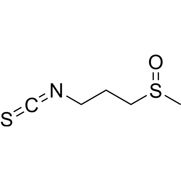
Iberin (NSC 321801), a sulfoxide analogue of sulforaphane, is a naturally occurring member of isothiocyanate family. Iberin inhibits cell survival with an IC50 of 2.3 μM in HL60 cell. Iberin induces apoptosis.
For research use only. We do not sell to patients.
- Purity : 98.0%
- CAS No.: 505-44-2
- Formula: C5H9NOS2
- Molecular Weight:163.26
-
Storage:
Solution, -20°C, 2 years

To place orders, for customer services and technical support, please contact: MedChemExpress USA
Tel: 609-228-6898 E-mail: [email protected] [email protected]


-
 Biological Activity
-
Chemical Information
- Protocols
- Purity & Documentation
- References
-
 Help & FAQs


-
HY-13755
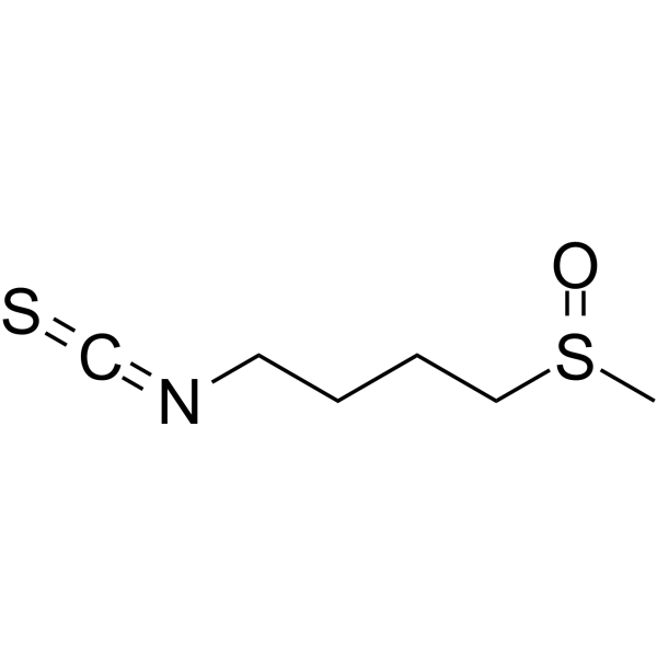
SulforaphaneKeap1/Nrf2/ARE Inducer -
HY-77813
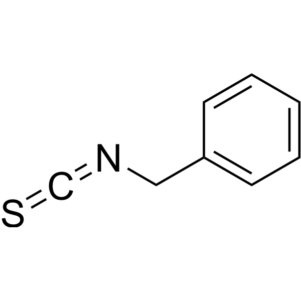
Benzyl isothiocyanateCancer Preventive Agent -
HY-121323
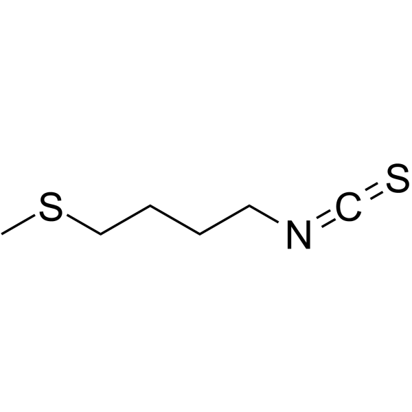
ErucinAnticancer Activity -
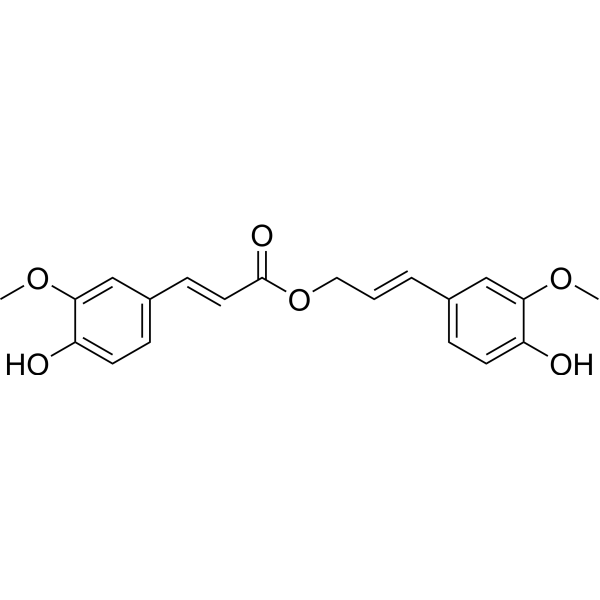
HY-N1916
Coniferyl ferulateGST Inhibitor -
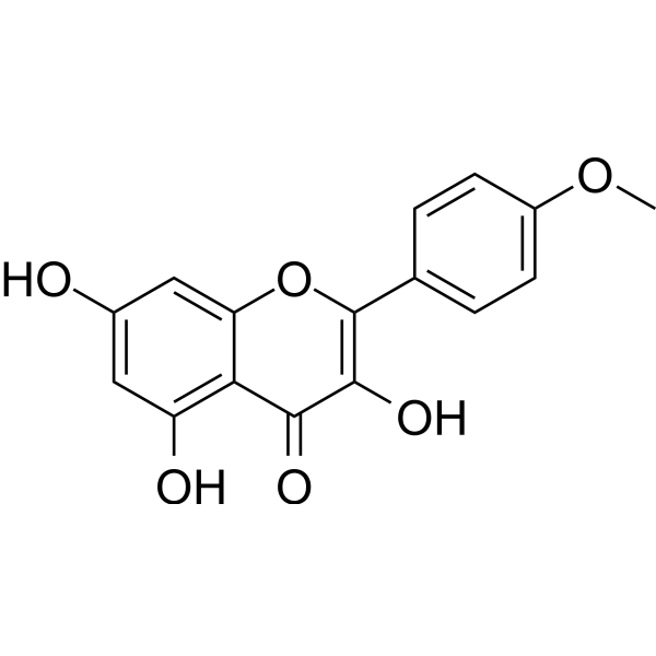
HY-15449
KaempferideAnti-cancer/Anti-inflammatory/Antioxidant -
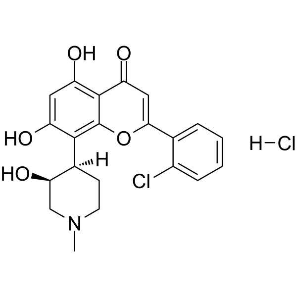
HY-10006
Flavopiridol HydrochlorideCDK Inhibitor -
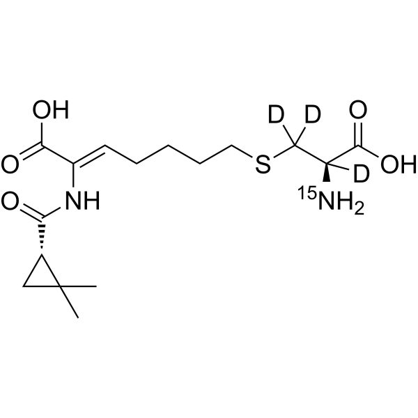
HY-A0166S
Cilastatin-15N,d3Stable Isotope -
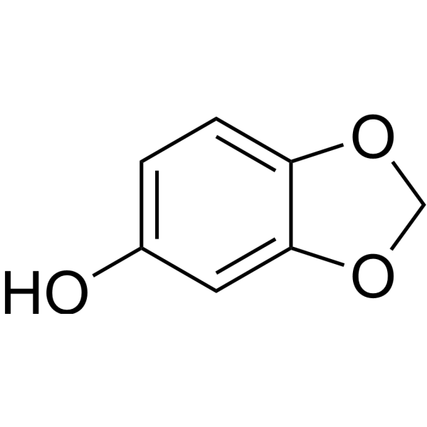
HY-N1417
SesamolApoptosis Inducer -
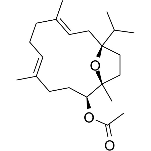
HY-N4098
Incensole AcetateApoptosis Modulator -
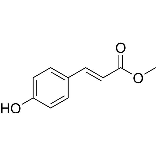
HY-N2492
(E)-Methyl 4-coumarateDerivative Of Cinnamic Acid -
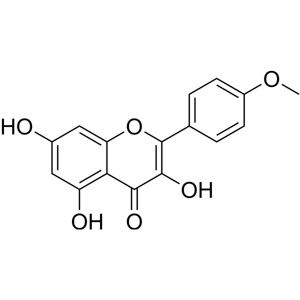
HY-15449R
Kaempferide (Standard) -

HY-N2492R
(E)-Methyl 4-coumarate (Standard)Standard Of (E)-Methyl 4-coumarate
Publications Citing Use of MedChemExpress (MCE) Iberin
MoreAll Endogenous Metabolite Isoforms
More

Biological Activity
Description
IC50 & Target
IC50: 2.3 μM (HL60 cell)[1]
In Vitro
Iberin inhibits the growth of neuroblastoma cells in a dose- and time-dependent manner. The iberin-induced cell cycle arrest in neuroblastoma cells is associated with inhibition of expression of cyclin-dependent kinase Cdk2, Cdk4, and Cdk6 proteins. There is an increase in apoptotic cell death in iberin treated cells as compared with control cells. The iberin-induced apoptosis is found to be associated with activation of caspase-9, caspase-3, and PARP[2]. Iberin inhibits growth of human glioblastoma cells in cell proliferation assays, enhances cytotoxicity, and induces apoptosis by activation of caspase-3 and caspase-9[3].
MedChemExpress (MCE) has not independently confirmed the accuracy of these methods. They are for reference only. Further protocols information, click here.
In Vivo
MedChemExpress (MCE) has not independently confirmed the accuracy of these methods. They are for reference only.


Chemical Information
-
CAS No. 505-44-2
-
Appearance Liquid
-
Molecular Weight 163.26
-
Formula C5H9NOS2
-
Color Colorless to light yellow
-
SMILES
O=S(CCCN=C=S)C
-
Synonyms
NSC 321801
-
Structure Classification
-
Initial Source
-
Shipping
Room temperature in continental US; may vary elsewhere.
-
Storage
Solution, -20°C, 2 years


Publications (2)
-
Journal Impact Factor
-
Most Recent
-
Nat Prod Res
Bioactivity-guided characterisation of iberin from Cakile edentula (Bigelow) Hook. inducing apoptosis in human melanoma cells. [Abstract]2025 Jun 5:1-7. PMID: 40470544 -
Biomed Pharmacother
Isothiocyanate Iberin inhibits cell proliferation and induces cell apoptosis in the progression of ovarian cancer by mediating ROS accumulation and GPX1 expression. [Abstract]2021 Oct:142:111533. PMID: 34148735

Protocols
-
Apoptosis
Apoptosis, also called programmed cell death, is generally characterized by distinct morphological characteristics.
-
TUNEL staining for apoptotic DNA fragmentation
TUNEL staining detects DNA strand breaks by using terminal deoxynucleotidyl transferase to add labeled nucleotides to exposed 3′-OH DNA termini, generating either microscopic staining in fixed cells or tissue sections, or fluorescence/cytometric signal in cell suspensions. TUNEL positivity reflects DNA fragmentation but should not be interpreted alone as definitive apoptosis, because TUNEL can also label necrotic, autolytic, mechanically damaged, or DNA-repair-associated DNA breaks.
-
Cell Cytotoxicity Assay
Cytotoxicity assays are usually based on the assessment of cell membrane damage, which can also be indirectly detected by measuring cell viability. Detection methods include MTT assay, CKK-8 assay, LDH assay and ATP assay, etc.
-
Mammalian live/dead viability and cytotoxicity staining
Live/dead viability and cytotoxicity staining assays are based on the simultaneous detection of intracellular esterase activity in metabolically active (viable) cells and membrane integrity loss in non-viable cells. In commonly used dual-staining approaches, membrane-permeant fluorogenic substrates are converted by intracellular esterases into fluorescent products in live cells, while impermeant DNA-binding dyes selectively enter cells with compromised plasma membranes and label nucleic acids in dead or dying cells, enabling discrimination between viable and non-viable populations by fluorescence microscopy or flow cytometry.
-
Annexin V plus membrane-impermeant dye apoptosis staining
Annexin V-based apoptosis assays rely on the detection of phosphatidylserine (PS) externalization from the inner leaflet of the plasma membrane to the outer leaflet, an early biochemical hallmark of apoptosis. Fluorescently labeled Annexin V binds PS in a calcium-dependent manner, enabling identification of early apoptotic cells by flow cytometry or fluorescence microscopy. When combined with a membrane-impermeant DNA-binding dye (e. g. , propidium iodide), this approach allows discrimination between viable (Annexin V−/dye−), early apoptotic (Annexin V+/dye−), and late apoptotic or necrotic (Annexin V+/dye+) cell populations by assessing membrane integrity and PS exposure.
-
Apoptosis Solutions
Apoptosis is a regulated, generally non-lytic cell-death pathway that removes unwanted, damaged, infected, or abnormal cells through coordinated morphological changes, caspase activation, DNA fragmentation, and membrane remodeling. The intrinsic apoptosis pathway is controlled mainly by mitochondrial outer membrane permeabilization, BCL-2 family proteins, cytochrome c release, apoptosome formation, caspase-9 activation, and downstream executioner caspase-3/7 activation. The extrinsic apoptosis pathway is initiated by death receptors such as Fas, TNFR, and TRAIL receptors, which recruit adaptor proteins and activate caspase-8 before engaging executioner caspases or mitochondrial amplification through BID cleavage. Apoptosis is linked to many phenotypes, including cancer cell killing, tissue homeostasis, immune regulation, neurodegeneration, infection response, and treatment-induced cytotoxicity; unresolved questions include how apoptosis interacts with necroptosis, pyroptosis, ferroptos
-
Cell Viability Determination by MTT Colorimetric Assay
The following protocol uses the MTT colorimetric assay as a classic literature-established method for assessing cell viability/metabolic activity in cultured mammalian cells. MTT[3-(4,5-dimethylthiazol-2-yl)-2,5-diphenyltetrazolium bromide] is reduced by metabolically active cells to a colored formazan product; the amount of formazan is quantified spectrophotometrically and provides an indirect measure of metabolically active viable cells. Importantly, MTT reduction reflects cellular oxidoreductase/metabolic activity rather than an absolute direct count of living cells, so changes in cellular metabolism can alter the signal independently of cell number.


Purity & Documentation
-
Data Sheet (272 KB)
-
SDS (565 KB)
- English - EN (565 KB)
- Français - FR (565 KB)
- Deutsch - DE (565 KB)
- Norwegian - NO (565 KB)
- Español - ES (565 KB)
- Swedish - SV (565 KB)
- Italian - IT (565 KB)
- Korean - KR (565 KB)
- Portuguese - PT (565 KB)
-
Handling Instructions (2659 KB)
References
[1]. Jakubikova J, et al. Isothiocyanates induce cell cycle arrest, apoptosis and mitochondrial potential depolarization in HL-60 and multidrug-resistant cell lines. Anticancer Res. 2005 Sep-Oct;25(5):3375-86. [Content Brief]
[2]. Jadhav U, et al. Iberin induces cell cycle arrest and apoptosis in human neuroblastoma cells. [Content Brief]
[3]. Jadhav U, et al. Dietary isothiocyanate iberin inhibits growth and induces apoptosis in human glioblastoma cells. J Pharmacol Sci. 2007 Feb;103(2):247-51. [Content Brief]
[4]. Jakobsen TH, et al. Food as a source for quorum sensing inhibitors: iberin from horseradish revealed as a quorumsensing inhibitor of Pseudomonas aeruginosa. Appl Environ Microbiol. 2012 Apr;78(7):2410-21. [Content Brief]
[5]. Munday R, et al. Induction of phase II detoxification enzymes in rats by plant-derived isothiocyanates: comparison of allyl isothiocyanate with sulforaphane and related compounds. J Agric Food Chem. 2004 Apr 7;52(7):1867-71. [Content Brief]

Calculators
Concentration (start) × Volume (start) = Concentration (final) × Volume (final)


Iberin Related Classifications
HY-101413 Related Classifications
 Powered by Bioz