



PF-562271 (besylate)
Based on 32 publication(s) in Google Scholar
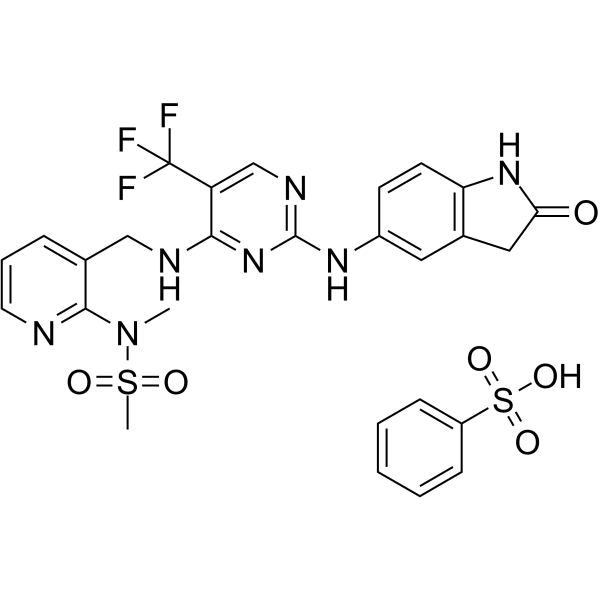
PF-562271 besylate (VS-6062 besylate) is an orally active, ATP-competitive, reversible FAK/Pyk2 inhibitor, with an IC50 of 1.5 nM for FAK and 13 nM for Pyk2. PF-562271 besylate induces tumor regression, and inhibits tumor growth, invasion and metastasis. PF-562271 besylate exerts no effect on tumor necrosis, angiogenesis or apoptosis in an orthotopic mouse model of pancreatic ductal adenocarcinoma. When combined with Sunitinib (HY-10255A), PF-562271 besylate reduces tumor vascularization, decreases serum alpha-fetoprotein levels and improves cachexia. PF-562271 besylate can be used in research related to prostate cancer, breast cancer, pancreatic cancer, colon cancer, glioblastoma, lung cancer, pancreatic ductal adenocarcinoma and hepatocellular carcinoma.
For research use only. We do not sell to patients.
- Purity : 99.17%
- CAS No.: 939791-38-5
- Formula: C27H26F3N7O6S2
- Molecular Weight:665.66
-
Storage:
4°C, sealed storage, away from moisture
* In solvent : -80°C, 6 months; -20°C, 1 month (sealed storage, away from moisture)

To place orders, for customer services and technical support, please contact: MedChemExpress USA
Tel: 609-228-6898 E-mail: [email protected] [email protected]


-
 Biological Activity
-
Chemical Information
-
Solvent & Solubility
- Protocols
- Purity & Documentation
- References
-
 Help & FAQs


-
HY-13917
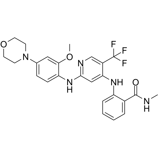
PND-1186FAK Inhibitor -
HY-12289
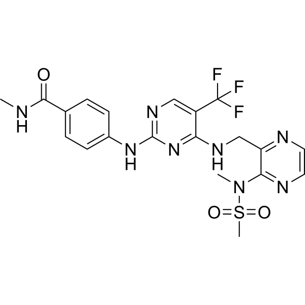
DefactinibFAK Inhibitor -
HY-10459
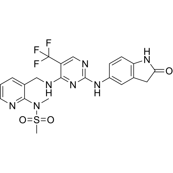
PF-562271FAK/Pyk2 Inhibitor -
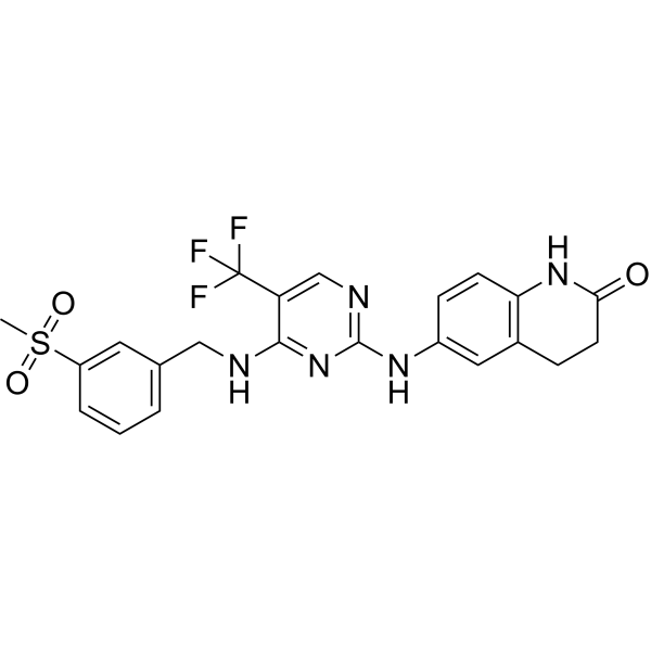
HY-10461
PF-573228FAK Inhibitor -
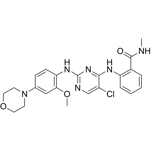
HY-13203
NVP-TAE 226Apoptosis Inducer -
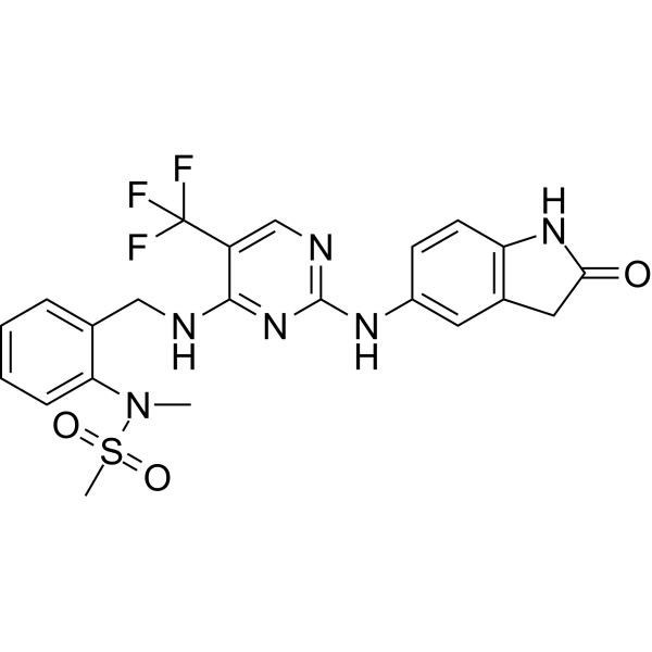
HY-10460
PF-431396Pyk2/FAK Inhibitor -
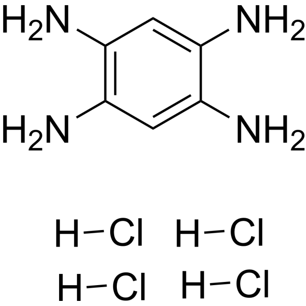
HY-12444
Y15FAK Inhibitor -
HY-20403
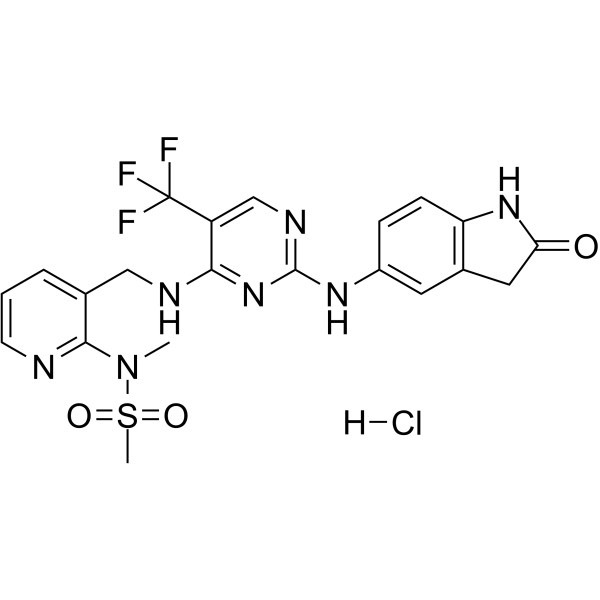
PF-562271 hydrochlorideFAK/Pyk2 Inhibitor -
HY-100498
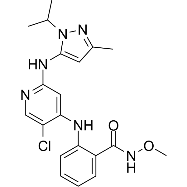
GSK2256098FAK Inhibitor -

HY-10458R
PF-562271 besylate (Standard)Reference Standard -

HY-13203R
NVP-TAE 226 (Standard)Apoptosis Inducer -
HY-109084
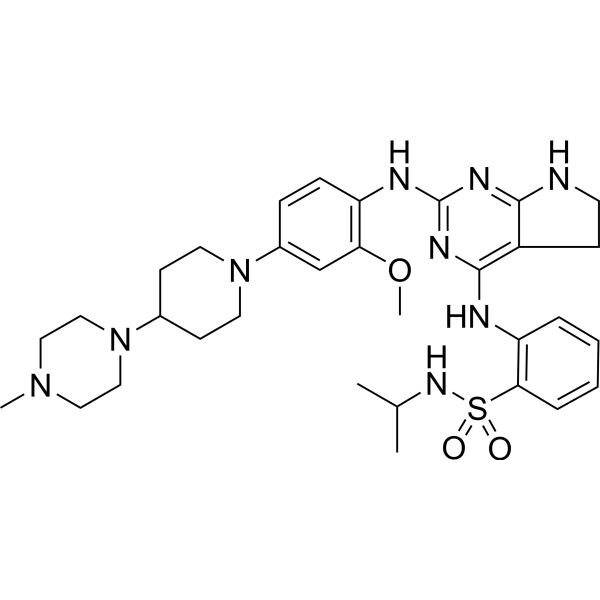
ConteltinibFAK Inhibitor
Publications Citing Use of MedChemExpress (MCE) PF-562271 (besylate)
More- Nat Biomed Eng. 2025 Nov 3. [Abstract]
- Cancer Res. 2013 May 1;73(9):2873-83. [Abstract]
- Autophagy. 2026 May 29:1-15. [Abstract]
- Cell Discov. 2022 Sep 6;8(1):84. [Abstract]
- Sci Transl Med. 2018 Jul 18;10(450):eaaq1093. [Abstract]
- Carbohydr Polym. 2024 Feb 15:326:121637. [Abstract]
- Cell Death Dis. 2023 Feb 24;14(2):157. [Abstract]
- Cell Death Dis. 2022 Sep 10;13(9):783. [Abstract]
- Clin Cancer Res. 2019 Jul 15;25(14):4552-4566. [Abstract]
- Int J Surg. 2025 Sep 17. [Abstract]
- Am J Chin Med. 2025;53(4):1225-1240. [Abstract]
- World J Gastroenterol. 2025 Jul 28;31(28):107361. [Abstract]
- Cell Rep. 2023 Oct 5;42(10):113213. [Abstract]
- Life Sci. 2021 Apr 1:270:119112. [Abstract]
- Breast Cancer Res. 2024 Mar 19;26(1):48. [Abstract]
- Sci Rep. 2018 May 8;8(1):7228. [Abstract]
- Int J Cancer. 2015 Oct 1;137(7):1549-59. [Abstract]
- Cell Signal. 2025 Sep 10:136:112117. [Abstract]
- Environ Toxicol Pharmacol. 2023 Nov:104:104301. [Abstract]
- Eur J Cell Biol. 2024 May 28;103(2):151427. [Abstract]
- Cancer Med. 2025 Sep;14(18):e71227. [Abstract]
- Mol Biol Rep. 2026 May 14;53(1):770. [Abstract]
- Mol Biol Rep. 2025 May 14;52(1):458. [Abstract]
- J Nat Med. 2020 Sep;74(4):732-740. [Abstract]
- Anticancer Drugs. 2024 Jan 1;35(1):46-54. [Abstract]
- Res Sq. 2025 Jul 11.
- University of Washington. 2025.
- bioRxiv. 2025 May 28.
- bioRxiv. 2025 Apr 7:2025.04.01.646098. [Abstract]
- Research Square Preprint. 2023 May 23.
- Research Square Preprint. 2021 Dec.
- Practical Oncology Journal. 2015, 29(5): 444-449.
 Customer Validation & Images

-
WB
-
WB
-
WB
-
WB


Biological Activity
Description
Cellular Effect
|
Cell Line

|
Type | Value | Description | References |
|---|---|---|---|---|
| A549 | GI50 |
2.1 μM
Compound: PF-562271
|
Antiproliferative activity against human A549 cells after 2 days by spectrophotometric analysis
Antiproliferative activity against human A549 cells after 2 days by spectrophotometric analysis
|
[PMID: 30340900] |
| HeLa | GI50 |
1.7 μM
Compound: PF-562271
|
Antiproliferative activity against human HeLa cells after 2 days by spectrophotometric analysis
Antiproliferative activity against human HeLa cells after 2 days by spectrophotometric analysis
|
[PMID: 30340900] |
| HepG2 | GI50 |
1.2 μM
Compound: PF-562271
|
Antiproliferative activity against human HepG2 cells after 2 days by spectrophotometric analysis
Antiproliferative activity against human HepG2 cells after 2 days by spectrophotometric analysis
|
[PMID: 30340900] |
| OVCAR-3 | GI50 |
1.8 μM
Compound: PF-562271
|
Antiproliferative activity against human OVCAR3 cells after 2 days by spectrophotometric analysis
Antiproliferative activity against human OVCAR3 cells after 2 days by spectrophotometric analysis
|
[PMID: 30340900] |
| U-87MG ATCC | GI50 |
3.7 μM
Compound: PF-562271
|
Antiproliferative activity against human U87MG cells after 2 days by spectrophotometric analysis
Antiproliferative activity against human U87MG cells after 2 days by spectrophotometric analysis
|
[PMID: 30340900] |
In Vitro
PF-562271 besylate potently inhibits purified recombinant FAK (with an IC50 of 1.5 nM) and recombinant Pyk2 (with an IC50 of 13 nM) via ATP-competitive reversible binding[1].
PF-562271 besylate exhibits over 100-fold selectivity against most tested non-target recombinant kinases, and only shows moderate activity against a subset of cyclin-dependent kinase complexes[1].
PF-562271 (starting concentration of 1 μM; 30 min) besylate potently and suppresses the autophosphorylation of FAKY397 in A431 epithelial cancer cells with an IC50 of 5 nM[1].
PF-562271 (1.1-3.3 μM; 48 h) besylate alters the cell cycle progression of PC-3M cells only after incubation at the high concentration of 3.3 μM for 48 h[1].
PF-562271 besylate is an ATP-competitive reversible inhibitor that potently inhibits the kinase activities of purified recombinant FAK (IC50 = 1.5 nM) and PYK2 (IC50 = 14 nM)[2].
PF-562271 (0.1 μM) besylate significantly inhibits IGF-I-stimulated chemotactic migration of the PDA cell line MPanc-96, but does not affect EGF-stimulated migration of this cell line[2].
MedChemExpress (MCE) has not independently confirmed the accuracy of these methods. They are for reference only. Further protocols information, click here.
-
Cell Line:Inducible stable A431 epithelial carcinoma clones expressing wild-type V5-tagged FAK
-
Concentration:1 μmol/L (starting concentration, serially diluted)
-
Incubation Time:30 min
-
Result:Inhibited FAKY397 autophosphorylation with an IC50 of 5 nmol/L (2.5 ng/mL).
Showed ~4-fold lower potency for Pyk2 in comparative cell assays.
-
Cell Line:PC-3M human prostate cancer cells
-
Concentration:1.1 μmol/L; 3.3 μmol/L
-
Incubation Time:48 h
-
Result:Reduced the percentage of cells in S and G2-M phases to levels comparable to serum-starved cells at 3.3 μmol/L.
Exerted no effect on cell cycle progression at 1.1 μmol/L.
In Vivo
PF-562271 (25-50 mg/kg; p.o.; once daily, twice daily; 15 days) besylate induces dose-dependent tumor growth inhibition in PC-3M xenografts. The efficacy of twice-daily administration is superior to that of once-daily administration at the same total daily dose, and the dose of 50 mg/kg twice daily achieves a TGI of 78% accompanied by partial tumor regression[1].
PF-562271 (50-100 mg/kg; p.o.; twice daily, once daily; continuous delivery via osmotic minipump) besylate induces significant tumor growth inhibition in BxPc3 xenografts, with the 50 mg/kg twice daily dose achieving 86% TGI and partial tumor regression[1].
PF-562271 (12.5-50 mg/kg; p.o.; twice daily; for 29 consecutive days) besylate induces dose-dependent tumor growth inhibition in BT474 xenografts, with the 50 mg/kg twice-daily dose achieving a 94% TGI accompanied by partial tumor regression[1].
PF-562271 (33 mg/kg; twice daily) besylate reduces tumor volume, decreases tumor cell proliferation rate, and significantly lowers the incidence of retroperitoneal invasion and metastasis of orthotopic MPanc-96 pancreatic ductal adenocarcinoma in athymic nude mice[2].
Combination treatment with PF-562271 (15 mg/kg; p.o.; twice daily; 7 days per week) besylate and Sunitinib (HY-10255A) administered at 20 mg/kg four times daily effectively inhibits hepatocellular carcinoma tumor growth in nude mice, reduces serum AFP and AST levels, impairs tumor angiogenesis, and induces tumor necrosis[3].
MedChemExpress (MCE) has not independently confirmed the accuracy of these methods. They are for reference only.
-
Animal Model:CD-1 Nu/Nu (female, ≈20 grams, U87MG human glioblastoma xenograft model)[1]
-
Dosage:3.3 mg/kg; 10 mg/kg; 33 mg/kg
-
Administration:p.o.; single dose
-
Result:Achieved 78% maximal inhibition of tumor phospho-FAK at 1 hour postdose, with >50% inhibition sustained for >4 hours (33 mg/kg dose).
Achieved 70% maximal inhibition of tumor phospho-FAK, with >50% inhibition sustained for <2 hours (10 mg/kg dose).
Calculated an EC50 of 93 ng/mL for total blood PF-562271 (mesylate) concentration required for half-maximal reduction of FAK phosphorylation.
-
Animal Model:CD-1 Nu/Nu (female, ≈20 grams, PC-3M human prostate xenograft model)[1]
-
Dosage:50 mg/kg (daily); 25 mg/kg (twice daily); 50 mg/kg (twice daily)
-
Administration:p.o.; daily; 15 days; p.o.; twice daily; 15 days
-
Result:Resulted in 45% tumor growth inhibition (TGI) and 37% inhibition of tumor phospho-FAK after 15 days, with a free Cₘₐₓ of 157 ng/mL (50 mg/kg daily dose).
Resulted in 61% TGI and 27% inhibition of tumor phospho-FAK after 15 days, with a free Cₘₐₓ of 25 ng/mL (25 mg/kg twice daily dose).
Resulted in 78% TGI, 52% inhibition of tumor phospho-FAK after 15 days, regressions in 3/7 mice, a free Cₘₐₓ of 96 ng/mL, and a free Cₐᵥₑ of 16 ng/mL (50 mg/kg twice daily dose).
Caused no weight loss, morbidity, or mortality.
-
Animal Model:CD-1 Nu/Nu (female, ≈20 grams, BxPc3 human pancreatic xenograft model)[1]
-
Dosage:50 mg/kg (twice daily); 100 mg/kg (daily); steady-state free concentration of 0.7 ng/mL (continuous delivery)
-
Administration:p.o.; twice daily; p.o.; daily; continuous delivery via osmotic mini-pump
-
Result:Resulted in 86% TGI, 31% inhibition of tumor phospho-FAK, regressions in 3/10 mice, and a free Cₘₐₓ of 273 ng/mL (50 mg/kg twice daily dose).
Resulted in 71% TGI and 59% inhibition of tumor phospho-FAK, with a free Cₘₐₓ of 705 ng/mL (100 mg/kg daily dose).
Resulted in 57% TGI and 37% inhibition of tumor phospho-FAK (continuous delivery via osmotic mini-pumps).
Caused no weight loss, morbidity, or mortality.
-
Animal Model:CD-1 Nu/Nu (female, ≈20 grams, BT474 human breast xenograft model)[1]
-
Dosage:12.5 mg/kg (twice daily); 25 mg/kg (twice daily); 50 mg/kg (twice daily)
-
Administration:p.o.; twice daily; 29 days
-
Result:Resulted in 37% TGI, 50% inhibition of tumor phospho-FAK, a free Cₘₐₓ of 29 ng/mL, and a free Cₐᵥₑ of 3 ng/mL (12.5 mg/kg twice daily dose).
Resulted in 59% TGI, 55% inhibition of tumor phospho-FAK, regressions in 3/8 mice, a free Cₘₐₓ of 116 ng/mL, and a free Cₐᵥₑ of 14 ng/mL (25 mg/kg twice daily dose).
Resulted in 94% TGI, 76% inhibition of tumor phospho-FAK, regressions in 4/8 mice, a free Cₘₐₓ of 180 ng/mL, and a free Cₐᵥₑ of 40 ng/mL (50 mg/kg twice daily dose).
Caused no weight loss, morbidity, or mortality.


Chemical Information
-
CAS No. 939791-38-5
-
Appearance Solid
-
Molecular Weight 665.66
-
Formula C27H26F3N7O6S2
-
Color Off-white to light yellow
-
SMILES
CS(=O)(N(C)C1=NC=CC=C1CNC2=NC(NC3=CC4=C(NC(C4)=O)C=C3)=NC=C2C(F)(F)F)=O.O=S(C5=CC=CC=C5)(O)=O
-
Synonyms
VS-6062 (besylate)
-
Shipping
Room temperature in continental US; may vary elsewhere.
-
Storage
4°C, sealed storage, away from moisture
* In solvent : -80°C, 6 months; -20°C, 1 month (sealed storage, away from moisture)


Publications (32)
-
Journal Impact Factor
-
Most Recent
-
Nat Biomed Eng
Nonexpansive biodegradable matrix promotes blood vessel organoid development for neurovascular repair and functional recovery in ischaemic stroke. [Abstract]2025 Nov 3. PMID: 41184604 -
Cancer Res
High-throughput tyrosine kinase activity profiling identifies FAK as a candidate therapeutic target in Ewing sarcoma. [Abstract]2013 May 1;73(9):2873-83. PMID: 23536552
PF-562271 (besylate) purchased from MedChemExpress. Usage Cited in: Cancer Res. 2013 May 1;73(9):2873-83. [Abstract]
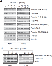
FAK inhibition downregulates the AKT/mTOR pathway and CAS activity. A, protein levels measured by Western immunoblotting for AKT/mTOR pathway proteins in A673 and TC32 cells serum-starved overnight, treated with PF-562271 for 6 hours, and then stimulated with IGF-1 for 2 hours. Vinculin is used as the loading control. B, Western immunoblots showing downregulation of phospho-CAS but not phospho-ERK in A673 and TC32 cells after treatment with PF-562271.
-
Autophagy
PTK2/FAK inhibition triggers TMED9-mediated protective autophagy in pancreatic cancer cell via enhancing ERGIC-ERES contact. [Abstract]2026 May 29:1-15. PMID: 42144738 -
Cell Discov
A positive mechanobiological feedback loop controls bistable switching of cardiac fibroblast phenotype. [Abstract]2022 Sep 6;8(1):84. PMID: 36068215 -
Sci Transl Med
PP2A inhibition is a druggable MEK inhibitor resistance mechanism in KRAS-mutant lung cancer cells. [Abstract]2018 Jul 18;10(450):eaaq1093. PMID: 30021885 -
Carbohydr Polym
Inulin-like polysaccharide ABWW may impede CCl4 induced hepatic stellate cell activation through mediating the FAK/PI3K/AKT signaling pathway in vitro & in vivo. [Abstract]2024 Feb 15:326:121637. PMID: 38142102 -
Cell Death Dis
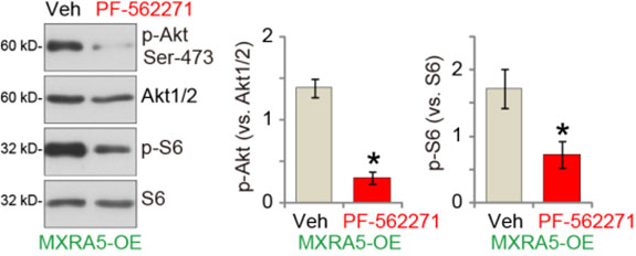
Identification of matrix-remodeling associated 5 as a possible molecular oncotarget of pancreatic cancer. [Abstract]2023 Feb 24;14(2):157. PMID: 36828810
PF-562271 (besylate) purchased from MedChemExpress. Usage Cited in: Cell Death Dis. 2023 Feb 24;14(2):157. [Abstract]
PF-562271 (250 nM; 6 h) largely inhibits Akt-S6 phosphorylation in OE-MXRA5 priPC-1 cells.
-
Cell Death Dis
P130cas-FAK interaction is essential for YAP-mediated radioresistance of non-small cell lung cancer. [Abstract]2022 Sep 10;13(9):783. PMID: 36088346 -
Clin Cancer Res
High-throughput Chemical Screening Identifies Focal Adhesion Kinase and Aurora Kinase B Inhibition as a Synergistic Treatment Combination in Ewing Sarcoma. [Abstract]2019 Jul 15;25(14):4552-4566. PMID: 30979745 -
Int J Surg
Supervising the recurrence of pancreatic ductal adenocarcinoma using CtDNA-based MIR129-2 methylation detection. [Abstract]2025 Sep 17. PMID: 40961228 -
Am J Chin Med
Evodiamine Suppresses Lung Cancer Progression Through Modulating FAK/STAT3/AKT Signaling Pathway. [Abstract]2025;53(4):1225-1240. PMID: 40582716 -
World J Gastroenterol
FBP1 as a key regulator of focal adhesion kinase-mediated hepatic stellate cell activation: Multi-omics and experimental validation. [Abstract]2025 Jul 28;31(28):107361. PMID: 40741470 -
Cell Rep
Mechanotransduction in response to ECM stiffening impairs cGAS immune signaling in tumor cells. [Abstract]2023 Oct 5;42(10):113213. PMID: 37804510 -
Life Sci
Harmine inhibits the proliferation and migration of glioblastoma cells via the FAK/AKT pathway. [Abstract]2021 Apr 1:270:119112. PMID: 33508300 -
Breast Cancer Res
TMEM120B strengthens breast cancer cell stemness and accelerates chemotherapy resistance via β1-integrin/FAK-TAZ-mTOR signaling axis by binding to MYH9. [Abstract]2024 Mar 19;26(1):48. PMID: 38504374 -
Sci Rep
The mechanical microenvironment regulates ovarian cancer cell morphology, migration, and spheroid disaggregation. [Abstract]2018 May 8;8(1):7228. PMID: 29740072 -
Int J Cancer
2015 Oct 1;137(7):1549-59. PMID: 25809490
PF-562271 (besylate) purchased from MedChemExpress. Usage Cited in: Int J Cancer. 2015 Oct 1;137(7):1549-59. [Abstract]
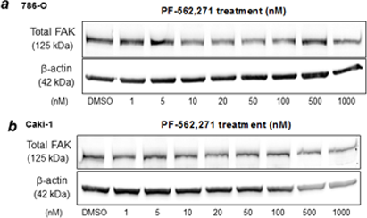
786-O (a) and Caki-1 (b) are treated for 24 and 48h with increasing concentrations of PF-562,271 and adherent cells were counted. Cell lysates are evaluated through immunoblotting for total FAK following treatment for 24h.
-
Cell Signal
Tenvermectin B, a novel macrocyclic lactone antibiotic, suppresses glioblastoma progression by targeting RhoJ. [Abstract]2025 Sep 10:136:112117. PMID: 40939916 -
Environ Toxicol Pharmacol
ZC3H4 Governs Epithelial Cell Migration through ROCK/p-PYK2/p-MLC2 Pathway in Silica-induced Pulmonary Fibrosis. [Abstract]2023 Nov:104:104301. PMID: 37866415 -
Eur J Cell Biol
The mechanical mechanism of angiotensin II induced activation of hepatic stellate cells promoting portal hypertension. [Abstract]2024 May 28;103(2):151427. PMID: 38820882 -
Cancer Med
Focal Adhesion Kinase Intersects With the BRD4-MYC Axis and YAP1 to Drive Tumor Cell Growth, Phenotypic Plasticity, Stemness, and Metastatic Potential in Colorectal Cancer. [Abstract]2025 Sep;14(18):e71227. PMID: 40959971 -
Mol Biol Rep
Piperlongumine inhibits the proliferation and migration of non-small cell lung cancer cells through the EGFR/FAK/STAT3 pathway. [Abstract]2026 May 14;53(1):770. PMID: 42133146 -
Mol Biol Rep
Ginkgetin inhibits the proliferation and migration of lung cancer cells via FAK/STAT3/AKT pathway. [Abstract]2025 May 14;52(1):458. PMID: 40366441 -
J Nat Med
2020 Sep;74(4):732-740. PMID: 32643027 -
Anticancer Drugs
2024 Jan 1;35(1):46-54. PMID: 37449977 -
-
-
-
bioRxiv
2025 Apr 7:2025.04.01.646098. PMID: 40291676 -
-
-
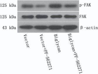
PF-562271 (besylate) purchased from MedChemExpress. Usage Cited in: Practical Oncology Journal. 2015, 29(5): 444-449.
Effects and mechanism of Biglycan and FAK signaling pathway on the invasion and metastasis of colon cancer cells.


Solvent & Solubility
In Vitro:
DMSO : 21.4 mg/mL (32.15 mM; Need ultrasonic and warming; Hygroscopic DMSO has a significant impact on the solubility of product, please use newly opened DMSO)
H2O : < 0.1 mg/mL (insoluble)
Please refer to the solubility information to select the appropriate solvent. Once prepared, please aliquot and store the solution to prevent product inactivation from repeated freeze-thaw cycles.
Storage method and period of stock solution: -80°C, 6 months; -20°C, 1 month (sealed storage, away from moisture). When stored at -80°C, please use it within 6 months. When stored at -20°C, please use it within 1 month.
Please refer to the solubility information to select the appropriate solvent. Once prepared, please aliquot and store the solution to prevent product inactivation from repeated freeze-thaw cycles.
Storage method and period of stock solution: -80°C, 6 months; -20°C, 1 month (sealed storage, away from moisture). When stored at -80°C, please use it within 6 months. When stored at -20°C, please use it within 1 month.
Concentration (start) × Volume (start) = Concentration (final) × Volume (final)
In Vivo:
Select the appropriate dissolution method based on your experimental animal and administration route.
- For the following dissolution methods, please ensure to first prepare a clear stock solution using an In Vitro approach and then sequentially add co-solvents:
- To ensure reliable experimental results, the clarified stock solution can be appropriately stored based on storage conditions. As for the working solution for In Vivo experiments, it is recommended to prepare freshly and use it on the same day.
- The percentages shown for the solvents indicate their volumetric ratio in the final prepared solution. If precipitation or phase separation occurs during preparation, heat and/or sonication can be used to aid dissolution.
Add each solvent one by one: 10% DMSO 40% PEG300 5% Tween-80 45% Saline
Solubility: ≥ 1.67 mg/mL (2.51 mM); Clear solution
This protocol yields a clear solution of ≥ 1.67 mg/mL (saturation unknown).
Taking 1 mL working solution as an example, add 100 μL DMSO stock solution (16.7 mg/mL) to 400 μL PEG300, and mix evenly; then add 50 μL Tween-80 and mix evenly; then add 450 μL Saline to adjust the volume to 1 mL.
Preparation of Saline: Dissolve 0.9 g sodium chloride in ddH₂O and dilute to 100 mL to obtain a clear Saline solution.
Add each solvent one by one: 10% DMSO 90% (20% SBE-β-CD in Saline)
Solubility: ≥ 1.67 mg/mL (2.51 mM); Clear solution
This protocol yields a clear solution of ≥ 1.67 mg/mL (saturation unknown).
Taking 1 mL working solution as an example, add 100 μL DMSO stock solution (16.7 mg/mL) to 900 μL 20% SBE-β-CD in Saline, and mix evenly.
Preparation of 20% SBE-β-CD in Saline (4°C, storage for one week): 2 g SBE-β-CD powder is dissolved in 10 mL Saline, completely dissolve until clear.
In Vivo Dissolution Calculator
Please enter the basic information of animal experiments:
-
-
-
-
Recommended: Prepare an additional quantity of animals to account for potential losses during experiments.
Please enter your animal formula composition:
-
%DMSO +
Recommended: Keep the proportion of DMSO in working solution below 2% if your animal is weak.
-
%+
-
+%Tween-80 + +
-
%Saline +
The co-solvents required include: DMSO, . All of co-solvents are available by MedChemExpress (MCE). , Tween 80. All of co-solvents are available by MedChemExpress (MCE).
Working solution concentration: 0.22 mg/mL
Method for preparing stock solution: mg drug dissolved in μL DMSO. Stock solution concentration: mg/mL. * In solvent : -80°C, 6 months; -20°C, 1 month (sealed storage, away from moisture)
1. Take μL DMSO stock solution;
2. Add μL .
μL , mix evenly;
3. Then add μL Tween 80, mix evenly;
4. Then add μL
Please ensure that the stock solution in the first step is dissolved to a clear state, and add co-solvents in sequence. You can use ultrasonic heating (ultrasonic cleaner, recommended frequency 20-40 kHz), vortexing, etc. to assist dissolution.

Protocols
-
Human pluripotent stem cell endothelial-cell differentiation
Human pluripotent stem cell endothelial differentiation is based on stepwise developmental patterning: early activation of WNT/GSK3β inhibition promotes mesodermal or vascular progenitor entry, followed by endothelial specification using VEGF-related signaling, BMP4, FGF2, Notch modulation, or cAMP depending on the published protocol. Endothelial differentiation is read out by acquisition of CD31, CD34, VE-cadherin/CD144, KDR/VEGFR2, vWF, Tie2, NOS3, acetylated LDL uptake, tube/network formation, barrier function, and in vivo vessel-forming capacity where tested.
-
Vascular/Branching Fractal Analysis
Vascular/branching fractal analysis quantifies the geometric complexity of vessel trees or vascular networks from segmented 2D images, commonly by converting vessels into binary and/or skeletonized maps and estimating fractal dimension using box-counting or related approaches. Fractal dimension is interpreted as an image-derived readout of vascular branching complexity, space filling, or density, and has been applied to retinal photographs, fluorescein angiography, OCT angiography, capillary perfusion maps, and in vitro Matrigel angiogenesis networks. The assay readout is generated from vessel-positive pixels after image preprocessing, vessel segmentation, binarization, and optional skeletonization; reported outputs include fractal dimension, vessel density, branchpoint density, endpoint density, vessel length density, tortuosity, and generation-based branching metrics when VESGEN-style analysis is used. The biological interpretation is limited to quantitative vascular patterning and s
-
Annexin V plus membrane-impermeant dye apoptosis staining
Annexin V-based apoptosis assays rely on the detection of phosphatidylserine (PS) externalization from the inner leaflet of the plasma membrane to the outer leaflet, an early biochemical hallmark of apoptosis. Fluorescently labeled Annexin V binds PS in a calcium-dependent manner, enabling identification of early apoptotic cells by flow cytometry or fluorescence microscopy. When combined with a membrane-impermeant DNA-binding dye (e. g. , propidium iodide), this approach allows discrimination between viable (Annexin V−/dye−), early apoptotic (Annexin V+/dye−), and late apoptotic or necrotic (Annexin V+/dye+) cell populations by assessing membrane integrity and PS exposure.
-
Patient-Derived Orthotopic Xenograft (PDOX)
Patient-derived orthotopic xenograft (PDOX) modeling implants fresh patient tumor tissue or patient-derived tumor cells into the anatomically corresponding organ or tissue site of immunodeficient mice, usually by surgical orthotopic implantation, to preserve patient tumor histology, local microenvironmental context, invasion, metastatic behavior, and treatment-response features better than subcutaneous implantation. PDOX readouts include tumor engraftment, orthotopic tumor growth, local invasion, metastasis, recurrence after resection, histologic similarity to the donor tumor, biomarker retention, molecular concordance, survival, and response or resistance to therapy. PDOX models are used for preclinical drug testing and individualized therapy evaluation, but engraftment success varies by tumor type and specimen quality.
-
Liver Cancer Modeling
Liver cancer can be classified into primary liver cancer and secondary liver cancer. Secondary liver cancer is the metastatic liver cancer. Primary liver cancer includes hepatocellular carcinoma (HCC), intrahepatic cholangiocarcinoma (ICC) and fibrolamellar HCC, of which HCC is the most common form, accounting for approximately 90% of primary liver cancers[1]. HCC mouse models include chemical agent-induced models, transplanted tumor models, and genetic engineered models.
-
Breast Cancer Modeling
Breast cancer is a heterogeneous cancer, and it has been distinguished into four subtypes: luminal A, luminal B, HER2-positive and basal-like. Molecular mutations, epigenetic alterations, hormone exposure and immune microenvironment are related to the progression of breast cancer.
-
How to Choose the Right Model Animal
Choosing the right model animal is a validity-driven decision in which the species, strain, sex, age, genetic background, disease-induction method, outcome measures, and welfare burden must match the scientific question rather than laboratory tradition or convenience. A model should be selected by judging face validity, construct validity, and predictive validity: whether it resembles the human phenotype, whether it reproduces relevant mechanisms, and whether results are likely to predict human biology or treatment response. Animal studies often fail to translate because of species differences, weak disease resemblance, poor experimental design, inadequate reporting, publication bias, and underuse of randomization, blinding, and sample-size justification. Unresolved questions include how to rank competing models objectively, how much human-disease complexity must be reproduced for a given objective, and when non-animal systems such as organoids, ex vivo tissue, or computational models
-
Apoptosis
Apoptosis, also called programmed cell death, is generally characterized by distinct morphological characteristics.
-
TUNEL staining for apoptotic DNA fragmentation
TUNEL staining detects DNA strand breaks by using terminal deoxynucleotidyl transferase to add labeled nucleotides to exposed 3′-OH DNA termini, generating either microscopic staining in fixed cells or tissue sections, or fluorescence/cytometric signal in cell suspensions. TUNEL positivity reflects DNA fragmentation but should not be interpreted alone as definitive apoptosis, because TUNEL can also label necrotic, autolytic, mechanically damaged, or DNA-repair-associated DNA breaks.
-
Cell Cytotoxicity Assay
Cytotoxicity assays are usually based on the assessment of cell membrane damage, which can also be indirectly detected by measuring cell viability. Detection methods include MTT assay, CKK-8 assay, LDH assay and ATP assay, etc.
-
Patient-Derived Xenograft (PDX)
Patient-derived xenograft (PDX) models are generated by engrafting primary human tumor tissue directly into immunodeficient mice, allowing in vivo propagation of patient tumor biology without initial in vitro adaptation. These models are used to preserve key histopathological and molecular characteristics of the original tumor and enable assessment of tumor growth dynamics and therapeutic response in a living organism. The biological readout is tumor engraftment and subsequent growth in the murine host, which reflects the ability of human tumor cells to survive, vascularize, and expand in an immunocompromised microenvironment.
-
Endothelial Tube Formation Assay
Endothelial tube formation assay evaluates the ability of endothelial cells to attach, migrate, align, and organize into capillary-like networks when cultured on gelled basement membrane extract or Matrigel; the readout is the morphology and quantity of tube-like networks, which reflects an in vitro endothelial morphogenesis step related to angiogenesis. Basement membrane extract/Matrigel provides laminin-rich extracellular matrix cues that support endothelial differentiation into capillary-like structures, but it can contain biologically active growth factors, so growth-factor-reduced matrix is preferred when testing defined angiogenic stimulators or inhibitors.
-
Apoptosis Solutions
Apoptosis is a regulated, generally non-lytic cell-death pathway that removes unwanted, damaged, infected, or abnormal cells through coordinated morphological changes, caspase activation, DNA fragmentation, and membrane remodeling. The intrinsic apoptosis pathway is controlled mainly by mitochondrial outer membrane permeabilization, BCL-2 family proteins, cytochrome c release, apoptosome formation, caspase-9 activation, and downstream executioner caspase-3/7 activation. The extrinsic apoptosis pathway is initiated by death receptors such as Fas, TNFR, and TRAIL receptors, which recruit adaptor proteins and activate caspase-8 before engaging executioner caspases or mitochondrial amplification through BID cleavage. Apoptosis is linked to many phenotypes, including cancer cell killing, tissue homeostasis, immune regulation, neurodegeneration, infection response, and treatment-induced cytotoxicity; unresolved questions include how apoptosis interacts with necroptosis, pyroptosis, ferroptos
-
Cell invasion
Cell invasion is the ability of cells to migrate from one area to another via the extracellular matrix. Cell invasion is the response of normal and cancer cells to chemical and mechanical stimuli. Before migrating to a new region, the extracellular matrix is degraded by proteases within the cell. Cell invasion often occurs during wound repair, vascularization and inflammation, abnormal tissue invasion, and tumor cell metastasis.
-
Subcutaneous Cell-Line-Derived Xenograft
Subcutaneous cell-line-derived xenograft (CDX) models are established by implanting cultured human cancer cell lines into immunodeficient mice, where the injected cells form localized tumors that can be monitored in vivo as a measure of tumorigenic potential, growth kinetics, and treatment response. These models are widely used in oncology research because they allow reproducible tumor formation and enable comparative assessment of tumor growth between different cell lines or genetic manipulations in a controlled in vivo microenvironment. Subcutaneous implantation of cancer cells in immunodeficient mice is a standard approach for evaluating tumor growth behavior and therapeutic response across multiple cancer types, including prostate, esophageal, pancreatic, and colon cancer models.
-
Orthotopic Cell-Line Xenograft
Orthotopic cell-line xenograft models involve implantation of human cancer cell lines into the anatomically corresponding organ of immunodeficient mice to reproduce tumor growth within a native microenvironment, enabling more clinically relevant tumor behavior compared with subcutaneous models. These models are widely used because orthotopic placement better recapitulates tumor progression, including invasion and metastatic spread, which are often underrepresented in heterotopic implantation systems. Compared with conventional xenografts, orthotopic implantation is described as more technically complex but provides improved simulation of tumor-microenvironment interactions and metastatic behavior, making it particularly valuable for translational oncology research. Surgical orthotopic implantation approaches have been emphasized as enabling faithful reproduction of clinical cancer features, including metastasis and disease progression patterns that align with the tumor’s organ of origi
-
Endothelial Cell Migration/Angiogenic Sprouting Assay
Endothelial cell migration and angiogenic sprouting assays are in vitro (and partially ex vivo-adapted) functional models that quantify the ability of endothelial cells to undergo coordinated migration, extracellular matrix invasion, and multicellular organization into capillary-like sprouts in response to pro-angiogenic stimuli such as VEGF, bFGF, or conditioned microenvironments. These assays are used to model early angiogenic events including tip-cell formation, directional migration, and lumen-like sprout extension, which collectively reflect angiogenic activation and vascular morphogenesis processes observed in vivo.


Purity & Documentation
-
Data Sheet (290 KB)
-
SDS (393 KB)
- English - EN (393 KB)
- Français - FR (393 KB)
- Deutsch - DE (393 KB)
- Norwegian - NO (393 KB)
- Español - ES (393 KB)
- Swedish - SV (393 KB)
- Italian - IT (393 KB)
- Korean - KR (393 KB)
- Portuguese - PT (393 KB)
-
Handling Instructions (2659 KB)
References
[1]. Roberts WG, et al. Antitumor activity and pharmacology of a selective focal adhesion kinase inhibitor, PF-562,271. Cancer research. 2008 Mar 15;68(6):1935-44. [Content Brief]
[2]. Stokes JB, et al. Inhibition of focal adhesion kinase by PF-562,271 inhibits the growth and metastasis of pancreatic cancer concomitant with altering the tumor microenvironment. Molecular cancer therapeutics. 2011 Nov;10(11):2135-45. [Content Brief]
[3]. Bagi CM, et al. Sunitinib and PF-562,271 (FAK/Pyk2 inhibitor) effectively block growth and recovery of human hepatocellular carcinoma in a rat xenograft model. Cancer biology & therapy. 2009 May;8(9):856-65. [Content Brief]


Complete Stock Solution Preparation Table
Please refer to the solubility information to select the appropriate solvent. Once prepared, please aliquot and store the solution to prevent product inactivation from repeated freeze-thaw cycles.
Storage method and period of stock solution: -80°C, 6 months; -20°C, 1 month (sealed storage, away from moisture). When stored at -80°C, please use it within 6 months. When stored at -20°C, please use it within 1 month.
| Optional Solvent | Concentration Solvent Mass | 1 mg | 5 mg | 10 mg | 25 mg |
|---|---|---|---|---|---|
| DMSO | 1 mM | 1.5023 mL | 7.5113 mL | 15.0227 mL | 37.5567 mL |
| 5 mM | 0.3005 mL | 1.5023 mL | 3.0045 mL | 7.5113 mL | |
| 10 mM | 0.1502 mL | 0.7511 mL | 1.5023 mL | 3.7557 mL | |
| 15 mM | 0.1002 mL | 0.5008 mL | 1.0015 mL | 2.5038 mL | |
| 20 mM | 0.0751 mL | 0.3756 mL | 0.7511 mL | 1.8778 mL | |
| 25 mM | 0.0601 mL | 0.3005 mL | 0.6009 mL | 1.5023 mL | |
| 30 mM | 0.0501 mL | 0.2504 mL | 0.5008 mL | 1.2519 mL |


PF-562271 (besylate) Related Classifications
HY-10458 Related Classifications
 Powered by Bioz