



PHA-665752
Based on 16 publication(s) in Google Scholar
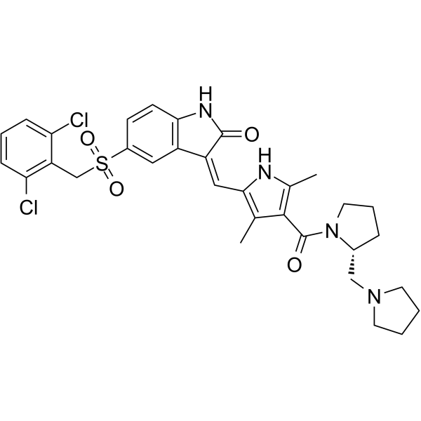
PHA-665752 is a selective, ATP-competitive, and active-site inhibitor of the catalytic activity of c-Met kinase (Ki=4 nM; IC50=9 nM). PHA-665752 exhibits >50-fold selectivity for c-Met compared with a panel of diverse tyrosine and serine-threonine kinases. PHA-665752 induces apoptosis and cell cycle arrest, and exhibits cytoreductive antitumor activity.
For research use only. We do not sell to patients.
- Purity : 99.71%
- CAS No.: 477575-56-7
- Formula: C32H34Cl2N4O4S
- Molecular Weight:641.61
-
Storage:Powder -20°C, 3 years , 4°C, 2 years ; In solvent -80°C, 2 years , -20°C, 1 year

To place orders, for customer services and technical support, please contact: MedChemExpress USA
Tel: 609-228-6898 E-mail: [email protected] [email protected]


-
 Biological Activity
-
Chemical Information
-
Solvent & Solubility
- Protocols
- Purity & Documentation
- References
-
 Help & FAQs


-
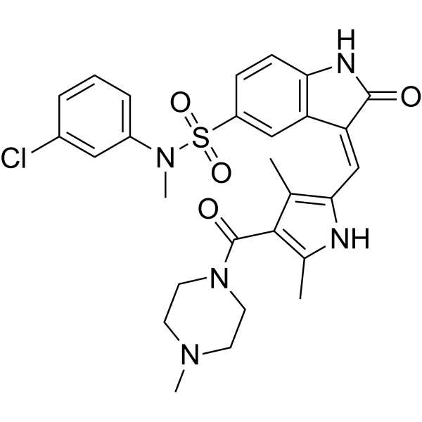
HY-12014
SU11274c-Met/HGFR Inhibitor -
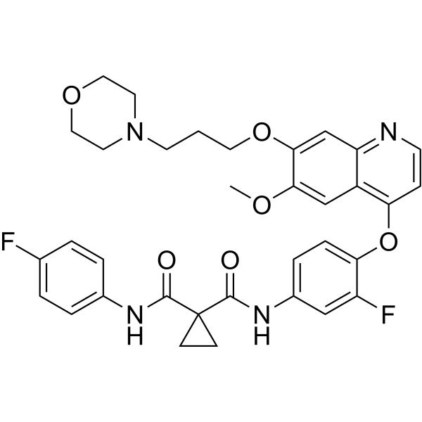
HY-10338
ForetinibMET/VEGFR2/KDR Inhibitor -
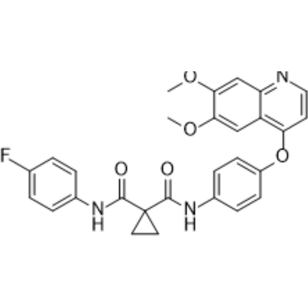
HY-13016
CabozantinibVEGFR2/MET Inhibitor -
HY-10255A
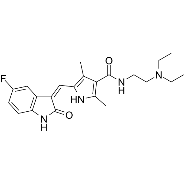
SunitinibMulti-targeted RTK Inhibitor -

HY-10431
SB-431542TGF-β Receptor Kinase Inhibitor -
HY-13404
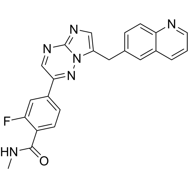
Capmatinibc-MET Kinase Inhibitor -
HY-10432
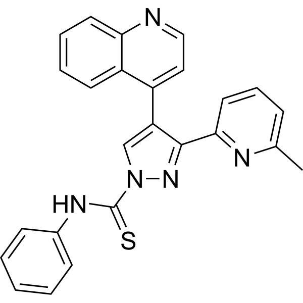
A 83-01ALK4/5/7 Inhibitor -
HY-10492
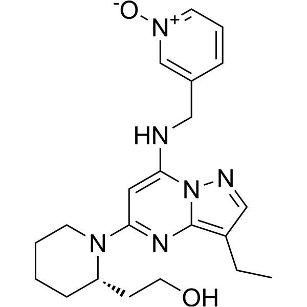
DinaciclibCDK2/5/9 Inhibitor -
HY-10221
VorinostatHDAC Inhibitor -
HY-13434
IonomycinCalcium Ionophore -

HY-15763
ErastinFerroptosis Inducer -
HY-10992
AZD-7762Chk Inhibitor
Publications Citing Use of MedChemExpress (MCE) PHA-665752
More- Cancer Res. 2025 Sep 18. [Abstract]
- J Exp Clin Cancer Res. 2024 Nov 20;43(1):308. [Abstract]
- Cell Death Dis. 2022 Apr 21;13(4):387. [Abstract]
- Cancer Lett. 2020 Dec 28:495:41-52. [Abstract]
- Adv Healthc Mater. 2025 Jan;14(2):e2402884. [Abstract]
- Mol Cancer Ther. 2018 Mar;17(3):603-613. [Abstract]
- Cell Biosci. 2026 Jun 20. [Abstract]
- Eur J Pharmacol. 2026 Apr 10:1020:178775. [Abstract]
- Respir Res. 2020 Aug 14;21(1):215. [Abstract]
- Int J Cancer. 2019 Aug 1;145(3):748-762. [Abstract]
- Med Oncol. 2020 Mar 12;37(4):24. [Abstract]
- Biomed Res Int. 2020 Mar 14:2020:3635787. [Abstract]
- FEBS Open Bio. 2021 Nov;11(11):3115-3125. [Abstract]
- Am J Transl Res. 2023 Feb 15;15(2):896-913. [Abstract]
- bioRxiv. 2026 Jul 6.
- Oncotarget. 2017 Jun 13;8(24):38717-38730. [Abstract]
 Customer Validation & Images

-
IF
-
WB
-
WB


Biological Activity
Description
IC50 & Target
Cellular Effect
|
Cell Line

|
Type | Value | Description | References |
|---|---|---|---|---|
| A549 | IC50 |
9 nM
Compound: 3, PHA-665752
|
Inhibition of human recombinant c-MET kinase expressed in A549 cells assessed as inhibition of HGF-induced autophosphorylation by ELISA method
Inhibition of human recombinant c-MET kinase expressed in A549 cells assessed as inhibition of HGF-induced autophosphorylation by ELISA method
|
[PMID: 21812414] |
| HCC827 | IC50 |
15.7 μM
Compound: PHA-665752
|
Cytotoxicity against erlotinib-sensitive human HCC827A cells assessed as reduction in cell viability after 72 hrs by MTT assay
Cytotoxicity against erlotinib-sensitive human HCC827A cells assessed as reduction in cell viability after 72 hrs by MTT assay
|
[PMID: 27563407] |
| HCC827 | IC50 |
8.9 μM
Compound: PHA-665752
|
Cytotoxicity against erlotinib-resistant human HCC827B cells expressing MET assessed as reduction in cell viability after 72 hrs by MTT assay
Cytotoxicity against erlotinib-resistant human HCC827B cells expressing MET assessed as reduction in cell viability after 72 hrs by MTT assay
|
[PMID: 27563407] |
| Sf9 | IC50 |
38 nM
Compound: 2, PHA-665752
|
Inhibition of GST-tagged c-Met expressed in Sf9 cells
Inhibition of GST-tagged c-Met expressed in Sf9 cells
|
[PMID: 19854051] |
| Sf9 | IC50 |
863 nM
Compound: 2, PHA-665752
|
Inhibition of GST-tagged VEGFR expressed in Sf9 cells
Inhibition of GST-tagged VEGFR expressed in Sf9 cells
|
[PMID: 19854051] |
| TAMH | IC50 |
8.9 μM
Compound: PHA-665752
|
Cytotoxicity against TAMH assessed as reduction in cell viability after 24 hrs by cell titer glo based luminescence assay
Cytotoxicity against TAMH assessed as reduction in cell viability after 24 hrs by cell titer glo based luminescence assay
|
[PMID: 27563407] |
In Vitro
PHA-665752 is a potent and ATP-competitive inhibitor of c-Met kinase activity with a Ki of 4 nM and an IC50 of 9 nM[1].
PHA-665752 exhibits >50-fold selectivity for c-Met enzyme compared with the majority of kinases evaluated[1].
PHA-665752 shows potent inhibition of c-Met RTK autophosphorylation in NIH3T3 cells engineered to express high levels of c-Met and hepatocyte growth factor (HGF)[1].
PHA-665752 inhibits HGF-stimulated or constitutive phosphorylation of mediators of downstream of c-Met such as Gab-1, ERK, Akt, STAT3, PLC-γ, and FAK in multiple tumor cell lines[1].
PHA-665752 (0-1.25 μM; 18 hours) potently inhibits HGF and c-Met-driven phenotypes such as cell growth (proliferation and survival), cell motility, invasion, and/or morphology of a variety of tumor cells[1].
PHA-665752 (0-1.25 μM; 72 hours) induces apoptosis in both the presence and absence of HGF at concentrations that inhibited tyrosine phosphorylation of c-Met in GTL-16 cells[1].
PHA-665752 (0.0125-0.2 μM; 4 hours) potent inhibits HGF-induced c-Met phosphorylation in A549 cells[1].
MedChemExpress (MCE) has not independently confirmed the accuracy of these methods. They are for reference only. Further protocols information, click here.
-
Cell Line:S114 cells, GTL-16 cells, NCI-H441 cells, or BxPC-3 cells
-
Concentration:0 μM, 0.002 μM, 0.01 μM, 0.05 μM, 0.25 μM, 1.25 μM
-
Incubation Time:18 hours
-
Result:Potently inhibited HGF and c-Met-driven cell growth.
-
Cell Line:GTL-16 cells
-
Concentration:0 μM, 0.002 μM, 0.01 μM, 0.05 μM, 0.25 μM, 1.25 μM
-
Incubation Time:72 hours
-
Result:Induced apoptosis in both the presence and absence of HGF at concentrations that inhibited tyrosine phosphorylation of c-Met in GTL-16 cells. Immunoblot Analysis.
-
Cell Line:A549 cells
-
Concentration:0.0125 μM, 0.025 μM,0.05 μM,0.1 μM,0.2 μM
-
Incubation Time:4 hours
-
Result:Potent inhibited HGF-induced c-Met phosphorylation in A549 cells.
In Vivo
PHA-665752 shows a potent cytoreductive activity in a gastric carcinoma xenograft model[1].
MedChemExpress (MCE) has not independently confirmed the accuracy of these methods. They are for reference only.
-
Animal Model:Female athymic mice (nu/nu, 8–12 weeks) bearing S114 or GTL-16 tumor xenografts[1]
-
Dosage:7.5 mg/kg/day, 15 mg/kg/day, 30 mg/kg/day
-
Administration:Intravenous injection; for 9 days
-
Result:Demonstrated statistically significant dose-dependent tumor growth inhibition.


Chemical Information
-
CAS No. 477575-56-7
-
Appearance Solid
-
Molecular Weight 641.61
-
Formula C32H34Cl2N4O4S
-
Color Yellow to orange
-
SMILES
O=C(C1=C(NC(/C=C2C(NC3=C\2C=C(C=C3)S(=O)(CC4=C(C=CC=C4Cl)Cl)=O)=O)=C1C)C)N5[C@H](CCC5)CN6CCCC6
-
Shipping
Room temperature in continental US; may vary elsewhere.
-
Storage
Powder -20°C 3 years 4°C 2 years In solvent -80°C 2 years -20°C 1 year


Publications (16)
-
Journal Impact Factor
-
Most Recent
-
Cancer Res
The Prolyl Isomerase PIN1 Impacts Fibroblast Differentiation States and Crosstalk in Pancreatic Cancer. [Abstract]2025 Sep 18. PMID: 40966318 -
J Exp Clin Cancer Res
Dual inhibition of HERs and PD-1 counteract resistance in KRASG12C-mutant head and neck cancer. [Abstract]2024 Nov 20;43(1):308. PMID: 39567998 -
Cell Death Dis
HGF/c-Met pathway facilitates the perineural invasion of pancreatic cancer by activating the mTOR/NGF axis. [Abstract]2022 Apr 21;13(4):387. PMID: 35449152 -
Cancer Lett
Loss of histone lysine methyltransferase EZH2 confers resistance to tyrosine kinase inhibitors in non-small cell lung cancer. [Abstract]2020 Dec 28:495:41-52. PMID: 32920200 -
Adv Healthc Mater
Antitumor Activity of a Bispecific Chimera Targeting EGFR and Met in Gefitinib-Resistant Non-Small Cell Lung Cancer. [Abstract]2025 Jan;14(2):e2402884. PMID: 39586988 -
Mol Cancer Ther
Afatinib Is a New Therapeutic Approach in Chordoma with a Unique Ability to Target EGFR and Brachyury. [Abstract]2018 Mar;17(3):603-613. PMID: 29237806 -
Cell Biosci
Hepatocyte growth factor suppresses melanogenesis via ERK-dependent nuclear exclusion of MITF. [Abstract]2026 Jun 20. PMID: 42323670 -
Eur J Pharmacol
Integrative Mendelian randomization and experimental validation unveil novel druggable targets in primary biliary cholangitis. [Abstract]2026 Apr 10:1020:178775. PMID: 41819524 -
Respir Res
HGF alleviates septic endothelial injury by inhibiting pyroptosis via the mTOR signalling pathway. [Abstract]2020 Aug 14;21(1):215. PMID: 32795289
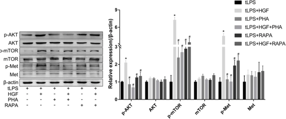
PHA-665752 purchased from MedChemExpress. Usage Cited in: Respir Res. 2020 Aug 14;21(1):215. [Abstract]
Immunoblotting reveals that c-Met in the plasma membrane is phosphorylated in response to the binding of HGF, and this c-Met phosphorylation is abolished by PHA-665752 in the LPS-transfected endothelial cells.
-
Int J Cancer
MET activation confers resistance to cetuximab, and prevents HER2 and HER3 upregulation in head and neck cancer. [Abstract]2019 Aug 1;145(3):748-762. PMID: 30694565 -
Med Oncol
Activation of the HGF/c-MET axis promotes lenvatinib resistance in hepatocellular carcinoma cells with high c-MET expression. [Abstract]2020 Mar 12;37(4):24. PMID: 32166604 -
Biomed Res Int
Upregulation of PDGF Mediates Robust Liver Regeneration after Nanosecond Pulsed Electric Field Ablation by Promoting the HGF/c-Met Pathway. [Abstract]2020 Mar 14:2020:3635787. PMID: 32258116 -
FEBS Open Bio
CXCR4 promotes gefitinib resistance of Huh7 cells by activating the c-Met signaling pathway. [Abstract]2021 Nov;11(11):3115-3125. PMID: 34555268 -
Am J Transl Res
2023 Feb 15;15(2):896-913. PMID: 36915773
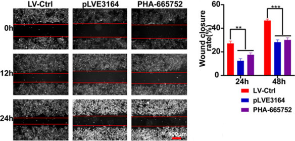
PHA-665752 purchased from MedChemExpress. Usage Cited in: Am J Transl Res. 2023 Feb 15;15(2):896-913. [Abstract]
PHA-665752 significantly decreases the migration rate at 24 h and 48 h in Huh-7R cells.
-
-
Oncotarget
Contributions of MET activation to BCR-ABL1 tyrosine kinase inhibitor resistance in chronic myeloid leukemia cells. [Abstract]2017 Jun 13;8(24):38717-38730. PMID: 28418880
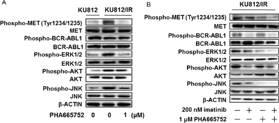
PHA-665752 purchased from MedChemExpress. Usage Cited in: Oncotarget. 2017 Jun 13;8(24):38717-38730. [Abstract]
KU812/IR cells are exposed to the indicated concentrations of Imatinib or PHA665752. After incubation for 48 h, the cytoplasmic fractions are extracted and then subjected to SDS-PAGE/immunoblotting with anti-phospho-MET, anti-phospho-ABL1, anti-phospho-ERK1/2, anti-phospho-AKT, anti-phospho-JNK, anti-MET, anti-ABL1, anti-ERK1/2, anti-AKT, and anti-JNK antibodies. Anti-β-ACTIN antibody is used as internal standards.


Solvent & Solubility
In Vitro:
DMSO : 25 mg/mL (38.96 mM; Need ultrasonic; Hygroscopic DMSO has a significant impact on the solubility of product, please use newly opened DMSO)
Please refer to the solubility information to select the appropriate solvent. Once prepared, please aliquot and store the solution to prevent product inactivation from repeated freeze-thaw cycles.
Storage method and period of stock solution: -80°C, 2 years; -20°C, 1 year. When stored at -80°C, please use it within 2 years. When stored at -20°C, please use it within 1 year.
Please refer to the solubility information to select the appropriate solvent. Once prepared, please aliquot and store the solution to prevent product inactivation from repeated freeze-thaw cycles.
Storage method and period of stock solution: -80°C, 2 years; -20°C, 1 year. When stored at -80°C, please use it within 2 years. When stored at -20°C, please use it within 1 year.
Concentration (start) × Volume (start) = Concentration (final) × Volume (final)
In Vivo:
Select the appropriate dissolution method based on your experimental animal and administration route.
- For the following dissolution methods, please ensure to first prepare a clear stock solution using an In Vitro approach and then sequentially add co-solvents:
- To ensure reliable experimental results, the clarified stock solution can be appropriately stored based on storage conditions. As for the working solution for In Vivo experiments, it is recommended to prepare freshly and use it on the same day.
- The percentages shown for the solvents indicate their volumetric ratio in the final prepared solution. If precipitation or phase separation occurs during preparation, heat and/or sonication can be used to aid dissolution.
Add each solvent one by one: 10% DMSO 40% PEG300 5% Tween-80 45% Saline
Solubility: ≥ 2.5 mg/mL (3.90 mM); Clear solution
This protocol yields a clear solution of ≥ 2.5 mg/mL (saturation unknown).
Taking 1 mL working solution as an example, add 100 μL DMSO stock solution (25.0 mg/mL) to 400 μL PEG300, and mix evenly; then add 50 μL Tween-80 and mix evenly; then add 450 μL Saline to adjust the volume to 1 mL.
Preparation of Saline: Dissolve 0.9 g sodium chloride in ddH₂O and dilute to 100 mL to obtain a clear Saline solution.
Add each solvent one by one: 10% DMSO 90% (20% SBE-β-CD in Saline)
Solubility: 2.5 mg/mL (3.90 mM); Suspended solution; Need ultrasonic
This protocol yields a suspended solution of 2.5 mg/mL. Suspended solution can be used for oral and intraperitoneal injection.
Taking 1 mL working solution as an example, add 100 μL DMSO stock solution (25.0 mg/mL) to 900 μL 20% SBE-β-CD in Saline, and mix evenly.
Preparation of 20% SBE-β-CD in Saline (4°C, storage for one week): 2 g SBE-β-CD powder is dissolved in 10 mL Saline, completely dissolve until clear.
In Vivo Dissolution Calculator
Please enter the basic information of animal experiments:
-
-
-
-
Recommended: Prepare an additional quantity of animals to account for potential losses during experiments.
Please enter your animal formula composition:
-
%DMSO +
Recommended: Keep the proportion of DMSO in working solution below 2% if your animal is weak.
-
%+
-
+%Tween-80 + +
-
%Saline +
The co-solvents required include: DMSO, . All of co-solvents are available by MedChemExpress (MCE). , Tween 80. All of co-solvents are available by MedChemExpress (MCE).
Working solution concentration: 0.22 mg/mL
Method for preparing stock solution: mg drug dissolved in μL DMSO. Stock solution concentration: mg/mL.
1. Take μL DMSO stock solution;
2. Add μL .
μL , mix evenly;
3. Then add μL Tween 80, mix evenly;
4. Then add μL
Please ensure that the stock solution in the first step is dissolved to a clear state, and add co-solvents in sequence. You can use ultrasonic heating (ultrasonic cleaner, recommended frequency 20-40 kHz), vortexing, etc. to assist dissolution.

Protocols
-
Apoptosis
Apoptosis, also called programmed cell death, is generally characterized by distinct morphological characteristics.
-
TUNEL staining for apoptotic DNA fragmentation
TUNEL staining detects DNA strand breaks by using terminal deoxynucleotidyl transferase to add labeled nucleotides to exposed 3′-OH DNA termini, generating either microscopic staining in fixed cells or tissue sections, or fluorescence/cytometric signal in cell suspensions. TUNEL positivity reflects DNA fragmentation but should not be interpreted alone as definitive apoptosis, because TUNEL can also label necrotic, autolytic, mechanically damaged, or DNA-repair-associated DNA breaks.
-
Cell Cytotoxicity Assay
Cytotoxicity assays are usually based on the assessment of cell membrane damage, which can also be indirectly detected by measuring cell viability. Detection methods include MTT assay, CKK-8 assay, LDH assay and ATP assay, etc.
-
Flow cytometric DNA-content cell-cycle staining
Flow cytometric DNA-content cell-cycle staining measures the fluorescence intensity of DNA-bound fluorochromes in single cells or nuclei to estimate DNA content distributions, allowing assignment of populations to G0/G1, S, and G2/M phases by DNA histogram deconvolution. Propidium iodide (PI) intercalates into DNA, and PI fluorescence is proportional to cellular DNA content when staining is performed under conditions that make DNA accessible and minimize non-DNA signal. Cells with G2/M DNA content are expected to show approximately twice the fluorescence intensity of G0/G1 cells, while S-phase cells occupy intermediate fluorescence values. PI-based DNA-content analysis can also detect cells with fractional DNA content, often reported as sub-G1, when DNA fragmentation and extraction during staining reduce retained DNA signal in apoptotic cells. DAPI is an alternative DNA fluorochrome for univariate DNA-content analysis, while bivariate approaches combining DNA content with proliferation
-
Annexin V plus membrane-impermeant dye apoptosis staining
Annexin V-based apoptosis assays rely on the detection of phosphatidylserine (PS) externalization from the inner leaflet of the plasma membrane to the outer leaflet, an early biochemical hallmark of apoptosis. Fluorescently labeled Annexin V binds PS in a calcium-dependent manner, enabling identification of early apoptotic cells by flow cytometry or fluorescence microscopy. When combined with a membrane-impermeant DNA-binding dye (e. g. , propidium iodide), this approach allows discrimination between viable (Annexin V−/dye−), early apoptotic (Annexin V+/dye−), and late apoptotic or necrotic (Annexin V+/dye+) cell populations by assessing membrane integrity and PS exposure.
-
BrdU Incorporation Assay
Bromodeoxyuridine (BrdU) incorporation assay is based on the principle that BrdU, a thymidine analog, is incorporated into newly synthesized DNA during the S phase of the cell cycle, thereby serving as a marker of DNA replication and cellular proliferation. Incorporated BrdU can be detected using anti-BrdU antibodies following DNA denaturation, enabling visualization or quantification of proliferating cells through immunochemical detection methods such as immunofluorescence or immunohistochemistry.
-
Apoptosis Solutions
Apoptosis is a regulated, generally non-lytic cell-death pathway that removes unwanted, damaged, infected, or abnormal cells through coordinated morphological changes, caspase activation, DNA fragmentation, and membrane remodeling. The intrinsic apoptosis pathway is controlled mainly by mitochondrial outer membrane permeabilization, BCL-2 family proteins, cytochrome c release, apoptosome formation, caspase-9 activation, and downstream executioner caspase-3/7 activation. The extrinsic apoptosis pathway is initiated by death receptors such as Fas, TNFR, and TRAIL receptors, which recruit adaptor proteins and activate caspase-8 before engaging executioner caspases or mitochondrial amplification through BID cleavage. Apoptosis is linked to many phenotypes, including cancer cell killing, tissue homeostasis, immune regulation, neurodegeneration, infection response, and treatment-induced cytotoxicity; unresolved questions include how apoptosis interacts with necroptosis, pyroptosis, ferroptos
-
Protocol for Cell Cycle
Cell-cycle analysis by flow cytometry measures DNA content in single cells to estimate the fraction of cells in G0/G1, S, and G2/M phases. Propidium iodide intercalates into DNA, and after RNA removal with RNase, fluorescence intensity reflects cellular DNA content: 2N cells are assigned to G0/G1, cells between 2N and 4N to S phase, and 4N cells to G2/M. DNA-content analysis alone cannot reliably separate G0 from G1 or G2 from M. Ki-67 can distinguish quiescent G0 cells from cycling cells, EdU or BrdU incorporation marks active DNA synthesis in S phase, and phospho-histone H3 staining identifies mitotic cells within the 4N population.
-
Subcutaneous Cell-Line-Derived Xenograft
Subcutaneous cell-line-derived xenograft (CDX) models are established by implanting cultured human cancer cell lines into immunodeficient mice, where the injected cells form localized tumors that can be monitored in vivo as a measure of tumorigenic potential, growth kinetics, and treatment response. These models are widely used in oncology research because they allow reproducible tumor formation and enable comparative assessment of tumor growth between different cell lines or genetic manipulations in a controlled in vivo microenvironment. Subcutaneous implantation of cancer cells in immunodeficient mice is a standard approach for evaluating tumor growth behavior and therapeutic response across multiple cancer types, including prostate, esophageal, pancreatic, and colon cancer models.


Purity & Documentation
-
Data Sheet (281 KB)
-
SDS (254 KB)
- English - EN (254 KB)
- Français - FR (254 KB)
- Deutsch - DE (254 KB)
- Norwegian - NO (254 KB)
- Español - ES (254 KB)
- Swedish - SV (254 KB)
- Italian - IT (254 KB)
- Korean - KR (254 KB)
- Portuguese - PT (254 KB)
-
Handling Instructions (2659 KB)
References
[1]. Christensen JG, et al. A selective small molecule inhibitor of c-Met kinase inhibits c-Met-dependent phenotypes in vitro and exhibits cytoreductive antitumor activity in vivo. Cancer Res. 2003 Nov 1;63(21):7345-55. [Content Brief]
[2]. Ma PC. et al. A selective small molecule c-MET Inhibitor, PHA665752, cooperates with rapamycin. Clin Cancer Res. 2005 Mar 15;11(6):2312-9. [Content Brief]


Complete Stock Solution Preparation Table
Please refer to the solubility information to select the appropriate solvent. Once prepared, please aliquot and store the solution to prevent product inactivation from repeated freeze-thaw cycles.
Storage method and period of stock solution: -80°C, 2 years; -20°C, 1 year. When stored at -80°C, please use it within 2 years. When stored at -20°C, please use it within 1 year.
| Optional Solvent | Concentration Solvent Mass | 1 mg | 5 mg | 10 mg | 25 mg |
|---|---|---|---|---|---|
| DMSO | 1 mM | 1.5586 mL | 7.7929 mL | 15.5858 mL | 38.9645 mL |
| 5 mM | 0.3117 mL | 1.5586 mL | 3.1172 mL | 7.7929 mL | |
| 10 mM | 0.1559 mL | 0.7793 mL | 1.5586 mL | 3.8964 mL | |
| 15 mM | 0.1039 mL | 0.5195 mL | 1.0391 mL | 2.5976 mL | |
| 20 mM | 0.0779 mL | 0.3896 mL | 0.7793 mL | 1.9482 mL | |
| 25 mM | 0.0623 mL | 0.3117 mL | 0.6234 mL | 1.5586 mL | |
| 30 mM | 0.0520 mL | 0.2598 mL | 0.5195 mL | 1.2988 mL |


PHA-665752 Related Classifications
HY-11107 Related Classifications
 Powered by Bioz