



Perhexiline maleate
Based on 7 publication(s) in Google Scholar
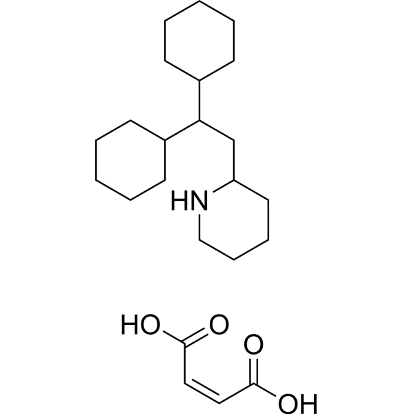
Perhexiline maleate is an orally active CPT1 and CPT2 inhibitor that reduces fatty acid metabolism. Perhexiline maleate induces mitochondrial dysfunction and apoptosis in hepatic cells. Perhexiline maleate can cross the blood brain barrier (BBB) and shows anti-tumor activity. Perhexiline maleate can be used in the research of cancers, and cardiovascular disease like angina.
For research use only. We do not sell to patients.
- Purity : 99.97%
- CAS No.: 6724-53-4
- Formula: C23H39NO4
- Molecular Weight:393.56
-
Storage:
4°C, sealed storage, away from moisture
* In solvent : -80°C, 6 months; -20°C, 1 month (sealed storage, away from moisture)

To place orders, for customer services and technical support, please contact: MedChemExpress USA
Tel: 609-228-6898 E-mail: [email protected] [email protected]


-
 Biological Activity
-
Chemical Information
-
Solvent & Solubility
- Protocols
- Purity & Documentation
- References
-
 Help & FAQs


-
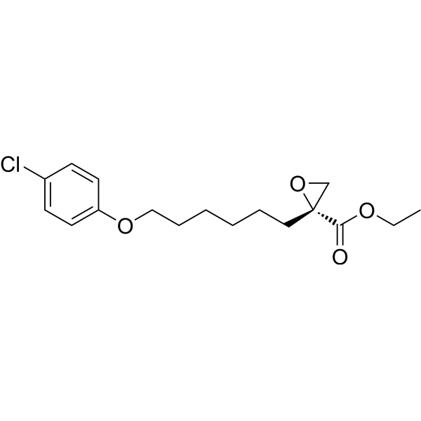
HY-50202
EtomoxirCPT1a Inhibitor -
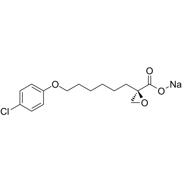
HY-50202A
Etomoxir sodium saltCPT1a Inhibitor -
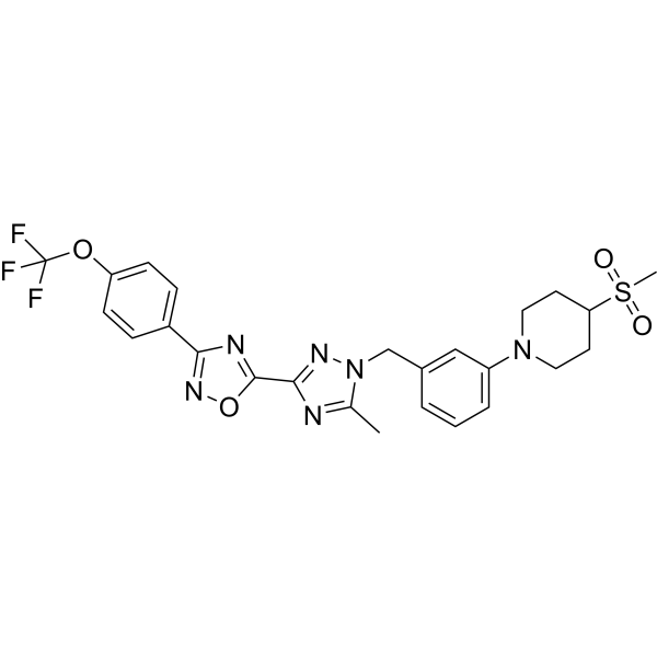
HY-112037
IACS-010759OXPHOS Inhibitor -
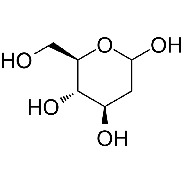
HY-13966
2-Deoxy-D-glucoseGlycolysis Inhibitor -
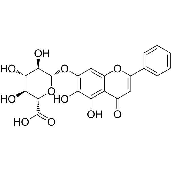
HY-N0197
BaicalinAutophagy Inducer -

HY-15763
ErastinFerroptosis Inducer -
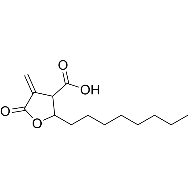
HY-12364
C75Fatty Acid Synthase Inhibitor -

HY-100558
Bafilomycin A1V-ATPase Inhibitor -
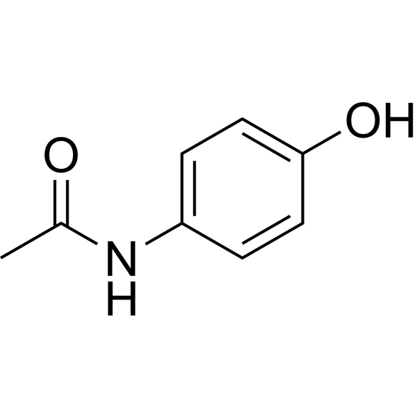
HY-66005
AcetaminophenCOX-2 Inhibitor -

HY-114118
SemaglutideGLP-1R Agonist -
HY-50935
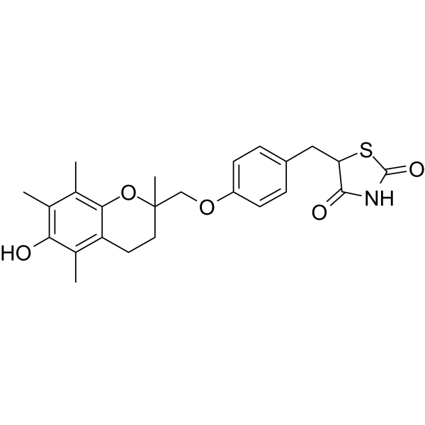
TroglitazonePPARγ Agonist -
HY-136408
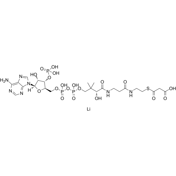
Malonyl CoA lithiumCPT1 Inhibitor
Publications Citing Use of MedChemExpress (MCE) Perhexiline maleate
More- Nat Genet. 2025 Mar;57(3):680-693. [Abstract]
- Acta Pharm Sin B. 2025 Jan;15(1):133-150. [Abstract]
- Adv Sci (Weinh). 2024 Jun;11(24):e2308945. [Abstract]
- EBioMedicine. 2026 Apr 27:127:106256. [Abstract]
- Cancer Metab. 2025 Mar 31;13(1):16. [Abstract]
- BMC Biol. 2024 Apr 12;22(1):83. [Abstract]
- Pestic Biochem Physiol. 2025 Dec 22;218:106908.
 Customer Validation & Images

-
WB
-
Cell Proliferation/Viability Assay
-
Histological Imaging/Staining
-
Cell Imaging/Staining
-
Bio/Physico-chemical Assay


Biological Activity
Description
IC50 & Target
|
CPT-1 |
CPT-2 |
In Vitro
Perhexiline (5-25 μM, 2-6 h) maleate reduces cell viability in HepG2 cells[2].
Perhexiline (5-25 μM, 2-6 h) maleate reduces cellular ATP content and Lactate dehydrogenase (LDH) release in HepG2 cells[2].
Perhexiline (20 μM, 2 h) maleate activates caspase 3/7 in HepG2 cells[2].
Perhexiline (5-25 μM, 4 h) maleate causes mitochondrial dysfunction in HepG2 cells[2].
Perhexiline (5 μM, 48 h) maleate selectively induces massive apoptosis in CLL cells (high expression of CPT)[3].
MedChemExpress (MCE) has not independently confirmed the accuracy of these methods. They are for reference only. Further protocols information, click here.
-
Cell Line:HepG2 cells
-
Concentration:5, 10, 15, 25 μM
-
Incubation Time:2, 4, 6 h
-
Result:Induced time- and concentration-dependent cytotoxicity in hepatic cells.
-
Cell Line:HepG2 cells
-
Concentration:5, 10, 15, 25 μM
-
Incubation Time:2 h
-
Result:Reduced Bcl-2 and Mcl-1 level, and increased Bad level.
In Vivo
Perhexiline (80 mg/kg, oral gavage, for 3 days) maleate demonstrates anti-tumor activity in glioblastoma mouse model[5].
MedChemExpress (MCE) has not independently confirmed the accuracy of these methods. They are for reference only.
-
Animal Model:Orthotopic glioblastoma mouse model[5]
-
Dosage:80 mg/kg
-
Administration:Oral gavage, for 3 days.
-
Result:Reduces tumor size (MR imaging) and improves in overall survival.


Chemical Information
-
CAS No. 6724-53-4
-
Appearance Solid
-
Molecular Weight 393.56
-
Formula C23H39NO4
-
Color White to off-white
-
SMILES
O=C(O)/C=C\C(O)=O.C1(CC(C2CCCCC2)C3CCCCC3)NCCCC1
-
Shipping
Room temperature in continental US; may vary elsewhere.
-
Storage
4°C, sealed storage, away from moisture
* In solvent : -80°C, 6 months; -20°C, 1 month (sealed storage, away from moisture)


Publications (7)
-
Journal Impact Factor
-
Most Recent
-
Nat Genet
2025 Mar;57(3):680-693. PMID: 40069506
Perhexiline maleate purchased from MedChemExpress. Usage Cited in: Nat Genet. 2025 Mar;57(3):680-693. [Abstract]
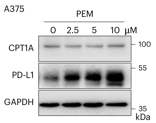
Inhibition of CPT1A activity using Perhexiline maleate (0-10 μM, 24 h) and PD-L1 detection by western blotting in A375 cell.
Perhexiline maleate purchased from MedChemExpress. Usage Cited in: Nat Genet. 2025 Mar;57(3):680-693. [Abstract]
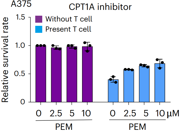
A375 cells treated with Perhexiline maleate (0-10 μM) for 24 h, then co-cultured with or without activated T cells for 24 h to assess the killing effect.
-
Acta Pharm Sin B
GSFM: A genome-scale functional module transformation to represent drug efficacy for in silico drug discovery. [Abstract]2025 Jan;15(1):133-150. PMID: 40041913
Perhexiline maleate purchased from MedChemExpress. Usage Cited in: Acta Pharm Sin B. 2025 Jan;15(1):133-150. [Abstract]
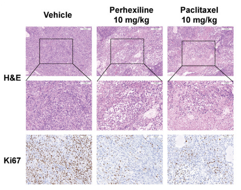
Results of H&E and Ki67 staining in representative A549 cells tumor sections of vehicle, perhexiline (10 mg/kg, ip, 3 days), and Perhexiline maleate (10 mg/kg) treated naked mice. Scale bar = 200 μm (H&E up).
Perhexiline maleate purchased from MedChemExpress. Usage Cited in: Acta Pharm Sin B. 2025 Jan;15(1):133-150. [Abstract]
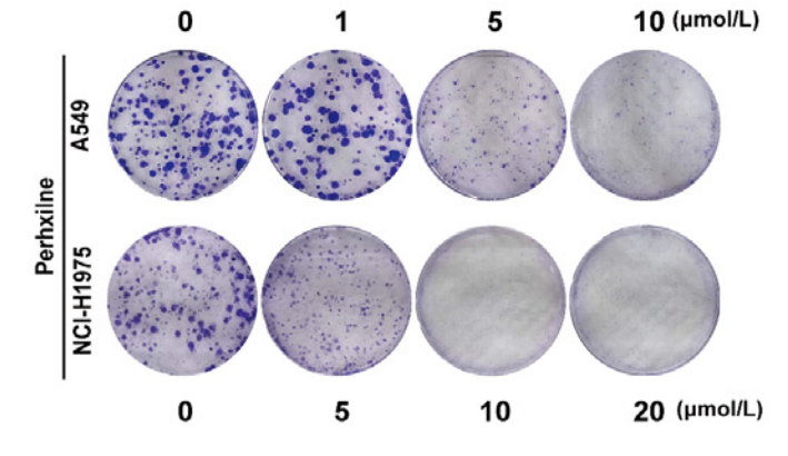
Perhexiline maleate (0-20 μM, 8 days) exhibited inhibition of colony formation in A549 and NCI-H1975 cells.
Perhexiline maleate purchased from MedChemExpress. Usage Cited in: Acta Pharm Sin B. 2025 Jan;15(1):133-150. [Abstract]
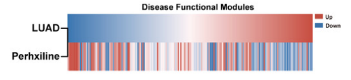
Reversal relationship between LUAD GSFM pattern and Perhexiline maleate GSFM pattern.
-
Adv Sci (Weinh)
Loss of OVOL2 in Triple-Negative Breast Cancer Promotes Fatty Acid Oxidation Fueling Stemness Characteristics. [Abstract]2024 Jun;11(24):e2308945. PMID: 38627980 -
EBioMedicine
Integrated spatial multi-omics delineates fatty acid degradation fuels malignant evolution at the tumour periphery in cervical squamous cell carcinoma. [Abstract]2026 Apr 27:127:106256. PMID: 42048995 -
Cancer Metab
Unveiling the powerhouse: ASCL1-driven small cell lung cancer is characterized by higher numbers of mitochondria and enhanced oxidative phosphorylation. [Abstract]2025 Mar 31;13(1):16. PMID: 40165271 -
BMC Biol
Targeting of REST with rationally-designed small molecule compounds exhibits synergetic therapeutic potential in human glioblastoma cells. [Abstract]2024 Apr 12;22(1):83. PMID: 38609948 -


Solvent & Solubility
In Vitro:
DMSO : 16.67 mg/mL (42.36 mM; ultrasonic and adjust pH to 5 with HCl; Hygroscopic DMSO has a significant impact on the solubility of product, please use newly opened DMSO)
Please refer to the solubility information to select the appropriate solvent. Once prepared, please aliquot and store the solution to prevent product inactivation from repeated freeze-thaw cycles.
Storage method and period of stock solution: -80°C, 6 months; -20°C, 1 month (sealed storage, away from moisture). When stored at -80°C, please use it within 6 months. When stored at -20°C, please use it within 1 month.
Please refer to the solubility information to select the appropriate solvent. Once prepared, please aliquot and store the solution to prevent product inactivation from repeated freeze-thaw cycles.
Storage method and period of stock solution: -80°C, 6 months; -20°C, 1 month (sealed storage, away from moisture). When stored at -80°C, please use it within 6 months. When stored at -20°C, please use it within 1 month.
Concentration (start) × Volume (start) = Concentration (final) × Volume (final)
In Vivo:
For the following dissolution methods, please prepare the working solution directly:
It is recommended to prepare fresh solutions and use them promptly within a short period of time.
The percentages shown for the solvents indicate their volumetric ratio in the final prepared solution. If precipitation or phase separation occurs during preparation, heat and/or sonication can be used to aid dissolution.
Add each solvent one by one: 0.5% CMC-Na/saline water
Solubility: 5 mg/mL (12.70 mM); Suspended solution; Need ultrasonic

Protocols
-
Apoptosis
Apoptosis, also called programmed cell death, is generally characterized by distinct morphological characteristics.
-
TUNEL staining for apoptotic DNA fragmentation
TUNEL staining detects DNA strand breaks by using terminal deoxynucleotidyl transferase to add labeled nucleotides to exposed 3′-OH DNA termini, generating either microscopic staining in fixed cells or tissue sections, or fluorescence/cytometric signal in cell suspensions. TUNEL positivity reflects DNA fragmentation but should not be interpreted alone as definitive apoptosis, because TUNEL can also label necrotic, autolytic, mechanically damaged, or DNA-repair-associated DNA breaks.
-
Annexin V plus membrane-impermeant dye apoptosis staining
Annexin V-based apoptosis assays rely on the detection of phosphatidylserine (PS) externalization from the inner leaflet of the plasma membrane to the outer leaflet, an early biochemical hallmark of apoptosis. Fluorescently labeled Annexin V binds PS in a calcium-dependent manner, enabling identification of early apoptotic cells by flow cytometry or fluorescence microscopy. When combined with a membrane-impermeant DNA-binding dye (e. g. , propidium iodide), this approach allows discrimination between viable (Annexin V−/dye−), early apoptotic (Annexin V+/dye−), and late apoptotic or necrotic (Annexin V+/dye+) cell populations by assessing membrane integrity and PS exposure.
-
Mitochondrial membrane-potential and mitochondrial mass staining
Mitochondrial membrane potential staining measures the electrochemical polarization across the mitochondrial inner membrane in live cells using lipophilic cationic fluorescent probes; early rhodamine-based work showed that selective mitochondrial dye accumulation is lost when the mitochondrial transmembrane potential is dissipated. JC-1 reports mitochondrial polarization by shifting from green monomer fluorescence to red J-aggregate fluorescence as dye concentration increases within energized mitochondria; therefore, the red/green fluorescence ratio is used as a relative readout of mitochondrial membrane potential. TMRE or TMRM staining provides a single-channel relative readout because these cationic rhodamine esters accumulate in polarized mitochondria, and lower fluorescence indicates reduced mitochondrial polarization when acquisition and dye-loading conditions are controlled. Mitochondrial mass staining is commonly performed with MitoTracker Green FM or related MitoTracker dyes as
-
Apoptosis Solutions
Apoptosis is a regulated, generally non-lytic cell-death pathway that removes unwanted, damaged, infected, or abnormal cells through coordinated morphological changes, caspase activation, DNA fragmentation, and membrane remodeling. The intrinsic apoptosis pathway is controlled mainly by mitochondrial outer membrane permeabilization, BCL-2 family proteins, cytochrome c release, apoptosome formation, caspase-9 activation, and downstream executioner caspase-3/7 activation. The extrinsic apoptosis pathway is initiated by death receptors such as Fas, TNFR, and TRAIL receptors, which recruit adaptor proteins and activate caspase-8 before engaging executioner caspases or mitochondrial amplification through BID cleavage. Apoptosis is linked to many phenotypes, including cancer cell killing, tissue homeostasis, immune regulation, neurodegeneration, infection response, and treatment-induced cytotoxicity; unresolved questions include how apoptosis interacts with necroptosis, pyroptosis, ferroptos
-
Research Protocol for Cardiovascular Diseases
Cardiovascular disease can be modeled as maladaptive cardiac remodeling, where ischemic injury or pressure overload activates inflammatory signaling, fibroblast activation, extracellular-matrix deposition, cardiomyocyte hypertrophy, vascular remodeling, and progressive ventricular dysfunction. The TGF-β/SMAD axis is a central profibrotic pathway after myocardial injury and pressure overload, while innate immune and cytokine pathways regulate leukocyte recruitment, scar formation, and adverse remodeling. Key unresolved questions include which inflammatory signals are reparative versus harmful, when fibrosis is protective versus maladaptive, and whether pathway inhibition improves function without weakening necessary infarct healing or compensatory remodeling.


Purity & Documentation
-
Data Sheet (277 KB)
-
SDS (393 KB)
- English - EN (393 KB)
- Français - FR (393 KB)
- Deutsch - DE (393 KB)
- Norwegian - NO (393 KB)
- Español - ES (393 KB)
- Swedish - SV (393 KB)
- Italian - IT (393 KB)
- Korean - KR (393 KB)
- Portuguese - PT (393 KB)
-
Handling Instructions (2659 KB)
References
[1]. E. Marc Jolicoeur, et al. 27 - Refractory Angina. Chronic Coronary Artery Disease, 2018, 412-431.
[2]. Zhen Ren, et al. Mitochondrial dysfunction and apoptosis underlie the hepatotoxicity of perhexiline. Toxicol In Vitro. 2020 Dec;69:104987. [Content Brief]
[3]. P-P Liu, et al. Elimination of chronic lymphocytic leukemia cells in stromal microenvironment by targeting CPT with an antiangina drug perhexiline. Oncogene. 2016 Oct 27;35(43):5663-5673. [Content Brief]
[4]. Giovanni Licari, et al. Enantioselectivity in the tissue distribution of perhexiline contributes to different effects on hepatic histology and peripheral neural function in rats. Pharmacol Res Perspect. 2018 Jun;6(3):e00406. [Content Brief]
[5]. Shiva Kant, et al. Perhexiline Demonstrates FYN-mediated Antitumor Activity in Glioblastoma. Mol Cancer Ther. 2020 Jul;19(7):1415-1422. [Content Brief]


Complete Stock Solution Preparation Table
Please refer to the solubility information to select the appropriate solvent. Once prepared, please aliquot and store the solution to prevent product inactivation from repeated freeze-thaw cycles.
Storage method and period of stock solution: -80°C, 6 months; -20°C, 1 month (sealed storage, away from moisture). When stored at -80°C, please use it within 6 months. When stored at -20°C, please use it within 1 month.
| Optional Solvent | Concentration Solvent Mass | 1 mg | 5 mg | 10 mg | 25 mg |
|---|---|---|---|---|---|
| DMSO | 1 mM | 2.5409 mL | 12.7045 mL | 25.4091 mL | 63.5227 mL |
| 5 mM | 0.5082 mL | 2.5409 mL | 5.0818 mL | 12.7045 mL | |
| 10 mM | 0.2541 mL | 1.2705 mL | 2.5409 mL | 6.3523 mL | |
| 15 mM | 0.1694 mL | 0.8470 mL | 1.6939 mL | 4.2348 mL | |
| 20 mM | 0.1270 mL | 0.6352 mL | 1.2705 mL | 3.1761 mL | |
| 25 mM | 0.1016 mL | 0.5082 mL | 1.0164 mL | 2.5409 mL | |
| 30 mM | 0.0847 mL | 0.4235 mL | 0.8470 mL | 2.1174 mL | |
| 40 mM | 0.0635 mL | 0.3176 mL | 0.6352 mL | 1.5881 mL |


Perhexiline maleate Related Classifications
HY-B1334A Related Classifications
 Powered by Bioz