



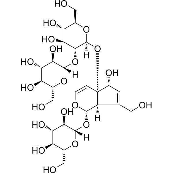
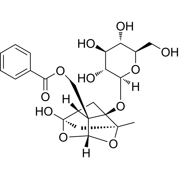
Rehmannioside D
Based on 2 publication(s) in Google Scholar
Rehmannioside D is an orally active Sirt7 modulator. Rehmannioside D upregulates Sirt7 expression, inhibits the level of acetylated p53, and blocks the activation of the p53 signaling pathway. Rehmannioside D alleviates liver injury, inflammatory response, collagen deposition and hepatocyte apoptosis. Rehmannioside D is applicable to research related to liver fibrosis.
연구목적의 판매만을 진행합니다. 환자를 대상으로 한 판매는 하지 않습니다.
- Purity : 99.89%
- CAS No.: 81720-08-3
- 화학식: C27H42O20
- 분자량:686.61
-
보관:Powder -20°C, 3 years , 4°C, 2 years ; In solvent -80°C, 2 years , -20°C, 1 year

To place orders, for customer services and technical support, please contact: MedChemExpress USA
Tel: 609-228-6898 E-mail: [email protected] [email protected]


-
 Biological Activity
-
Chemical Information
-
용액&용해도
- Protocol
- 순도&문서
- References
-
 Help & FAQs



Publications Citing Use of MedChemExpress (MCE) Rehmannioside D
MoreAll Caspase Isoforms
More

Biological Activity
제품 설명
In Vitro
Rehmannioside D (5-10 μM; 24 h) alleviates H2O2-induced apoptosis of AML12 cells by upregulating Sirt7 expression[1].
MedChemExpress (MCE) has not independently confirmed the accuracy of these methods. They are for reference only. Further protocols information, click here.
-
Cell Line:H2O2-injured AML12 hepatocytes
-
Concentration:5 μM; 10 μM
-
Incubation Time:24 h (co-incubation with H2O2)
-
Result:Significantly reduced the proportion of apoptotic AML12 cells induced by H2O2, with both 5 μM and 10 μM doses yielding statistically significant effects.
Increased nuclear Sirt7 protein expression, decreased nuclear acetyl-p53 levels, reduced the Bax/Bcl2 ratio, and lowered Cleaved-caspase 3 levels compared to H2O2-only treated cells.
In Vivo
Rehmannioside D (10-40 mg/kg; p.o.; daily; 2 weeks) attenuates DMN-induced liver fibrosis in male Wistar rats by upregulating Sirt7 expression, inhibiting acetyl-p53-mediated hepatocyte apoptosis, reducing hepatic injury, inflammation, and collagen deposition[1].
Rehmannioside D (19-76 mg/kg; i.g.; daily; 2 weeks) has an ameliorative effect in a rat model of impaired ovarian reserve[3].
MedChemExpress (MCE) has not independently confirmed the accuracy of these methods. They are for reference only.
-
Animal Model:C57BL/6J (male, 22-24 g, CCl4-induced liver fibrosis model)[1]
-
Dosage:10 mg/kg; 40 mg/kg
-
Administration:i.g.; daily; 3 weeks
-
Result:Significantly reduced serum ALT and AST activities compared to CCl4-induced controls.
Ameliorated liver histopathological damage, including reduced lobule structural distortion, hepatocellular swelling/necrosis, and inflammatory cell infiltration.
Decreased F4/80-positive macrophage area and downregulated hepatic mRNA expression of inflammatory genes (Adgre1, Tnfα, Il1β, Ccl2) compared to CCl4-induced controls.
Reduced collagen deposition.
Reduced the number of TUNEL+/HNF4α+ hepatocytes compared to CCl4-induced controls.
Restored hepatic Sirt7 expression, reduced nuclear acetyl-p53 levels, lowered the Bax/Bcl2 ratio, and decreased Cleaved-caspase 3 levels compared to CCl4-induced controls.
-
Animal Model:SD rats (female, 8 weeks old, Cyclophosphamide (HY-17420) induced DOR)[3]
-
Dosage:19 mg/kg, 38 mg/kg, 76 mg/kg
-
Administration:i.g.; daily; 2 weeks
-
Result:Notably recovered disordered estrous cycles and increased ovarian index of DOR rats.
Elevated the quantity of primordial and mature follicles while reducing atretic follicle count via ovarian histomorphology observation.
Downregulated abnormally high FSH and LH concentrations and upregulated declined E2 level.
Decreased granulosa cell apoptotic rate and upregulated FOXO1 expression.


Chemical Information
-
CAS No. 81720-08-3
-
Appearance Solid
-
분자량 686.61
-
화학식 C27H42O20
-
Color White to off-white
-
SMILES
O[C@H]1[C@@]([C@](C(CO)=C1)([H])[C@@H]2O[C@]([C@@H]([C@@H](O)[C@@H]3O)O)([H])O[C@@H]3CO)(C=CO2)O[C@@](O[C@H](CO)[C@@H](O)[C@@H]4O)([H])[C@@H]4O[C@]([C@@H]([C@@H](O)[C@@H]5O)O)([H])O[C@@H]5CO
-
Structure Classification
-
선적
Room temperature in continental US; may vary elsewhere.
-
보관
Powder -20°C 3 years 4°C 2 years In solvent -80°C 2 years -20°C 1 year


Publications (2)
-
Journal Impact Factor
-
Most Recent
-
Phytomedicine
Rehmannioside D prevents estrogen-deficiency induced osteoporosis by interacting with c-Jun to dismantle the AP-1 complex and suppress MAPK/NF-κB signaling. [Abstract]2026 Aug:158:158371. PMID: 42251792 -
J Ethnopharmacol
Protecting the brain from stroke: Huoxue Rongluo formula (HXRLF) targets ferroptosis for neuroprotection. [Abstract]2025 Jul 29;353(Pt A):120329. PMID: 40744419


용액&용해도
In Vitro:
H2O : 100 mg/mL (145.64 mM; Need ultrasonic)
Please refer to the solubility information to select the appropriate solvent. Once prepared, please aliquot and store the solution to prevent product inactivation from repeated freeze-thaw cycles.
Storage method and period of stock solution: -80°C, 2 years; -20°C, 1 year. When stored at -80°C, please use it within 2 years. When stored at -20°C, please use it within 1 year.
* Note: If you choose water as the stock solution, please dilute it to the working solution, then filter and sterilize it with a 0.22 μm filter before use.
Please refer to the solubility information to select the appropriate solvent. Once prepared, please aliquot and store the solution to prevent product inactivation from repeated freeze-thaw cycles.
Storage method and period of stock solution: -80°C, 2 years; -20°C, 1 year. When stored at -80°C, please use it within 2 years. When stored at -20°C, please use it within 1 year.
* Note: If you choose water as the stock solution, please dilute it to the working solution, then filter and sterilize it with a 0.22 μm filter before use.
Concentration (start) × Volume (start) = Concentration (final) × Volume (final)

Protocol
-
Fibrosis/Collagen Morphometry
Fibrosis and collagen morphometry is based on the quantitative visualization of fibrillar collagen deposition in tissue sections using histochemical stains such as Sirius Red (Picrosirius Red) or Masson's trichrome, followed by image-based or polarization-enhanced analysis to estimate collagen proportional area as a surrogate of extracellular matrix accumulation during fibrotic remodeling. Sirius Red combined with polarized light microscopy enhances detection of collagen fibers due to birefringence properties, enabling more specific visualization of collagen type I and III fibrils compared to conventional bright-field histology, while whole-section or region-restricted digital morphometry reduces field-selection bias in fibrosis assessment. Alternative quantitative approaches include second harmonic generation (SHG) and two-photon excited fluorescence microscopy, which enable label-free detection of fibrillar collagen and have been validated against histological staining and biochemica
-
RNA extraction experimental
By lysing cells, releasing RNA, and removing impurities such as proteins and DNA, high-purity RNA products are finally obtained. The commonly used traditional method is the guanidine isothiocyanate/phenol/chloroform method (Trizol), which is suitable for a variety of animal materials including animal tissues, microorganisms, cultured cells, etc., and most plant materials.
-
Collagen: Sirius Red Staining
Sirius Red or picrosirius red staining is a histochemical method for visualizing collagen-rich extracellular matrix in tissue sections, and collagen fibers are detected as red-stained structures under bright-field microscopy with enhanced birefringence under polarized light. Picrosirius red is useful for assessing total collagen organization, distribution, and fibrosis burden, but polarized color should not be interpreted as a definitive collagen type I versus type III readout because color is affected by fiber orientation, thickness, and packing.
-
Apoptosis
Apoptosis, also called programmed cell death, is generally characterized by distinct morphological characteristics.
-
TUNEL staining for apoptotic DNA fragmentation
TUNEL staining detects DNA strand breaks by using terminal deoxynucleotidyl transferase to add labeled nucleotides to exposed 3′-OH DNA termini, generating either microscopic staining in fixed cells or tissue sections, or fluorescence/cytometric signal in cell suspensions. TUNEL positivity reflects DNA fragmentation but should not be interpreted alone as definitive apoptosis, because TUNEL can also label necrotic, autolytic, mechanically damaged, or DNA-repair-associated DNA breaks.
-
Annexin V plus membrane-impermeant dye apoptosis staining
Annexin V-based apoptosis assays rely on the detection of phosphatidylserine (PS) externalization from the inner leaflet of the plasma membrane to the outer leaflet, an early biochemical hallmark of apoptosis. Fluorescently labeled Annexin V binds PS in a calcium-dependent manner, enabling identification of early apoptotic cells by flow cytometry or fluorescence microscopy. When combined with a membrane-impermeant DNA-binding dye (e. g. , propidium iodide), this approach allows discrimination between viable (Annexin V−/dye−), early apoptotic (Annexin V+/dye−), and late apoptotic or necrotic (Annexin V+/dye+) cell populations by assessing membrane integrity and PS exposure.
-
Connective Tissue: Masson's Trichrome/Collagen Trichrome Staining
Masson’s Trichrome (collagen/trichrome staining) is a histological technique that differentially stains tissue compartments using sequential acidic dyes to distinguish collagen from muscle and cytoplasmic components based on dye affinity and tissue permeability differences, enabling visualization of fibrosis and connective tissue architecture in histological sections. The classical formulation typically uses Weigert's iron hematoxylin for nuclear staining, Biebrich scarlet-acid fuchsin for cytoplasm and muscle, and aniline blue (or light green variants) for collagen, producing a characteristic blue/green collagen signal contrasted against red cytoplasm and dark nuclei. The staining principle relies on selective displacement of smaller dye molecules by larger anionic dyes in collagen-rich regions under controlled acidified conditions, which enhances collagen-specific dye retention. This property makes the method widely used for fibrosis assessment in organs such as heart, liver, lung, a
-
Apoptosis Solutions
Apoptosis is a regulated, generally non-lytic cell-death pathway that removes unwanted, damaged, infected, or abnormal cells through coordinated morphological changes, caspase activation, DNA fragmentation, and membrane remodeling. The intrinsic apoptosis pathway is controlled mainly by mitochondrial outer membrane permeabilization, BCL-2 family proteins, cytochrome c release, apoptosome formation, caspase-9 activation, and downstream executioner caspase-3/7 activation. The extrinsic apoptosis pathway is initiated by death receptors such as Fas, TNFR, and TRAIL receptors, which recruit adaptor proteins and activate caspase-8 before engaging executioner caspases or mitochondrial amplification through BID cleavage. Apoptosis is linked to many phenotypes, including cancer cell killing, tissue homeostasis, immune regulation, neurodegeneration, infection response, and treatment-induced cytotoxicity; unresolved questions include how apoptosis interacts with necroptosis, pyroptosis, ferroptos
-
Research Protocol for Inflammation-related Diseases
The NLRP3 inflammasome is a cytosolic innate immune signaling platform that integrates priming signals and danger-signal activation to promote caspase-1 activation, maturation of IL-1β and IL-18, and gasdermin D-mediated pyroptotic cell death. The core experimental logic is to determine whether inflammatory disease phenotypes are driven by increased NLRP3 expression, ASC-containing inflammasome assembly, caspase-1 cleavage, GSDMD cleavage, and extracellular release of IL-1β/IL-18 rather than by nonspecific cell injury alone. The pathway is strongly linked to inflammation-related disease phenotypes because monosodium urate crystals activate NALP3/NLRP3 inflammasome signaling in gout-like crystal inflammation, cholesterol crystals activate NLRP3 inflammasomes in atherogenesis models, and DSS-induced intestinal inflammation has been reported to involve NLRP3 inflammasome activity. However, experimental colitis studies also show context-dependent protective effects of NLRP3 inflammasome co
-
Liver Histomorphometry
Liver histomorphometry is a quantitative histological approach used to measure structural alterations in hepatic tissue, including parenchymal loss, steatosis, fibrosis, and vascular remodeling, by combining stained tissue section analysis with stereological or computerized image-based measurements. Classical morphometric frameworks quantify volume fractions of liver compartments and fibrotic regions using systematic sampling and image analysis, enabling objective comparison of pathological changes across experimental groups. These approaches are widely applied in liver cirrhosis and fibrosis studies to reduce subjectivity in histological scoring and improve reproducibility of tissue evaluation. Recent methodological advances integrate automated image analysis and radiomics-based extraction of histological features from standard liver stains (e. g. , H&E and fibrotic stains), enabling quantitative correlation between morphometric features and fibrosis stages in non-alcoholic fatty live


순도&문서
-
Data Sheet (287 KB)
-
SDS (392 KB)
- English - EN (392 KB)
- Français - FR (392 KB)
- Deutsch - DE (392 KB)
- Norwegian - NO (392 KB)
- Español - ES (392 KB)
- Swedish - SV (392 KB)
- Italian - IT (392 KB)
- Korean - KR (392 KB)
- Portuguese - PT (392 KB)
-
Handling Instructions (2659 KB)
References
[1]. Liang Y, et al. Rehmannioside D ameliorates hepatocyte apoptosis and liver fibrosis by suppressing Sirt7/p53 axis. J Ethnopharmacol. 2026;357:120863. [Content Brief]
[2]. Liu W, et al. Phytochemical Profiles and Antioxidant Activity of Rehmannia glutinosa from Different Production Locations. Chem Biodivers. 2020;17(8):e2000341. [Content Brief]


Complete Stock Solution Preparation Table
Please refer to the solubility information to select the appropriate solvent. Once prepared, please aliquot and store the solution to prevent product inactivation from repeated freeze-thaw cycles.
Storage method and period of stock solution: -80°C, 2 years; -20°C, 1 year. When stored at -80°C, please use it within 2 years. When stored at -20°C, please use it within 1 year.
| Optional Solvent | Concentration Solvent Mass | 1 mg | 5 mg | 10 mg | 25 mg |
|---|---|---|---|---|---|
| H2O | 1 mM | 1.4564 mL | 7.2822 mL | 14.5643 mL | 36.4108 mL |
| 5 mM | 0.2913 mL | 1.4564 mL | 2.9129 mL | 7.2822 mL | |
| 10 mM | 0.1456 mL | 0.7282 mL | 1.4564 mL | 3.6411 mL | |
| 15 mM | 0.0971 mL | 0.4855 mL | 0.9710 mL | 2.4274 mL | |
| 20 mM | 0.0728 mL | 0.3641 mL | 0.7282 mL | 1.8205 mL | |
| 25 mM | 0.0583 mL | 0.2913 mL | 0.5826 mL | 1.4564 mL | |
| 30 mM | 0.0485 mL | 0.2427 mL | 0.4855 mL | 1.2137 mL | |
| 40 mM | 0.0364 mL | 0.1821 mL | 0.3641 mL | 0.9103 mL | |
| 50 mM | 0.0291 mL | 0.1456 mL | 0.2913 mL | 0.7282 mL | |
| 60 mM | 0.0243 mL | 0.1214 mL | 0.2427 mL | 0.6068 mL | |
| 80 mM | 0.0182 mL | 0.0910 mL | 0.1821 mL | 0.4551 mL | |
| 100 mM | 0.0146 mL | 0.0728 mL | 0.1456 mL | 0.3641 mL |
* Note: If you choose water as the stock solution, please dilute it to the working solution, then filter and sterilize it with a 0.22 μm filter before use.


Rehmannioside D Related Classifications
HY-N0912 Related Classifications
 Powered by Bioz