



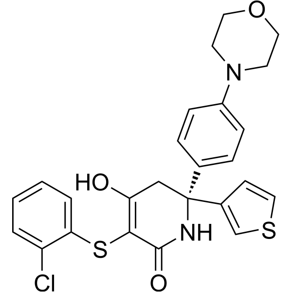
(R)-GNE-140
Based on 20 publication(s) in Google Scholar
(R)-GNE-140 is a potent inhibitor of lactate dehydrogenase (LDH) A, B and C, with IC50 values of 3, 5 and 5 nM against LDHA, LDHB, LDHC, respectively. (R)-GNE-140 blocks the conversion of pyruvate to lactate, reduces lactate production and histone lysine lactylation, and inhibits glycolysis. (R)-GNE-140 attenuates cardiac hypertrophy, alleviates PM2.5-induced pulmonary inflammation and fibrosis, blocks MRSA-induced Arg1 expression. (R)-GNE-140 is applicable to research related to pathological cardiac hypertrophy, pulmonary fibrosis, MRSA infection and pancreatic cancer.
For research use only. We do not sell to patients.
- Purity : 99.73%
- CAS No.: 2003234-63-5
- Formula: C25H23ClN2O3S2
- Molecular Weight:499.04
-
Storage:Powder -20°C, 3 years , 4°C, 2 years ; In solvent -80°C, 1 year , -20°C, 6 months

To place orders, for customer services and technical support, please contact: MedChemExpress USA
Tel: 609-228-6898 E-mail: [email protected] [email protected]


-
 Biological Activity
-
Chemical Information
-
Solvent & Solubility
- Protocols
- Purity & Documentation
- References
-
 Help & FAQs


-
HY-100681
GSK2837808ALDHA Inhibitor -
HY-W013032A
Oxamic acid sodiumLDH-A Inhibitor -
HY-13966
2-Deoxy-D-glucoseGlycolysis Inhibitor -
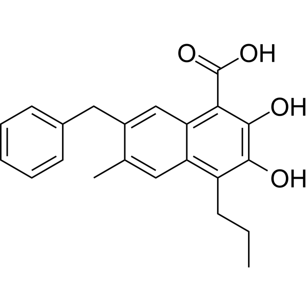
HY-16214
FX-11LDHA Inhibitor -
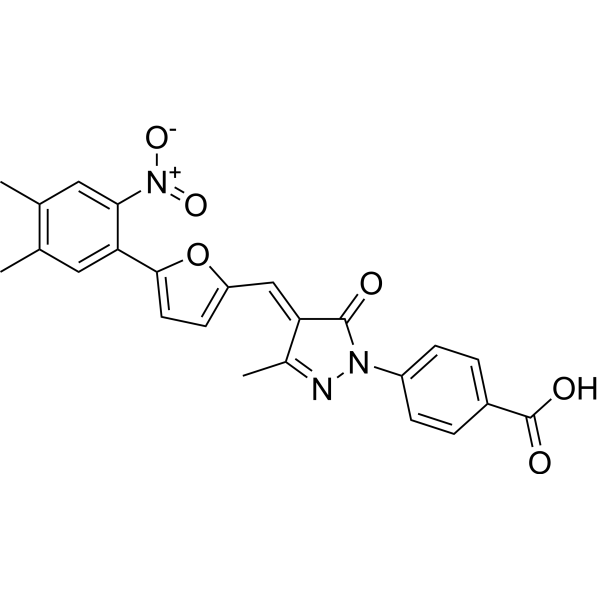
HY-13823
C646p300/CBP HAT Inhibitor -
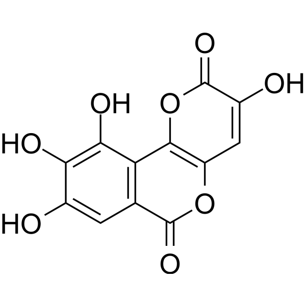
HY-W040118
GalloflavinLactate Dehydrogenase Inhibitor -
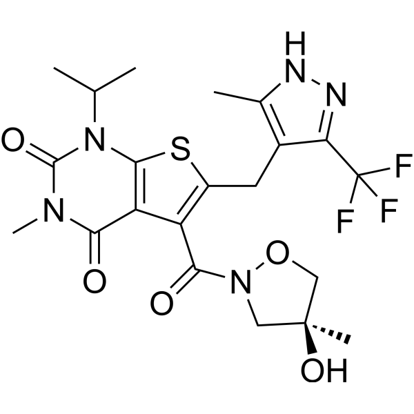
HY-12750
AZD3965MCT1 Inhibitor -
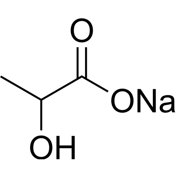
HY-B2227B
Lactic acid sodium (60% w/w in water)Glycolysis Production -
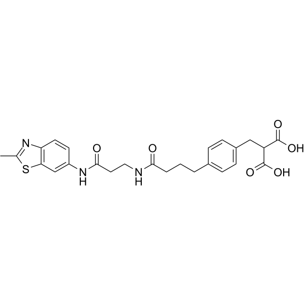
HY-18968
AZ-33LDHA Inhibitor -
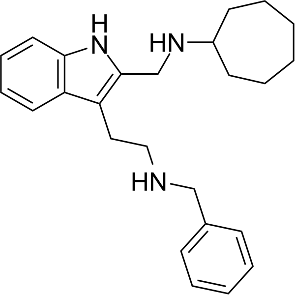
HY-147216
AXKO-0046LDHB Inhibitor -
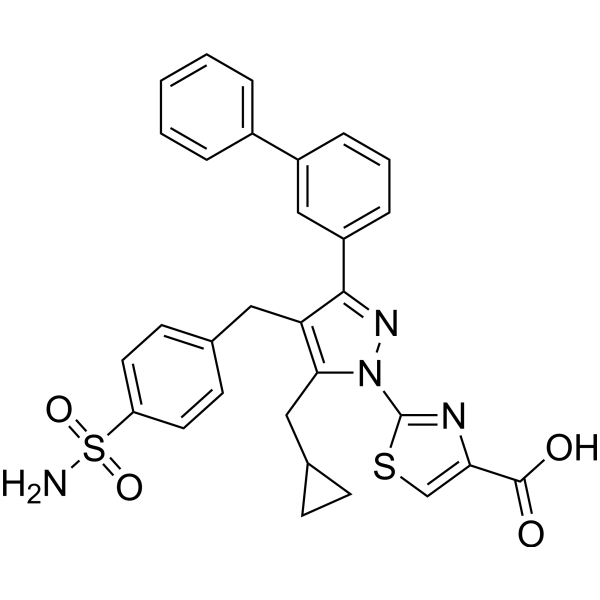
HY-111108
LDH-IN-1LDH Inhibitor -
HY-B2227
Lactic acidGlycolysis Production
Publications Citing Use of MedChemExpress (MCE) (R)-GNE-140
More- Cell Metab. 2021 Jan 5;33(1):51-64.e9. [Abstract]
- Cancer Discov. 2023 Aug 4;13(8):1884-1903. [Abstract]
- Nat Chem Biol. 2025 Jan;21(1):91-99. [Abstract]
- J Exp Clin Cancer Res. 2025 Aug 23;44(1):252. [Abstract]
- Adv Sci (Weinh). 2025 Dec 5:e20444. [Abstract]
- Adv Sci (Weinh). 2025 Nov 26:e08645. [Abstract]
- Sci Adv. 2025 Jul 18;11(29):eadw5228. [Abstract]
- Cell Death Dis. 2023 Nov 30;14(11):784. [Abstract]
- Acta Biomater. 2024 Oct 15:188:79-92. [Abstract]
- Fundam Res. 2024 Oct 18.
- Mol Cancer Ther. 2021 May;20(5):816-832. [Abstract]
- Sci Rep. 2026 Jan 21;16(1):5921. [Abstract]
- Biochim Biophys Acta Mol Cell Res. 2023 Apr;1870(4):119434. [Abstract]
- J Biol Chem. 2020 Jan 3;295(1):83-98. [Abstract]
- Bone. 2024 Aug:185:117132. [Abstract]
- Mol Immunol. 2022 Jun;146:69-77. [Abstract]
- Theriogenology. 2026 Jan 15:250:117706. [Abstract]
- SSRN. 2026 Apr 14.
- bioRxiv. 2024 November 05.
- bioRxiv. 2023 Sep 30:2023.09.30.560315. [Abstract]
 Customer Validation & Images

-
Histological Imaging/Staining
-
In Vivo Efficacy Study
-
Cell Proliferation/Viability Assay
-
Cell Proliferation/Viability Assay
-
Bio/Physico-chemical Assay


Biological Activity
Description
IC50 & Target
|
LDHA 3 nM (IC50) |
LDHB 5 nM (IC50) |
Cellular Effect
|
Cell Line

|
Type | Value | Description | References |
|---|---|---|---|---|
| A549 | IC50 |
0.36 μM
Compound: Gen140
|
Antiproliferative activity against human A549 cells assessed as reduction in cell viability at incubated for 72 hrs by MTT assay
Antiproliferative activity against human A549 cells assessed as reduction in cell viability at incubated for 72 hrs by MTT assay
|
[PMID: 31129449] |
| A673 | IC50 |
2.63 μM
Compound: 3
|
Cytotoxicity against human A673 cells after 48 hrs by CellTiter-Glo assay
Cytotoxicity against human A673 cells after 48 hrs by CellTiter-Glo assay
|
[PMID: 29120638] |
| MG-63 | IC50 |
0.66 μM
Compound: 1
|
Antiproliferative activity against human MG63 cells after 72 hrs by MTT assay
Antiproliferative activity against human MG63 cells after 72 hrs by MTT assay
|
[PMID: 29861142] |
| MIA PaCa-2 | IC50 |
0.48 μM
Compound: Gen140
|
Antiproliferative activity against human MIAPaCa2 cells assessed as reduction in cell viability incubated for 72 hrs by MTT assay
Antiproliferative activity against human MIAPaCa2 cells assessed as reduction in cell viability incubated for 72 hrs by MTT assay
|
[PMID: 31129449] |
| MIA PaCa-2 | IC50 |
1.24 μM
Compound: 3
|
Cytotoxicity against human MIAPaCa2 cells after 48 hrs by CellTiter-Glo assay
Cytotoxicity against human MIAPaCa2 cells after 48 hrs by CellTiter-Glo assay
|
[PMID: 29120638] |
In Vitro
(R)-GNE-140 (10 μM; 48 h) reduces intracellular lactate levels, decreases the surface area of neonatal mouse cardiomyocytes, downregulates the expression of cardiac hypertrophy markers, and inhibits multiple histone lysine lactylation modifications, thereby alleviating angiotensin II (Ang II)-induced hypertrophy of neonatal mouse cardiomyocytes (NMCM)[1].
Pretreatment of mouse RAW264.7 macrophages with (R)-GNE-140 (10 μM; 4 h) inhibits PM2.5-induced glycolysis, histone lactylation, pro-fibrotic gene expression and cytokine secretion, and partially reverses macrophage-mediated epithelial-mesenchymal transition (EMT) in MLE-12 cells[2].
(R)-GNE-140 (6-12 h) inhibits MSM-induced Arg1 mRNA expression in MRSA-infected THP1 cells at 6 h and 12 h post-infection[3].
(R)-GNE-140 (6-12 h) blocks MSM-induced Arg1 protein expression in THP1 cells infected with MRSA at 6 h and 12 h post-infection[3].
(R)-GNE-140 (Compound 29-R) can inhibit lactate production in human pancreatic cancer MiaPaca2 cells, with its IC50 being 0.67 μM[4].
MedChemExpress (MCE) has not independently confirmed the accuracy of these methods. They are for reference only. Further protocols information, click here.
In Vivo
MedChemExpress (MCE) has not independently confirmed the accuracy of these methods. They are for reference only.
-
Animal Model:C57BL/6 J (male, intratracheal instillation of PM2.5 once every other day for 4 weeks)[2]
-
Dosage:5 mg/kg
-
Administration:i.t.; once every other day; 4 weeks (administered 4 h prior to each PM2.5 dose)
-
Result:Significantly reduced PM2.5-induced peribronchial inflammation scores by 41%.
Reduced collagenous fiber area by 31%.
Reduced macrophage infiltration by 39%.
Decreased lung collagen content by 28%.
Decreased LDH activity by 31%.
Decreased lactate content by 55%.
Attenuated PM2.5-induced up-regulation of histone lactylation.
Restored the expression of epithelial (ZO-1, E-cadherin) and mesenchymal (N-cadherin, vimentin) markers to near control levels.


Chemical Information
-
CAS No. 2003234-63-5
-
Appearance Solid
-
Molecular Weight 499.04
-
Formula C25H23ClN2O3S2
-
Color Off-white to yellow
-
SMILES
O=C1N[C@@](C2=CSC=C2)(C3=CC=C(N4CCOCC4)C=C3)CC(O)=C1SC5=CC=CC=C5Cl
-
Shipping
Room temperature in continental US; may vary elsewhere.
-
Storage
Powder -20°C 3 years 4°C 2 years In solvent -80°C 1 year -20°C 6 months


Publications (20)
-
Journal Impact Factor
-
Most Recent
-
Cell Metab
Metabolic-Pathway-Based Subtyping of Triple-Negative Breast Cancer Reveals Potential Therapeutic Targets. [Abstract]2021 Jan 5;33(1):51-64.e9. PMID: 33181091 -
Cancer Discov
Mitochondrial-Encoded Complex I Impairment Induces a Targetable Dependency on Aerobic Fermentation in Hürthle Cell Carcinoma of the Thyroid. [Abstract]2023 Aug 4;13(8):1884-1903. PMID: 37262072
(R)-GNE-140 purchased from MedChemExpress. Usage Cited in: Cancer Discov. 2023 Aug 4;13(8):1884-1903. [Abstract]
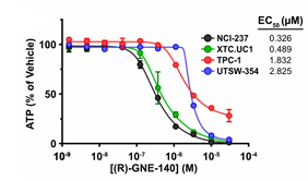
Viability assay for cells treated with (R)-GNE-140 for 3 days was performed.
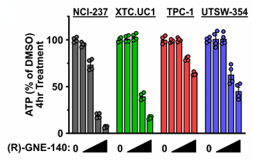
(R)-GNE-140 purchased from MedChemExpress. Usage Cited in: Cancer Discov. 2023 Aug 4;13(8):1884-1903. [Abstract]
ATP levels in cells treated with (R)-GNE-140 for 4 hours were measured. Concentrations were 0.041, 0.37, 3.33, and 30 μM (R)-GNE-140.
-
Nat Chem Biol
2025 Jan;21(1):91-99. PMID: 39030363 -
J Exp Clin Cancer Res
H4K79 and H4K91 histone lactylation, newly identified lactylation sites enriched in breast cancer. [Abstract]2025 Aug 23;44(1):252. PMID: 40849487 -
Adv Sci (Weinh)
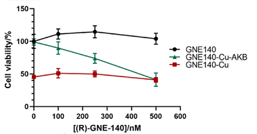
NADH-Reductive Stress Induced by Dihydrolipoamide Dehydrogenase Activation Contributes to Cuproptosis. [Abstract]2025 Dec 5:e20444. PMID: 41346305
(R)-GNE-140 purchased from MedChemExpress. Usage Cited in: Adv Sci (Weinh). 2025 Dec 5:e20444. [Abstract]
Viability of SH-SY5Y cells after 24 h treatment with 1 mM AKB, 500 μM CuSO4, and (R)-GNE-140 was measured.
-
Adv Sci (Weinh)
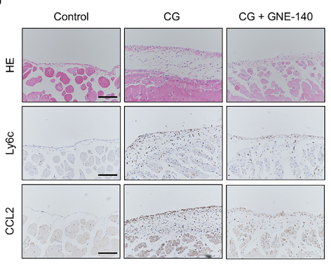
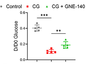
Mesenchymal Stem Cell-Derived Extracellular Vesicles Modulate the Course of Peritoneal Inflammation Through Metabolic and Epigenetic Regulation. [Abstract]2025 Nov 26:e08645. PMID: 41293953
(R)-GNE-140 purchased from MedChemExpress. Usage Cited in: Adv Sci (Weinh). 2025 Nov 26:e08645. [Abstract]
Mice received intraperitoneal injections of 0.1% CG or saline, with or without GNE-140 treatment (5 μg/g body weight, three times weekly), starting on day 7. Representative images of H&E, CCL2, and Ly6c immunostaining in peritoneal tissues were shown.
(R)-GNE-140 purchased from MedChemExpress. Usage Cited in: Adv Sci (Weinh). 2025 Nov 26:e08645. [Abstract]
Mice received intraperitoneal injections of 0.1% CG or saline, with or without GNE-140 treatment (5 μg/g body weight, three times weekly), starting on day 7. The D/D0 glucose ratio was measured.
-
Sci Adv
2025 Jul 18;11(29):eadw5228. PMID: 40668928 -
Cell Death Dis
The mitochondrial NADH shuttle system is a targetable vulnerability for Group 3 medulloblastoma in a hypoxic microenvironment. [Abstract]2023 Nov 30;14(11):784. PMID: 38036520 -
Acta Biomater
Enhancing melanoma therapy by modulating the immunosuppressive microenvironment with an MMP-2 sensitive and nHA/GNE co-encapsulated hydrogel. [Abstract]2024 Oct 15:188:79-92. PMID: 39241819 -
-
Mol Cancer Ther
Identification and Characterization of IMD-0354 as a Glutamine Carrier Protein Inhibitor in Melanoma. [Abstract]2021 May;20(5):816-832. PMID: 33632871 -
Sci Rep
Deploying the high-throughput virtual screening (HTVS) approach for the identification of new lactate dehydrogenase (LDH) inhibitors with anticancer assets. [Abstract]2026 Jan 21;16(1):5921. PMID: 41565942 -
Biochim Biophys Acta Mol Cell Res
Cytoskeletal orchestration of glucose uptake in Sertoli cell to support efferocytosis of apoptotic germ cells. [Abstract]2023 Apr;1870(4):119434. PMID: 36716822 -
J Biol Chem
2020 Jan 3;295(1):83-98. PMID: 31690627 -
Bone
PGC-1α/LDHA signaling facilitates glycolysis initiation to regulate mechanically induced bone remodeling under inflammatory microenvironment. [Abstract]2024 Aug:185:117132. PMID: 38789096 -
Mol Immunol
Methylsulfonylmethane protects against lethal dose MRSA-induced sepsis through promoting M2 macrophage polarization. [Abstract]2022 Jun;146:69-77. PMID: 35461144 -
Theriogenology
Lactate metabolism inhibition disrupts goat oocyte meiosis via H3K18la-mediated epigenetic dysregulation. [Abstract]2026 Jan 15:250:117706. PMID: 41086577 -
-
-
bioRxiv
Forward genetic screens identify mechanisms of resistance to small molecule lactate dehydrogenase inhibitors. [Abstract]2023 Sep 30:2023.09.30.560315. PMID: 37808702


Solvent & Solubility
In Vitro:
DMSO : ≥ 50 mg/mL (100.19 mM; Hygroscopic DMSO has a significant impact on the solubility of product, please use newly opened DMSO)
* "≥" means soluble, but saturation unknown.
Please refer to the solubility information to select the appropriate solvent. Once prepared, please aliquot and store the solution to prevent product inactivation from repeated freeze-thaw cycles.
Storage method and period of stock solution: -80°C, 1 year; -20°C, 6 months. When stored at -80°C, please use it within 1 year. When stored at -20°C, please use it within 6 months.
Please refer to the solubility information to select the appropriate solvent. Once prepared, please aliquot and store the solution to prevent product inactivation from repeated freeze-thaw cycles.
Storage method and period of stock solution: -80°C, 1 year; -20°C, 6 months. When stored at -80°C, please use it within 1 year. When stored at -20°C, please use it within 6 months.
Concentration (start) × Volume (start) = Concentration (final) × Volume (final)
In Vivo:
Select the appropriate dissolution method based on your experimental animal and administration route.
- For the following dissolution methods, please ensure to first prepare a clear stock solution using an In Vitro approach and then sequentially add co-solvents:
- To ensure reliable experimental results, the clarified stock solution can be appropriately stored based on storage conditions. As for the working solution for In Vivo experiments, it is recommended to prepare freshly and use it on the same day.
- The percentages shown for the solvents indicate their volumetric ratio in the final prepared solution. If precipitation or phase separation occurs during preparation, heat and/or sonication can be used to aid dissolution.
Add each solvent one by one: 10% DMSO 40% PEG300 5% Tween-80 45% Saline
Solubility: ≥ 2.5 mg/mL (5.01 mM); Clear solution
This protocol yields a clear solution of ≥ 2.5 mg/mL (saturation unknown).
Taking 1 mL working solution as an example, add 100 μL DMSO stock solution (25.0 mg/mL) to 400 μL PEG300, and mix evenly; then add 50 μL Tween-80 and mix evenly; then add 450 μL Saline to adjust the volume to 1 mL.
Preparation of Saline: Dissolve 0.9 g sodium chloride in ddH₂O and dilute to 100 mL to obtain a clear Saline solution.
Add each solvent one by one: 10% DMSO 90% (20% SBE-β-CD in Saline)
Solubility: 2.5 mg/mL (5.01 mM); Suspended solution; Need ultrasonic
This protocol yields a suspended solution of 2.5 mg/mL. Suspended solution can be used for oral and intraperitoneal injection.
Taking 1 mL working solution as an example, add 100 μL DMSO stock solution (25.0 mg/mL) to 900 μL 20% SBE-β-CD in Saline, and mix evenly.
Preparation of 20% SBE-β-CD in Saline (4°C, storage for one week): 2 g SBE-β-CD powder is dissolved in 10 mL Saline, completely dissolve until clear.
In Vivo Dissolution Calculator
Please enter the basic information of animal experiments:
-
-
-
-
Recommended: Prepare an additional quantity of animals to account for potential losses during experiments.
Please enter your animal formula composition:
-
%DMSO +
Recommended: Keep the proportion of DMSO in working solution below 2% if your animal is weak.
-
%+
-
+%Tween-80 + +
-
%Saline +
The co-solvents required include: DMSO, . All of co-solvents are available by MedChemExpress (MCE). , Tween 80. All of co-solvents are available by MedChemExpress (MCE).
Working solution concentration: 0.22 mg/mL
Method for preparing stock solution: mg drug dissolved in μL DMSO. Stock solution concentration: mg/mL.
1. Take μL DMSO stock solution;
2. Add μL .
μL , mix evenly;
3. Then add μL Tween 80, mix evenly;
4. Then add μL
Please ensure that the stock solution in the first step is dissolved to a clear state, and add co-solvents in sequence. You can use ultrasonic heating (ultrasonic cleaner, recommended frequency 20-40 kHz), vortexing, etc. to assist dissolution.

Protocols
-
Fibrosis/Collagen Morphometry
Fibrosis and collagen morphometry is based on the quantitative visualization of fibrillar collagen deposition in tissue sections using histochemical stains such as Sirius Red (Picrosirius Red) or Masson's trichrome, followed by image-based or polarization-enhanced analysis to estimate collagen proportional area as a surrogate of extracellular matrix accumulation during fibrotic remodeling. Sirius Red combined with polarized light microscopy enhances detection of collagen fibers due to birefringence properties, enabling more specific visualization of collagen type I and III fibrils compared to conventional bright-field histology, while whole-section or region-restricted digital morphometry reduces field-selection bias in fibrosis assessment. Alternative quantitative approaches include second harmonic generation (SHG) and two-photon excited fluorescence microscopy, which enable label-free detection of fibrillar collagen and have been validated against histological staining and biochemica
-
Collagen: Sirius Red Staining
Sirius Red or picrosirius red staining is a histochemical method for visualizing collagen-rich extracellular matrix in tissue sections, and collagen fibers are detected as red-stained structures under bright-field microscopy with enhanced birefringence under polarized light. Picrosirius red is useful for assessing total collagen organization, distribution, and fibrosis burden, but polarized color should not be interpreted as a definitive collagen type I versus type III readout because color is affected by fiber orientation, thickness, and packing.
-
RNA extraction experimental
By lysing cells, releasing RNA, and removing impurities such as proteins and DNA, high-purity RNA products are finally obtained. The commonly used traditional method is the guanidine isothiocyanate/phenol/chloroform method (Trizol), which is suitable for a variety of animal materials including animal tissues, microorganisms, cultured cells, etc., and most plant materials.
-
Research Protocol for Infectious Diseases
Infectious-disease experiments test how pathogens interact with host barriers, innate immune receptors, inflammatory signaling, pathogen replication, and tissue injury; pattern-recognition receptors such as TLRs, RIG-I-like receptors, NOD-like receptors, and inflammasomes detect microbial molecules and activate NF-κB, interferon, and cytokine responses. The central hypothesis is that infection severity reflects the balance between pathogen burden and host response: protective inflammation restricts pathogen growth, whereas excessive or mislocalized inflammation contributes to tissue damage and disease phenotype. Unresolved questions include which host pathways are protective versus pathogenic, why some infection models fail to translate to human disease, and which combined readouts best predict clinically relevant infection outcomes.
-
Cell Cytotoxicity Assay
Cytotoxicity assays are usually based on the assessment of cell membrane damage, which can also be indirectly detected by measuring cell viability. Detection methods include MTT assay, CKK-8 assay, LDH assay and ATP assay, etc.
-
Connective Tissue: Masson's Trichrome/Collagen Trichrome Staining
Masson’s Trichrome (collagen/trichrome staining) is a histological technique that differentially stains tissue compartments using sequential acidic dyes to distinguish collagen from muscle and cytoplasmic components based on dye affinity and tissue permeability differences, enabling visualization of fibrosis and connective tissue architecture in histological sections. The classical formulation typically uses Weigert's iron hematoxylin for nuclear staining, Biebrich scarlet-acid fuchsin for cytoplasm and muscle, and aniline blue (or light green variants) for collagen, producing a characteristic blue/green collagen signal contrasted against red cytoplasm and dark nuclei. The staining principle relies on selective displacement of smaller dye molecules by larger anionic dyes in collagen-rich regions under controlled acidified conditions, which enhances collagen-specific dye retention. This property makes the method widely used for fibrosis assessment in organs such as heart, liver, lung, a
-
Research Protocol for Inflammation-related Diseases
The NLRP3 inflammasome is a cytosolic innate immune signaling platform that integrates priming signals and danger-signal activation to promote caspase-1 activation, maturation of IL-1β and IL-18, and gasdermin D-mediated pyroptotic cell death. The core experimental logic is to determine whether inflammatory disease phenotypes are driven by increased NLRP3 expression, ASC-containing inflammasome assembly, caspase-1 cleavage, GSDMD cleavage, and extracellular release of IL-1β/IL-18 rather than by nonspecific cell injury alone. The pathway is strongly linked to inflammation-related disease phenotypes because monosodium urate crystals activate NALP3/NLRP3 inflammasome signaling in gout-like crystal inflammation, cholesterol crystals activate NLRP3 inflammasomes in atherogenesis models, and DSS-induced intestinal inflammation has been reported to involve NLRP3 inflammasome activity. However, experimental colitis studies also show context-dependent protective effects of NLRP3 inflammasome co
-
Cardiac Morphometry
Cardiac morphometry is based on quantitative histological and stereological assessment of myocardial structure, including cardiomyocyte size, number, and extracellular matrix composition, to evaluate cardiac growth and remodeling under physiological or pathological conditions. Design-based stereology is considered a reference framework for obtaining unbiased estimates of structural parameters such as cardiomyocyte number, volume, and tissue architecture, enabling quantitative comparison across experimental groups. Histological image-based morphometry further enables measurement of cardiomyocyte cross-sectional area and collagen deposition using microscopy combined with image analysis software, allowing assessment of hypertrophy and fibrosis in cardiac remodeling models. These morphometric readouts reflect underlying biological processes such as cardiomyocyte hypertrophy, loss, or structural reorganization during disease progression or experimental stress.


Purity & Documentation
-
Data Sheet (287 KB)
-
SDS (396 KB)
- English - EN (396 KB)
- Français - FR (396 KB)
- Deutsch - DE (396 KB)
- Norwegian - NO (396 KB)
- Español - ES (396 KB)
- Swedish - SV (396 KB)
- Italian - IT (396 KB)
- Korean - KR (396 KB)
- Portuguese - PT (396 KB)
-
Handling Instructions (2659 KB)
References
[1]. Zhao SS, et al. Lactate regulates pathological cardiac hypertrophy via histone lactylation modification. J Cell Mol Med. 2024;28(16):e70022. [Content Brief]
[2]. Li J, et al. Urban airborne PM2.5 induces pulmonary fibrosis through triggering glycolysis and subsequent modification of histone lactylation in macrophages. Ecotoxicol Environ Saf. 2024;273:116162. [Content Brief]
[3]. Ma W, et al. Methylsulfonylmethane protects against lethal dose MRSA-induced sepsis through promoting M2 macrophage polarization. Mol Immunol. 2022;146:69-77. [Content Brief]
[4]. Purkey HE, et al. Cell Active Hydroxylactam Inhibitors of Human Lactate Dehydrogenase with Oral Bioavailability in Mice. ACS Med Chem Lett. 2016 Aug 26;7(10):896-901. [Content Brief]


Complete Stock Solution Preparation Table
Please refer to the solubility information to select the appropriate solvent. Once prepared, please aliquot and store the solution to prevent product inactivation from repeated freeze-thaw cycles.
Storage method and period of stock solution: -80°C, 1 year; -20°C, 6 months. When stored at -80°C, please use it within 1 year. When stored at -20°C, please use it within 6 months.
| Optional Solvent | Concentration Solvent Mass | 1 mg | 5 mg | 10 mg | 25 mg |
|---|---|---|---|---|---|
| DMSO | 1 mM | 2.0038 mL | 10.0192 mL | 20.0385 mL | 50.0962 mL |
| 5 mM | 0.4008 mL | 2.0038 mL | 4.0077 mL | 10.0192 mL | |
| 10 mM | 0.2004 mL | 1.0019 mL | 2.0038 mL | 5.0096 mL | |
| 15 mM | 0.1336 mL | 0.6679 mL | 1.3359 mL | 3.3397 mL | |
| 20 mM | 0.1002 mL | 0.5010 mL | 1.0019 mL | 2.5048 mL | |
| 25 mM | 0.0802 mL | 0.4008 mL | 0.8015 mL | 2.0038 mL | |
| 30 mM | 0.0668 mL | 0.3340 mL | 0.6679 mL | 1.6699 mL | |
| 40 mM | 0.0501 mL | 0.2505 mL | 0.5010 mL | 1.2524 mL | |
| 50 mM | 0.0401 mL | 0.2004 mL | 0.4008 mL | 1.0019 mL | |
| 60 mM | 0.0334 mL | 0.1670 mL | 0.3340 mL | 0.8349 mL | |
| 80 mM | 0.0250 mL | 0.1252 mL | 0.2505 mL | 0.6262 mL | |
| 100 mM | 0.0200 mL | 0.1002 mL | 0.2004 mL | 0.5010 mL |


(R)-GNE-140 Related Classifications
HY-100742A Related Classifications
 Powered by Bioz