



β-Amyloid (22-35)
β-Amyloid 22-35 (Amyloid β-Protein 22-35), the residues 22-35 fragment ofβ-amyloid protein, has a cytotoxic effect on cultured neurons from the rat hippocampus in serum-free medium. β-Amyloid 22-35 forms aggregates and typical amyloid fibrils resembling those of the β-amyloid protein in neutral buffer solution).
For research use only. We do not sell to patients.
- CAS No.: 144189-71-9
- Formula: C59H102N16O21S
- Molecular Weight:1403.62
-
Storage:
Please store the product under the recommended conditions in the Certificate of Analysis.


To place orders, for customer services and technical support, please contact: MedChemExpress USA
Tel: 609-228-6898 E-mail: [email protected] [email protected]


-
 Biological Activity
-
Chemical Information
- Protocols
- Purity & Documentation
- References
-
 Help & FAQs


-
HY-P1388
β-Amyloid (1-42), (rat/mouse)β-Amyloid (1-42) Peptide -
HY-P1362
β-Amyloid (42-1), humanInactive Form of Aβ(1-42) -

HY-P1363A
β-Amyloid (1-42), humanAmyloid β Peptide -

HY-P1363B
β-Amyloid (1-42), human, HFIP-treatedβ-Amyloid (1-42) Peptide -
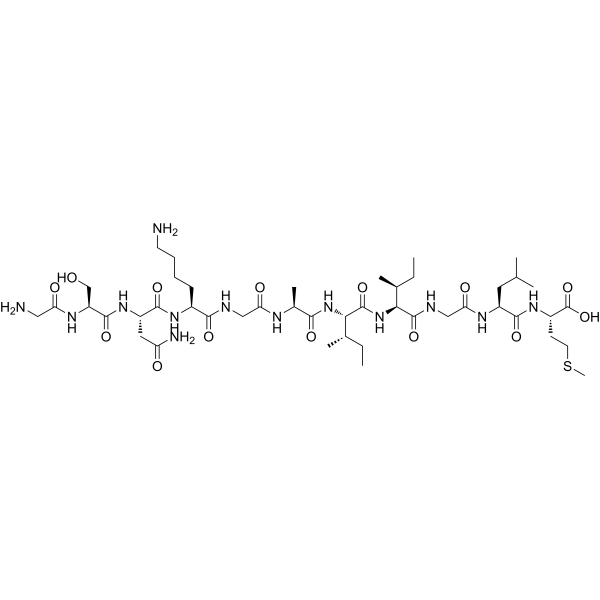
HY-P0128
β-Amyloid (25-35)Amyloid β Peptide -
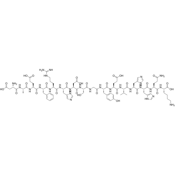
HY-P1466
β-Amyloid (1-16)β-Amyloid -
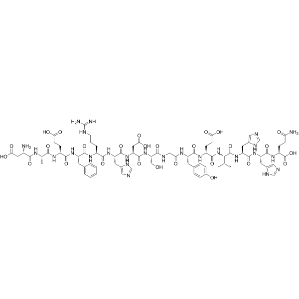
HY-P1046
β-Amyloid (1-15)Peptide Fragment -
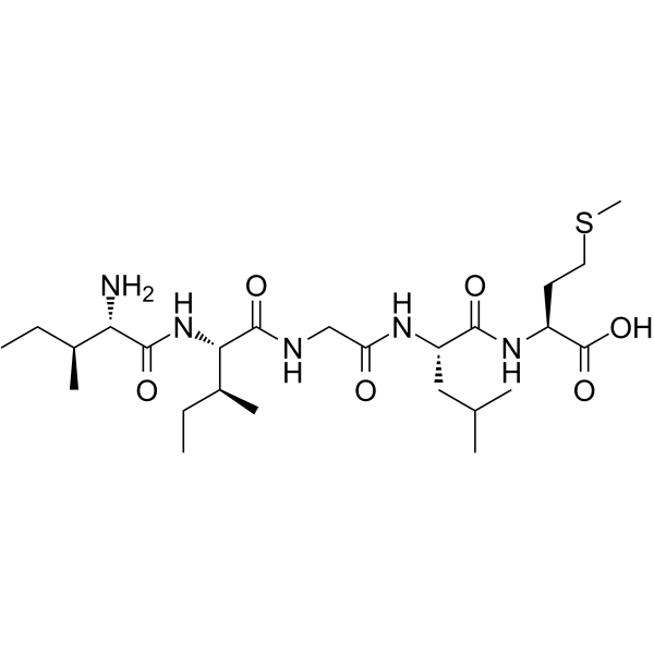
HY-P1517
β-Amyloid (31-35)β-Amyloid Peptide Fragment -

HY-P0128A
β-Amyloid (25-35), HFIP-treatedAmyloid β Peptide -
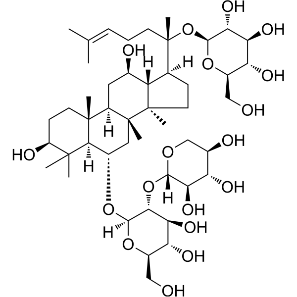
HY-N0615
Notoginsenoside R1Apoptosis Inhibitor -
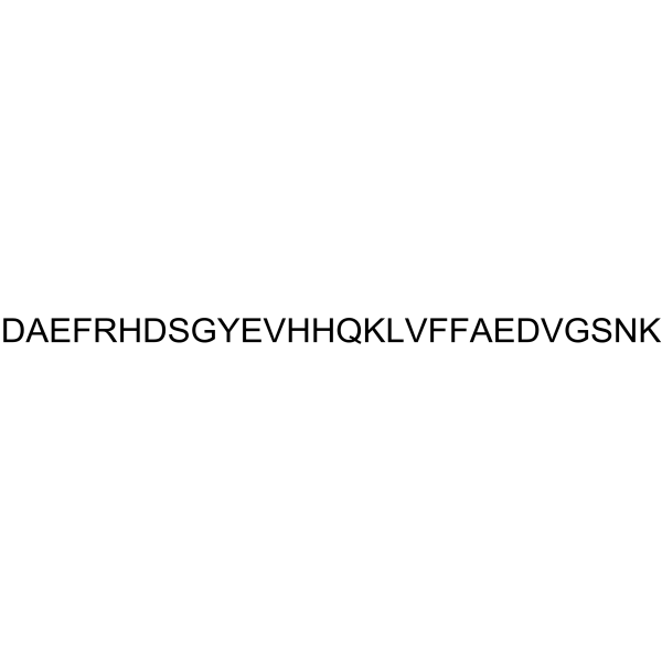
HY-P1468
β-Amyloid (1-28)Amyloid-β Peptide -

HY-P1522
β-Amyloid (29-40)Amyloid-β Peptide Fragment


Biological Activity
Description
IC50 & Target
Amyloid-β[1]
In Vitro
β-Amyloid 22-35 shows significant cytotoxicity on the rat hippocampal neurons at 40 μg/mL, but exhibits no cytotoxic effect on the glial cells[1].
MedChemExpress (MCE) has not independently confirmed the accuracy of these methods. They are for reference only. Further protocols information, click here.


Chemical Information
-
CAS No. 144189-71-9
-
Molecular Weight 1403.62
-
Formula C59H102N16O21S
-
Synonyms
Amyloid β-Protein (22-35)
-
Sequence
Glu-Asp-Val-Gly-Ser-Asn-Lys-Gly-Ala-Ile-Ile-Gly-Leu-Met
-
Sequence Shortening
EDVGSNKGAIIGLM
-
Shipping
Room temperature in continental US; may vary elsewhere.
-
Storage
Please store the product under the recommended conditions in the Certificate of Analysis.

Protocols
-
Amyloid: Congo Red Amyloid Staining
Congo red amyloid staining is a histochemical method used to detect extracellular amyloid deposits in tissue sections based on the affinity of Congo red dye for β-pleated sheet-rich protein aggregates. When bound to amyloid, Congo red produces characteristic apple-green birefringence under polarized light microscopy, which is widely regarded as a diagnostic feature of amyloid deposition in histopathology. The diagnostic principle relies on the combination of dye binding (congophilia) and optical anisotropy under polarized illumination, which distinguishes amyloid from most non-amyloid eosinophilic extracellular deposits in routine histological evaluation. Amyloid identification by Congo red staining remains a cornerstone in diagnostic pathology despite the availability of adjunct methods such as immunohistochemistry and mass spectrometry, particularly because of its ability to localize deposits directly within tissue architecture. The specificity of Congo red-positive deposits is incre
-
Cell Viability Determination by MTT Colorimetric Assay
The following protocol uses the MTT colorimetric assay as a classic literature-established method for assessing cell viability/metabolic activity in cultured mammalian cells. MTT[3-(4,5-dimethylthiazol-2-yl)-2,5-diphenyltetrazolium bromide] is reduced by metabolically active cells to a colored formazan product; the amount of formazan is quantified spectrophotometrically and provides an indirect measure of metabolically active viable cells. Importantly, MTT reduction reflects cellular oxidoreductase/metabolic activity rather than an absolute direct count of living cells, so changes in cellular metabolism can alter the signal independently of cell number.


Purity & Documentation
References
[1]. Takadera T, et al. Toxic effect of a beta-amyloid peptide (beta 22-35) on the hippocampal neuron and its prevention. Neurosci Lett. 1993 Oct 14;161(1):41-4. [Content Brief]
[2]. Zhou WW, et al. Decreasing oxidative stress and neuroinflammation with a multifunctional peptide rescues memory deficits in mice with Alzheimer disease. Free Radic Biol Med. 2014;74:50-63. [Content Brief]

Calculators
Concentration (start) × Volume (start) = Concentration (final) × Volume (final)


β-Amyloid (22-35) Related Classifications
HY-P1474 Related Classifications
 Powered by Bioz