



ADH-6 TFA

Based on 1 Customer Validation
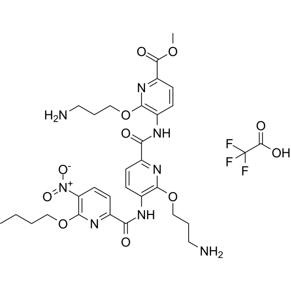
ADH-6 TFA is a tripyridylamide compound. ADH-6 abrogates self-assembly of the aggregation-nucleating subdomain of mutant p53 DBD. ADH-6 TFA targets and dissociates mutant p53 aggregates in human cancer cells, which restores p53's transcriptional activity, leading to cell cycle arrest and apoptosis. ADH-6 TFA has the potential for the research of cancer diseases.
For research use only. We do not sell to patients.
- Purity : 99.67%
- CAS No.: 2990065-87-5
- Formula: C31H37F3N8O11
- Molecular Weight:754.67
-
Storage:
4°C, sealed storage, away from moisture
* In solvent : -80°C, 6 months; -20°C, 1 month (sealed storage, away from moisture)

To place orders, for customer services and technical support, please contact: MedChemExpress USA
Tel: 609-228-6898 E-mail: [email protected] [email protected]


-
 Biological Activity
-
Chemical Information
-
Solvent & Solubility
- Protocols
- Purity & Documentation
- References
-
 Help & FAQs




Biological Activity
Description
In Vitro
ADH-6 (25 μM, 10 h) TFA inhibits aggregation of pR248W (indicated by dot blot assay)[1].
ADH-6 (5 μM, 6 h) TFA dissociates intracellular mutant p53 aggregates in MIA PaCa-2 cells[1].
ADH-6 (0-10 μM, 24 or 48 h) TFA causes selective cytotoxicity in cancer cells bearing mutant p53 (MIA PaCa-2)[1].
ADH-6 (5 μM, 24 h) TFA specifically targets and reactivates aggregation-prone mutant p53 in MIA PaCa-2 cells[1].
MedChemExpress (MCE) has not independently confirmed the accuracy of these methods. They are for reference only.
-
Cell Line:MIA PaCa-2 (mutant R248W p53), SK-BR-3 (mutant R175H p53)
-
Concentration:0, 2.5, 5, 7.5, 10 μM
-
Incubation Time:24, 48 h
-
Result:Caused death of cancer cells bearing mutant, but not WT, p53.
-
Cell Line:MIA PaCa-2 cells
-
Concentration:5 μM
-
Incubation Time:24 h
-
Result:Increased expression of p53-inducible MDM2 and proapoptotic Bax.
In Vivo
MedChemExpress (MCE) has not independently confirmed the accuracy of these methods. They are for reference only.
-
Animal Model:MIA PaCa-2 xenografts[1]
-
Dosage:716.4 µM in 0.02% DMSO
-
Administration:Intraperitoneal injection, every 2 days, for a total of 12 doses
-
Result:Reduced tumor growth relative to the saline-treated control group.
Reduced mutant p53 levels and shrinked xenografts harboring aggregation-prone mutant p53.
-
Animal Model:MIA PaCa-2 xenografts (pharmacokinetics assay)[1]
-
Dosage:15 mg/kg
-
Administration:Intraperitoneal injection, for a single dose
-
Result:Cmax: 21 µg/mL, T1/2: 3.6 h


Chemical Information
-
CAS No. 2990065-87-5
-
Appearance Solid
-
Molecular Weight 754.67
-
Formula C31H37F3N8O11
-
Color Off-white to light yellow
-
SMILES
O=C(C1=NC(OCCCN)=C(NC(C2=NC(OCCCN)=C(NC(C3=NC(OCCCC)=C([N+]([O-])=O)C=C3)=O)C=C2)=O)C=C1)OC.OC(C(F)(F)F)=O
-
Shipping
Room temperature in continental US; may vary elsewhere.
-
Storage
4°C, sealed storage, away from moisture
* In solvent : -80°C, 6 months; -20°C, 1 month (sealed storage, away from moisture)


Solvent & Solubility
In Vitro:
DMSO : 100 mg/mL (132.51 mM; Need ultrasonic; Hygroscopic DMSO has a significant impact on the solubility of product, please use newly opened DMSO)
Please refer to the solubility information to select the appropriate solvent. Once prepared, please aliquot and store the solution to prevent product inactivation from repeated freeze-thaw cycles.
Storage method and period of stock solution: -80°C, 6 months; -20°C, 1 month (sealed storage, away from moisture). When stored at -80°C, please use it within 6 months. When stored at -20°C, please use it within 1 month.
Please refer to the solubility information to select the appropriate solvent. Once prepared, please aliquot and store the solution to prevent product inactivation from repeated freeze-thaw cycles.
Storage method and period of stock solution: -80°C, 6 months; -20°C, 1 month (sealed storage, away from moisture). When stored at -80°C, please use it within 6 months. When stored at -20°C, please use it within 1 month.
Concentration (start) × Volume (start) = Concentration (final) × Volume (final)


Purity & Documentation
-
Data Sheet (275 KB)
-
SDS (252 KB)
- English - EN (252 KB)
- Français - FR (252 KB)
- Deutsch - DE (252 KB)
- Norwegian - NO (252 KB)
- Español - ES (252 KB)
- Swedish - SV (252 KB)
- Italian - IT (252 KB)
- Korean - KR (252 KB)
- Portuguese - PT (252 KB)
-
Handling Instructions (2659 KB)
References


Complete Stock Solution Preparation Table
Please refer to the solubility information to select the appropriate solvent. Once prepared, please aliquot and store the solution to prevent product inactivation from repeated freeze-thaw cycles.
Storage method and period of stock solution: -80°C, 6 months; -20°C, 1 month (sealed storage, away from moisture). When stored at -80°C, please use it within 6 months. When stored at -20°C, please use it within 1 month.
| Optional Solvent | Concentration Solvent Mass | 1 mg | 5 mg | 10 mg | 25 mg |
|---|---|---|---|---|---|
| DMSO | 1 mM | 1.3251 mL | 6.6254 mL | 13.2508 mL | 33.1271 mL |
| 5 mM | 0.2650 mL | 1.3251 mL | 2.6502 mL | 6.6254 mL | |
| 10 mM | 0.1325 mL | 0.6625 mL | 1.3251 mL | 3.3127 mL | |
| 15 mM | 0.0883 mL | 0.4417 mL | 0.8834 mL | 2.2085 mL | |
| 20 mM | 0.0663 mL | 0.3313 mL | 0.6625 mL | 1.6564 mL | |
| 25 mM | 0.0530 mL | 0.2650 mL | 0.5300 mL | 1.3251 mL | |
| 30 mM | 0.0442 mL | 0.2208 mL | 0.4417 mL | 1.1042 mL | |
| 40 mM | 0.0331 mL | 0.1656 mL | 0.3313 mL | 0.8282 mL | |
| 50 mM | 0.0265 mL | 0.1325 mL | 0.2650 mL | 0.6625 mL | |
| 60 mM | 0.0221 mL | 0.1104 mL | 0.2208 mL | 0.5521 mL | |
| 80 mM | 0.0166 mL | 0.0828 mL | 0.1656 mL | 0.4141 mL | |
| 100 mM | 0.0133 mL | 0.0663 mL | 0.1325 mL | 0.3313 mL |


ADH-6 TFA Related Classifications
HY-145785A Related Classifications
 Powered by Bioz