



Allethrin

Based on 1 Customer Validation
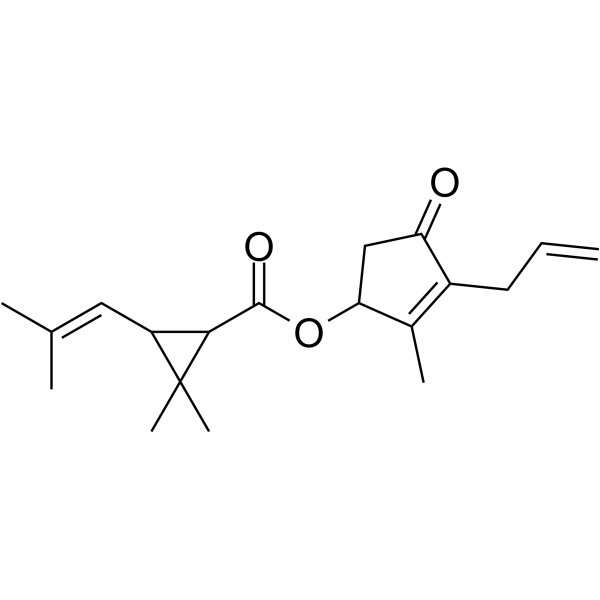
Allethrin, a pyrethroid insecticide is a major mosquito repellent agent. Allethrin induces oxidative stress, apoptosis and calcium release in rat testicular carcinoma cells (LC540). Allethrin induces BCL-2, caspase-3 activation and release of intracellular calcium.
For research use only. We do not sell to patients.
- Purity : 98.04%
- CAS No.: 584-79-2
- Formula: C19H26O3
- Molecular Weight:302.41
-
Storage:
4°C, sealed storage, away from moisture and light
* In solvent : -80°C, 6 months; -20°C, 1 month (sealed storage, away from moisture and light)

To place orders, for customer services and technical support, please contact: MedChemExpress USA
Tel: 609-228-6898 E-mail: [email protected] [email protected]


-
 Biological Activity
-
Chemical Information
-
Solvent & Solubility
- Protocols
- Purity & Documentation
- References
-
 Help & FAQs


-

HY-B1559R
Allethrin (Standard)Pyrethroid Insecticide -
HY-122062
FenoxycarbInsect Growth Regulator -

HY-21200
Perfluoroheptanesulfonic acidPerfluoroalkyl Compounds -
HY-125326
Spinosyn DSpinosad Component -

HY-113958
DifethialoneAnti-coagulant Rodenticide -

HY-W517092
1,2,3,4,7,8,9-HeptachlorodibenzofuranCYP1A1 Inducer -

HY-122062R
Fenoxycarb (Standard)Insect Growth Regulator -

HY-113958R
Difethialone (Standard)Anti-coagulant Rodenticide


Biological Activity
Description
In Vitro
Allethrin (0.001-250 μM) induces cytotoxicity and oxidative stress. Allethrin is cytotoxic to isolated Leydig cells and testicular cancer cells. Cytotoxicity is due to free radical generation and altered antioxidant status[1].
Morphological analyses of LC540 cells treated with Allethrin (125 μM) reveals the presence of apoptotic bodies[1].
Allethrin (125 μM) induces BCL-2, caspase-3 activation and release of intracellular calcium[1].
Allethrin (IC50≈85 μM) is toxic to human corneal epithelial (HCE) cells causing death through mitochondrial pathway[2].
MedChemExpress (MCE) has not independently confirmed the accuracy of these methods. They are for reference only. Further protocols information, click here.
-
Cell Line:LC540 cells (derived from rat Leydig cell tumor)
-
Concentration:0.001-250 μM
-
Incubation Time:24 hours
-
Result:At low concentrations did not display appreciable cell killing activity up to 50 μM when incubated for 24 h.
At concentrations above 100 μM, cell killing was observed. Based on the results obtained, the IC50 was 125 μM.
-
Cell Line:LC540 cells
-
Concentration:125 μM
-
Incubation Time:24 hours
-
Result:Revealed the presence of apoptotic bodies. The percentage of cells displaying early apoptotic features increased significantly.
-
Cell Line:LC540 cells
-
Concentration:125 μM
-
Incubation Time:0, 3, 6, 9, 12, 24 hours
-
Result:BCL-2, pro-Caspase-3 and PARP-1 protein expression decreased significantly with an increase in cleaved PARP-1 levels.
In Vivo
MedChemExpress (MCE) has not independently confirmed the accuracy of these methods. They are for reference only.
-
Animal Model:Male Wistar rats aged 90 days[3]
-
Dosage:25, 50, 100, and 150 mg/kg
-
Administration:Orally administered every day for 60 days
-
Result:Increased levels of LPO products were observed in the caput, cauda, and testes of allethrin treated rats.
In the caput, increased levels of NO was observed at all the doses tested, when compared with the vehicle treated control.
Significant increase in catalase activity was observed in the cauda obtained from 50, 100, and 150 mg/kg.
GPx activity was significantly increased in the caput obtained from 150 mg/kg treated rats. In the cauda, it was found to be increased significantly in the 50 and 100 mg/kg treated groups. In contrast, the activity of GPx activity was significantly decreased in the testes of rats treated with 150 mg/kg.
GST activity was found to be increased significantly in a dose dependent manner in the caput and cauda of allethrin treated rats.
In the caput, significant increase in the activity of SOD was observed in the 150 mg/kg body treated rats. In the cauda and testes, treatment resulted in increase of SOD activity at all the doses tested.


Chemical Information
-
CAS No. 584-79-2
-
Appearance Liquid (Density: 1.01 g/cm3)
-
Molecular Weight 302.41
-
Formula C19H26O3
-
Color Colorless to light yellow
-
SMILES
O=C(C1C(C)(C)C1/C=C(C)\C)OC2C(C)=C(CC=C)C(C2)=O
-
Shipping
Room temperature in continental US; may vary elsewhere.
-
Storage
4°C, sealed storage, away from moisture and light
* In solvent : -80°C, 6 months; -20°C, 1 month (sealed storage, away from moisture and light)


Solvent & Solubility
In Vitro:
DMSO : 100 mg/mL (330.68 mM; Need ultrasonic; Hygroscopic DMSO has a significant impact on the solubility of product, please use newly opened DMSO)
Please refer to the solubility information to select the appropriate solvent. Once prepared, please aliquot and store the solution to prevent product inactivation from repeated freeze-thaw cycles.
Storage method and period of stock solution: -80°C, 6 months; -20°C, 1 month (sealed storage, away from moisture and light). When stored at -80°C, please use it within 6 months. When stored at -20°C, please use it within 1 month.
Please refer to the solubility information to select the appropriate solvent. Once prepared, please aliquot and store the solution to prevent product inactivation from repeated freeze-thaw cycles.
Storage method and period of stock solution: -80°C, 6 months; -20°C, 1 month (sealed storage, away from moisture and light). When stored at -80°C, please use it within 6 months. When stored at -20°C, please use it within 1 month.
Concentration (start) × Volume (start) = Concentration (final) × Volume (final)

Protocols
-
Apoptosis
Apoptosis, also called programmed cell death, is generally characterized by distinct morphological characteristics.
-
TUNEL staining for apoptotic DNA fragmentation
TUNEL staining detects DNA strand breaks by using terminal deoxynucleotidyl transferase to add labeled nucleotides to exposed 3′-OH DNA termini, generating either microscopic staining in fixed cells or tissue sections, or fluorescence/cytometric signal in cell suspensions. TUNEL positivity reflects DNA fragmentation but should not be interpreted alone as definitive apoptosis, because TUNEL can also label necrotic, autolytic, mechanically damaged, or DNA-repair-associated DNA breaks.
-
Annexin V plus membrane-impermeant dye apoptosis staining
Annexin V-based apoptosis assays rely on the detection of phosphatidylserine (PS) externalization from the inner leaflet of the plasma membrane to the outer leaflet, an early biochemical hallmark of apoptosis. Fluorescently labeled Annexin V binds PS in a calcium-dependent manner, enabling identification of early apoptotic cells by flow cytometry or fluorescence microscopy. When combined with a membrane-impermeant DNA-binding dye (e. g. , propidium iodide), this approach allows discrimination between viable (Annexin V−/dye−), early apoptotic (Annexin V+/dye−), and late apoptotic or necrotic (Annexin V+/dye+) cell populations by assessing membrane integrity and PS exposure.
-
ROS/oxidative-stress fluorescent staining
ROS/oxidative-stress fluorescent staining uses cell-permeant fluorogenic probes that become fluorescent after oxidation inside cells or tissues; commonly used examples include DCFH-DA/DCFDA for broad cellular oxidant detection, DHE for superoxide-related signal detection, MitoSOX for mitochondrial superoxide-related signal detection, and CellROX probes for oxidative-stress-associated fluorescence readouts. The assay detects probe oxidation rather than a single ROS species unless the probe and analysis method have been chemically validated for that species. DCFH-DA enters cells, is deacetylated by intracellular esterases to DCFH, and produces fluorescent DCF after oxidation, so the readout is used as an operational measure of total cellular oxidative stress rather than a species-specific ROS measurement. DHE and MitoSOX can report superoxide-related oxidation, but red fluorescence alone can include non-specific ethidium-like oxidation products; HPLC or optimized spectral approaches are
-
Apoptosis Solutions
Apoptosis is a regulated, generally non-lytic cell-death pathway that removes unwanted, damaged, infected, or abnormal cells through coordinated morphological changes, caspase activation, DNA fragmentation, and membrane remodeling. The intrinsic apoptosis pathway is controlled mainly by mitochondrial outer membrane permeabilization, BCL-2 family proteins, cytochrome c release, apoptosome formation, caspase-9 activation, and downstream executioner caspase-3/7 activation. The extrinsic apoptosis pathway is initiated by death receptors such as Fas, TNFR, and TRAIL receptors, which recruit adaptor proteins and activate caspase-8 before engaging executioner caspases or mitochondrial amplification through BID cleavage. Apoptosis is linked to many phenotypes, including cancer cell killing, tissue homeostasis, immune regulation, neurodegeneration, infection response, and treatment-induced cytotoxicity; unresolved questions include how apoptosis interacts with necroptosis, pyroptosis, ferroptos
-
Calcium Spark Assay
Calcium sparks are localized, transient increases in intracellular calcium concentration ([Ca2+]i) that occur in cardiac myocytes and represent elementary events underlying excitation-contraction coupling. These events are generated by the coordinated opening of clusters of ryanodine receptors (RyRs) on the sarcoplasmic reticulum membrane, leading to a brief release of Ca2+ into the cytosol. The detection and analysis of calcium sparks provide insights into the mechanisms of calcium handling and signaling in cardiac cells. Imaging techniques using fluorescent calcium indicators such as Fluo-3 are employed to visualize these subcellular calcium transients with high spatial and temporal resolution. The protocol is based on established methodologies described in primary literature for both experimental measurement and automated analysis of calcium sparks.
-
Ca2+ Staining Technique
Ca2+ staining is an experimental technique that utilizes specific fluorescent probes (such as Fluo-4 AM, Fura-2, etc.) to qualitatively or quantitatively detect dynamic changes in intracellular Ca2+ concentrations; this is achieved by monitoring the changes in fluorescent signals generated when these probes bind to free intracellular calcium ions. The underlying principle relies primarily on the presence of chelating groups within the probe's molecular structure that possess high affinity for calcium ions.


Purity & Documentation
-
Data Sheet (276 KB)
-
SDS (597 KB)
- English - EN (597 KB)
- Français - FR (597 KB)
- Deutsch - DE (597 KB)
- Norwegian - NO (597 KB)
- Español - ES (597 KB)
- Swedish - SV (597 KB)
- Italian - IT (597 KB)
- Korean - KR (597 KB)
- Portuguese - PT (597 KB)
-
Handling Instructions (2659 KB)
References
[1]. Golla Madhubabu, et al. Allethrin induces oxidative stress, apoptosis and calcium release in rat testicular carcinoma cells (LC540). Toxicol In Vitro. 2014 Dec;28(8):1386-95. [Content Brief]
[2]. Geetika Gupta, et al. Allethrin toxicity on human corneal epithelial cells involves mitochondrial pathway mediated apoptosis. Toxicol In Vitro. 2013 Dec;27(8):2242-8. [Content Brief]
[3]. Golla Madhubabu, et al. Allethrin induced toxicity in the male reproductive tract of rats contributes to disruption in the transcription of genes involved in germ cell production. Environ Toxicol. 2014 Nov;29(11):1330-45. [Content Brief]


Complete Stock Solution Preparation Table
Please refer to the solubility information to select the appropriate solvent. Once prepared, please aliquot and store the solution to prevent product inactivation from repeated freeze-thaw cycles.
Storage method and period of stock solution: -80°C, 6 months; -20°C, 1 month (sealed storage, away from moisture and light). When stored at -80°C, please use it within 6 months. When stored at -20°C, please use it within 1 month.
| Optional Solvent | Concentration Solvent Mass | 1 mg | 5 mg | 10 mg | 25 mg |
|---|---|---|---|---|---|
| DMSO | 1 mM | 3.3068 mL | 16.5338 mL | 33.0677 mL | 82.6692 mL |
| 5 mM | 0.6614 mL | 3.3068 mL | 6.6135 mL | 16.5338 mL | |
| 10 mM | 0.3307 mL | 1.6534 mL | 3.3068 mL | 8.2669 mL | |
| 15 mM | 0.2205 mL | 1.1023 mL | 2.2045 mL | 5.5113 mL | |
| 20 mM | 0.1653 mL | 0.8267 mL | 1.6534 mL | 4.1335 mL | |
| 25 mM | 0.1323 mL | 0.6614 mL | 1.3227 mL | 3.3068 mL | |
| 30 mM | 0.1102 mL | 0.5511 mL | 1.1023 mL | 2.7556 mL | |
| 40 mM | 0.0827 mL | 0.4133 mL | 0.8267 mL | 2.0667 mL | |
| 50 mM | 0.0661 mL | 0.3307 mL | 0.6614 mL | 1.6534 mL | |
| 60 mM | 0.0551 mL | 0.2756 mL | 0.5511 mL | 1.3778 mL | |
| 80 mM | 0.0413 mL | 0.2067 mL | 0.4133 mL | 1.0334 mL | |
| 100 mM | 0.0331 mL | 0.1653 mL | 0.3307 mL | 0.8267 mL |


Allethrin Related Classifications
HY-B1559 Related Classifications
 Powered by Bioz