



alpha-Hederin
Based on 4 publication(s) in Google Scholar
alpha-Hederin (α-Hederin), a monodesmosidic triterpenoid saponin, exhibits promising antitumor potential against a variety of human cancer cell lines. alpha-Hederin could inhibit the proliferation and induce apoptosis of gastric cancer accompanied by glutathione decrement and reactive oxygen species generation via activating mitochondrial dependent pathway.
For research use only. We do not sell to patients.
- Purity : 98.10%
- CAS No.: 27013-91-8
- Formula: C41H66O12
- Molecular Weight:750.96
-
Storage:Powder -20°C, 3 years , 4°C, 2 years ; In solvent -80°C, 2 years , -20°C, 1 year

To place orders, for customer services and technical support, please contact: MedChemExpress USA
Tel: 609-228-6898 E-mail: [email protected] [email protected]


-
 Biological Activity
-
Chemical Information
-
Solvent & Solubility
- Protocols
- Purity & Documentation
- References
-
 Help & FAQs


-
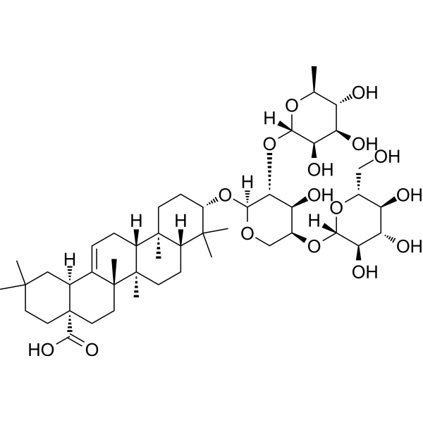
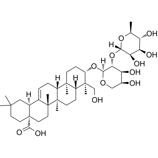
HY-N6950
Hederacolchiside A1Antischistosomal Agent -
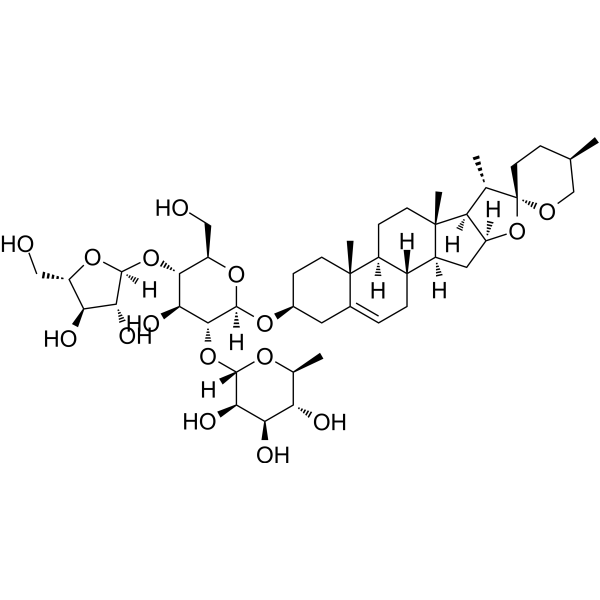
HY-N0047
Polyphyllin IAutophagy Inducer -
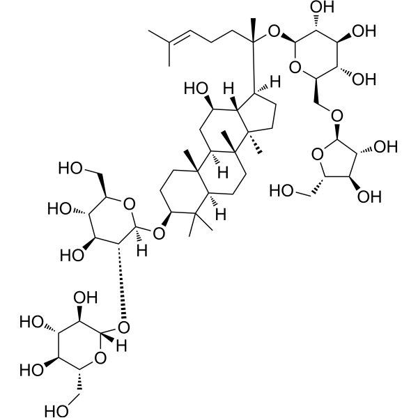
HY-N0042
Ginsenoside RcTNF Receptor Inhibitor -
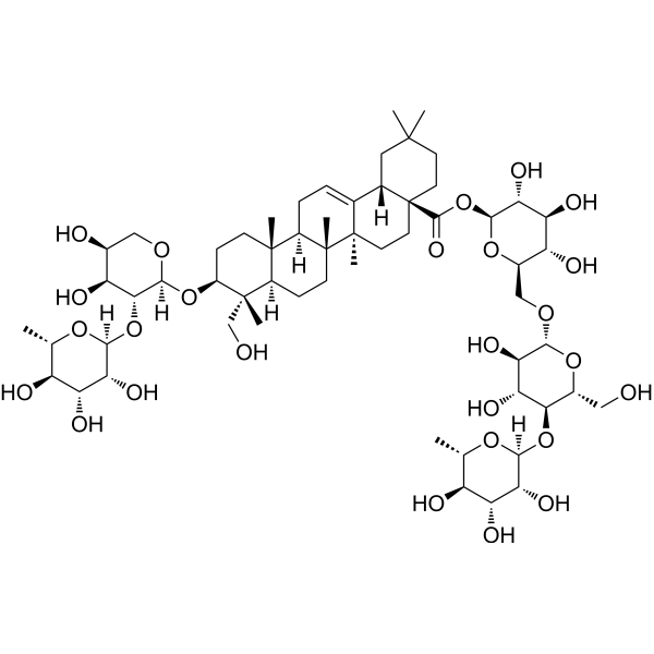
HY-N0253
Hederacoside CAnti-inflammatory agent -
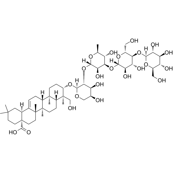
HY-N5008
Macranthoside BAnticancer Agent -

HY-N0255R
alpha-Hederin (Standard)Apoptosis Inducer -
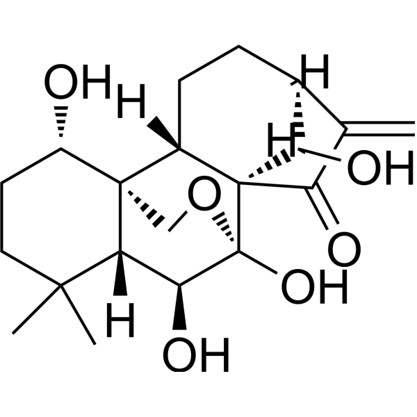
HY-N0004
OridoninBacterial Inhibitor -
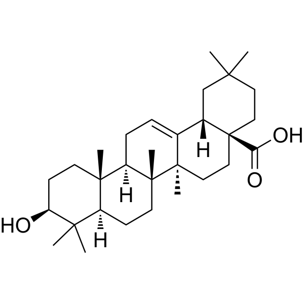
HY-N0156
Oleanolic acidAutophagy Inducer -
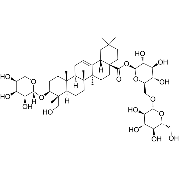
HY-N0265
Asperosaponin VISaponin -
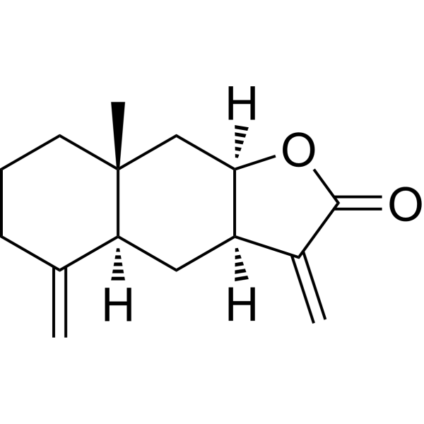
HY-N0780
IsoalantolactoneBacterial Inhibitor -
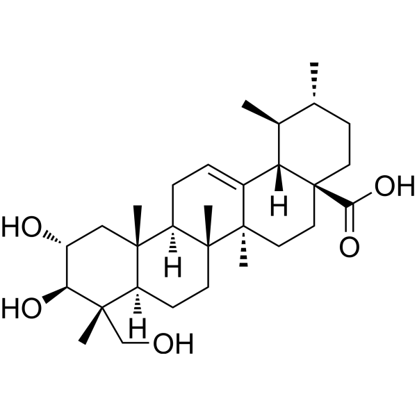
HY-N0194
Asiatic acidApoptosis Inducer -
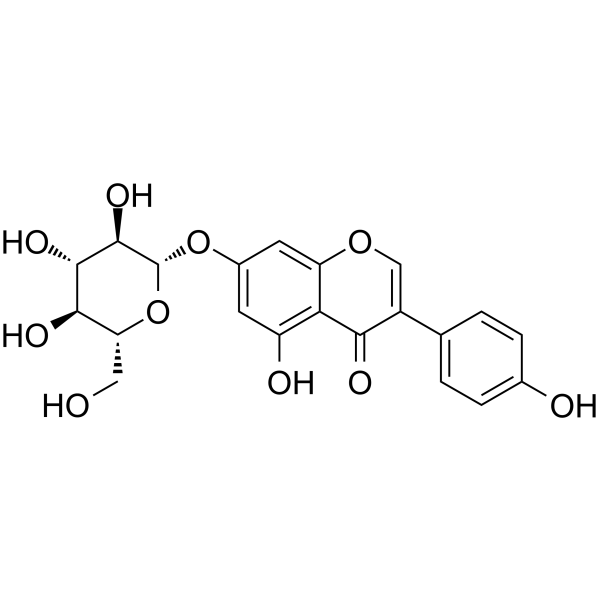
HY-N0595
GenistinAnti-Adipogenic/Anti-Lipogenic Agent
Publications Citing Use of MedChemExpress (MCE) alpha-Hederin
More Customer Validation & Images

-
Cell Proliferation/Viability Assay
-
IF
-
Cell Imaging/Staining
-
Flow Cytometry
-
WB


Biological Activity
Description
Cellular Effect
|
Cell Line

|
Type | Value | Description | References |
|---|---|---|---|---|
| A549 | IC50 |
12.29 μM
Compound: 12a
|
Cytotoxicity against human A549 cells assessed as cell viability after 72 hrs by MTT assay
Cytotoxicity against human A549 cells assessed as cell viability after 72 hrs by MTT assay
|
[PMID: 23992864] |
| A549 | IC50 |
15.3 μM
Compound: 1, PSA
|
Cytotoxicity against human A549 cells assessed as inhibition of cell growth after 24 hrs by MTT assay
Cytotoxicity against human A549 cells assessed as inhibition of cell growth after 24 hrs by MTT assay
|
[PMID: 25958248] |
| A549 | IC50 |
28 μM
Compound: 24
|
Cell membrane permeabilization in human A549 cells assessed as drug level causing decrease in calcein fluorescence
Cell membrane permeabilization in human A549 cells assessed as drug level causing decrease in calcein fluorescence
|
[PMID: 19200744] |
| A549 | IC50 |
33 μM
Compound: 24
|
Cytotoxicity against human A549 cells after 48 hrs by resazurin reduction test
Cytotoxicity against human A549 cells after 48 hrs by resazurin reduction test
|
[PMID: 19200744] |
| Bel-7402 | IC50 |
13.2 μM
Compound: 1, PSA
|
Cytotoxicity against human Bel7402 cells assessed as inhibition of cell growth after 24 hrs by MTT assay
Cytotoxicity against human Bel7402 cells assessed as inhibition of cell growth after 24 hrs by MTT assay
|
[PMID: 25958248] |
| DLD-1 | IC50 |
38 μM
Compound: 24
|
Cell membrane permeabilization in human DLD1 cells assessed as drug level causing decrease in calcein fluorescence
Cell membrane permeabilization in human DLD1 cells assessed as drug level causing decrease in calcein fluorescence
|
[PMID: 19200744] |
| DLD-1 | IC50 |
60 μM
Compound: 24
|
Cytotoxicity against human DLD1 cells after 48 hrs by resazurin reduction test
Cytotoxicity against human DLD1 cells after 48 hrs by resazurin reduction test
|
[PMID: 19200744] |
| ECV-304 | IC50 |
29 μM
Compound: alpha-hederin
|
Membranolytic activity in human ECV304 cells assessed as leakage of intracellular lactate dehydrogenase after 2 hrs by spectrophotometry
Membranolytic activity in human ECV304 cells assessed as leakage of intracellular lactate dehydrogenase after 2 hrs by spectrophotometry
|
[PMID: 22503361] |
| ECV-304 | IC50 |
29 μM
Compound: Alpha-hederin
|
Membrane toxicity against human ECV304 cells after 2 hrs by LDH release assay
Membrane toxicity against human ECV304 cells after 2 hrs by LDH release assay
|
[PMID: 24084294] |
| ECV-304 | IC50 |
35 μM
Compound: alpha-hederin
|
Cytotoxicity against human ECV304 cells after 72 hrs by Hoechst 33258 staining based fluorescence assay
Cytotoxicity against human ECV304 cells after 72 hrs by Hoechst 33258 staining based fluorescence assay
|
[PMID: 22503361] |
| ECV-304 | IC50 |
35 μM
Compound: Alpha-hederin
|
Cytotoxicity against human ECV304 cells after 72 hrs by MTT assay
Cytotoxicity against human ECV304 cells after 72 hrs by MTT assay
|
[PMID: 24084294] |
| HeLa | IC50 |
21.18 μM
Compound: 12a
|
Cytotoxicity against human HeLa cells assessed as cell viability after 72 hrs by MTT assay
Cytotoxicity against human HeLa cells assessed as cell viability after 72 hrs by MTT assay
|
[PMID: 23992864] |
| HepG2 | IC50 |
16.34 μM
Compound: 12a
|
Cytotoxicity against human HepG2 cells assessed as cell viability after 72 hrs by MTT assay
Cytotoxicity against human HepG2 cells assessed as cell viability after 72 hrs by MTT assay
|
[PMID: 23992864] |
| HepG2 | IC50 |
5.5 μM
Compound: 8
|
Inhibition of TNF-alpha-induced NFkappaB activation in human HepG2 cells after 1 hr by luciferase reporter assay
Inhibition of TNF-alpha-induced NFkappaB activation in human HepG2 cells after 1 hr by luciferase reporter assay
|
[PMID: 21870831] |
| HL-60 | IC50 |
11.78 μM
Compound: 12a
|
Cytotoxicity against human HL60 cells assessed as cell viability after 72 hrs by MTT assay
Cytotoxicity against human HL60 cells assessed as cell viability after 72 hrs by MTT assay
|
[PMID: 23992864] |
| HL-60 | IC50 |
7.1 μg/mL
Compound: 2
|
Cytotoxicity against human HL-60 cells after 72 hrs by MTT assay
Cytotoxicity against human HL-60 cells after 72 hrs by MTT assay
|
[PMID: 10514313] |
| HL-60 | IC50 |
7.1 μg/mL
Compound: page 1228, R18C1
|
Cytotoxicity against human HL60 cells by MTT assay
Cytotoxicity against human HL60 cells by MTT assay
|
[PMID: 11575962] |
| L02 | IC50 |
11.5 μM
Compound: 1, PSA
|
Cytotoxicity against human HL7702 cells assessed as inhibition of cell growth after 24 hrs by MTT assay
Cytotoxicity against human HL7702 cells assessed as inhibition of cell growth after 24 hrs by MTT assay
|
[PMID: 25958248] |
| L02 | IC50 |
20.98 μM
Compound: BD
|
Cytotoxicity against human L02 cells by MTT assay
Cytotoxicity against human L02 cells by MTT assay
|
[PMID: 33964439] |
| RAW264.7 | IC50 |
1.1 μM
Compound: 10
|
Antiinflammatory activity in mouse RAW264.7 cells assessed as inhibition of LPS-induced nitirc oxide production preincubated for 1 hr followed by LPS-stimulation and measured after 18 hrs by Griess assay
Antiinflammatory activity in mouse RAW264.7 cells assessed as inhibition of LPS-induced nitirc oxide production preincubated for 1 hr followed by LPS-stimulation and measured after 18 hrs by Griess assay
|
[PMID: 31301930] |
| SMMC-7721 | IC50 |
10.8 μM
Compound: 1, PSA
|
Cytotoxicity against human SMMC7721 cells assessed as inhibition of cell growth after 24 hrs by MTT assay
Cytotoxicity against human SMMC7721 cells assessed as inhibition of cell growth after 24 hrs by MTT assay
|
[PMID: 25958248] |
| U-87MG ATCC | IC50 |
17.34 μM
Compound: 12a
|
Cytotoxicity against human U87MG cells assessed as cell viability after 72 hrs by MTT assay
Cytotoxicity against human U87MG cells assessed as cell viability after 72 hrs by MTT assay
|
[PMID: 23992864] |
In Vitro
alpha-Hederin (α-Hederin) is cytotoxic and inhibits proliferation in both cel lines at rather low concentrations. alpha-Hederin (α-Hederin)reduces the mitotic activity in treated cells[2].
MedChemExpress (MCE) has not independently confirmed the accuracy of these methods. They are for reference only. Further protocols information, click here.
In Vivo
MedChemExpress (MCE) has not independently confirmed the accuracy of these methods. They are for reference only.


Chemical Information
-
CAS No. 27013-91-8
-
Appearance Solid
-
Molecular Weight 750.96
-
Formula C41H66O12
-
Color White to off-white
-
SMILES
OC[C@@]1(C)[C@]2([H])CC[C@@]3(C)[C@]4(C)CC[C@@]5(C(O)=O)CCC(C)(C)C[C@@]5([H])C4=CC[C@]3([H])[C@@]2(C)CC[C@@H]1O[C@]6([H])OC[C@H](O)[C@H](O)[C@H]6O[C@@]7([H])[C@H](O)[C@H](O)[C@@H](O)[C@H](C)O7
-
Synonyms
α-Hederin
-
Structure Classification
-
Shipping
Room temperature in continental US; may vary elsewhere.
-
Storage
Powder -20°C 3 years 4°C 2 years In solvent -80°C 2 years -20°C 1 year


Publications (4)
-
Journal Impact Factor
-
Most Recent
-
Adv Sci (Weinh)
Discovery of Natural Compound α-Hederin via Large-Scale Screening as a Targeted JAK/STAT3 Inhibitor for Ovarian Cancer Therapy. [Abstract]2025 Jul 16:e17278. PMID: 40667652
alpha-Hederin purchased from MedChemExpress. Usage Cited in: Adv Sci (Weinh). 2025 Jul 16:e17278. [Abstract]
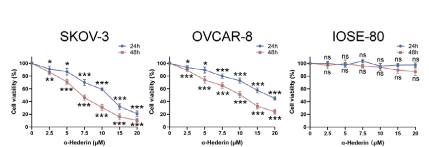
Cell viability of SKOV‐3, OVCAR‐8, and IOSE‐80 cells treated with increasing concentrations of α‐Hederin (2.5, 5, 10, 15, 20 μM) for 24 and 48 h, measured by the CCK‐8 assay.
alpha-Hederin purchased from MedChemExpress. Usage Cited in: Adv Sci (Weinh). 2025 Jul 16:e17278. [Abstract]
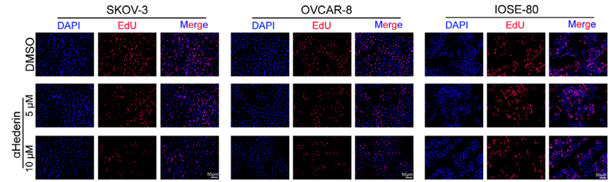
Representative images of EdU staining in cells treated with 0, 5, or 10 µM α‐Hederin for 48 h to assess proliferation.
alpha-Hederin purchased from MedChemExpress. Usage Cited in: Adv Sci (Weinh). 2025 Jul 16:e17278. [Abstract]
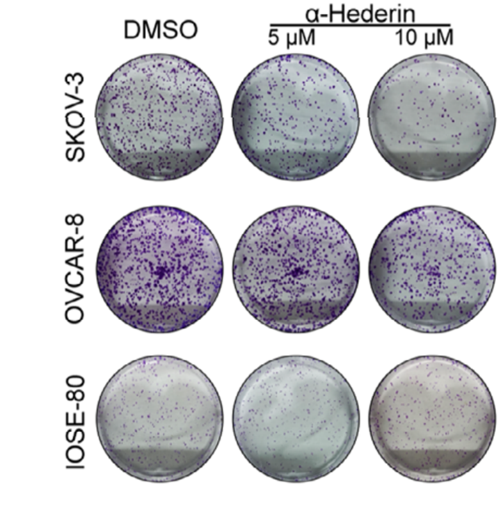
Representative images of colony formation in SKOV‐3, OVCAR‐8, and IOSE‐80 cells treated with α‐Hederin (5, 10 μM).
alpha-Hederin purchased from MedChemExpress. Usage Cited in: Adv Sci (Weinh). 2025 Jul 16:e17278. [Abstract]
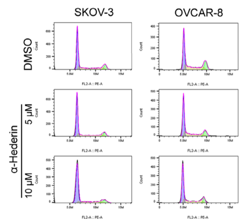
Representative flow cytometry plots of the cell cycle distribution in SKOV‐3 and OVCAR‐8 cells following treatment with 0, 5, or 10 µM α‐Hederin for 48h.
alpha-Hederin purchased from MedChemExpress. Usage Cited in: Adv Sci (Weinh). 2025 Jul 16:e17278. [Abstract]
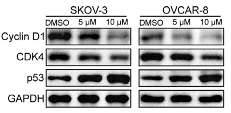
Western blot analysis of Cyclin D1, CDK4, and p53 expression in response to α‐Hederin at 5 or 10 µM for 48 h.
-
Phytomedicine
α-Hederin causes ferroptosis in triple-negative breast cancer through modulating IRF1 to suppress GPX4. [Abstract]2025 Jun:141:156611. PMID: 40153970 -
Biomedicines
α-Hederin Alleviates Endoplasmic Reticulum Stress by Upregulating TRIM38 Expression, Thereby Inhibiting Hepatic Stellate Cell Activation and Liver Fibrosis. [Abstract]2026 Apr 5;14(4):829. PMID: 42072369 -
Naunyn Schmiedebergs Arch Pharmacol
α-Hederin inhibited pancreatic cancer cell malignant progression by inhibiting LDHA-mediated glycolysis. [Abstract]2025 Feb 19. PMID: 39969605


Solvent & Solubility
In Vitro:
DMSO : ≥ 100 mg/mL (133.16 mM; Hygroscopic DMSO has a significant impact on the solubility of product, please use newly opened DMSO)
H2O : < 0.1 mg/mL (insoluble)
* "≥" means soluble, but saturation unknown.
Please refer to the solubility information to select the appropriate solvent. Once prepared, please aliquot and store the solution to prevent product inactivation from repeated freeze-thaw cycles.
Storage method and period of stock solution: -80°C, 2 years; -20°C, 1 year. When stored at -80°C, please use it within 2 years. When stored at -20°C, please use it within 1 year.
Please refer to the solubility information to select the appropriate solvent. Once prepared, please aliquot and store the solution to prevent product inactivation from repeated freeze-thaw cycles.
Storage method and period of stock solution: -80°C, 2 years; -20°C, 1 year. When stored at -80°C, please use it within 2 years. When stored at -20°C, please use it within 1 year.
Concentration (start) × Volume (start) = Concentration (final) × Volume (final)
In Vivo:
Select the appropriate dissolution method based on your experimental animal and administration route.
- For the following dissolution methods, please ensure to first prepare a clear stock solution using an In Vitro approach and then sequentially add co-solvents:
- To ensure reliable experimental results, the clarified stock solution can be appropriately stored based on storage conditions. As for the working solution for In Vivo experiments, it is recommended to prepare freshly and use it on the same day.
- The percentages shown for the solvents indicate their volumetric ratio in the final prepared solution. If precipitation or phase separation occurs during preparation, heat and/or sonication can be used to aid dissolution.
Add each solvent one by one: 10% DMSO 40% PEG300 5% Tween-80 45% Saline
Solubility: ≥ 2.5 mg/mL (3.33 mM); Clear solution
This protocol yields a clear solution of ≥ 2.5 mg/mL (saturation unknown).
Taking 1 mL working solution as an example, add 100 μL DMSO stock solution (25.0 mg/mL) to 400 μL PEG300, and mix evenly; then add 50 μL Tween-80 and mix evenly; then add 450 μL Saline to adjust the volume to 1 mL.
Preparation of Saline: Dissolve 0.9 g sodium chloride in ddH₂O and dilute to 100 mL to obtain a clear Saline solution.
Add each solvent one by one: 10% DMSO 90% (20% SBE-β-CD in Saline)
Solubility: ≥ 2.5 mg/mL (3.33 mM); Clear solution
This protocol yields a clear solution of ≥ 2.5 mg/mL (saturation unknown).
Taking 1 mL working solution as an example, add 100 μL DMSO stock solution (25.0 mg/mL) to 900 μL 20% SBE-β-CD in Saline, and mix evenly.
Preparation of 20% SBE-β-CD in Saline (4°C, storage for one week): 2 g SBE-β-CD powder is dissolved in 10 mL Saline, completely dissolve until clear.
In Vivo Dissolution Calculator
Please enter the basic information of animal experiments:
-
-
-
-
Recommended: Prepare an additional quantity of animals to account for potential losses during experiments.
Please enter your animal formula composition:
-
%DMSO +
Recommended: Keep the proportion of DMSO in working solution below 2% if your animal is weak.
-
%+
-
+%Tween-80 + +
-
%Saline +
The co-solvents required include: DMSO, . All of co-solvents are available by MedChemExpress (MCE). , Tween 80. All of co-solvents are available by MedChemExpress (MCE).
Working solution concentration: 0.22 mg/mL
Method for preparing stock solution: mg drug dissolved in μL DMSO. Stock solution concentration: mg/mL.
1. Take μL DMSO stock solution;
2. Add μL .
μL , mix evenly;
3. Then add μL Tween 80, mix evenly;
4. Then add μL
Please ensure that the stock solution in the first step is dissolved to a clear state, and add co-solvents in sequence. You can use ultrasonic heating (ultrasonic cleaner, recommended frequency 20-40 kHz), vortexing, etc. to assist dissolution.

Protocols
-
Apoptosis
Apoptosis, also called programmed cell death, is generally characterized by distinct morphological characteristics.
-
TUNEL staining for apoptotic DNA fragmentation
TUNEL staining detects DNA strand breaks by using terminal deoxynucleotidyl transferase to add labeled nucleotides to exposed 3′-OH DNA termini, generating either microscopic staining in fixed cells or tissue sections, or fluorescence/cytometric signal in cell suspensions. TUNEL positivity reflects DNA fragmentation but should not be interpreted alone as definitive apoptosis, because TUNEL can also label necrotic, autolytic, mechanically damaged, or DNA-repair-associated DNA breaks.
-
Annexin V plus membrane-impermeant dye apoptosis staining
Annexin V-based apoptosis assays rely on the detection of phosphatidylserine (PS) externalization from the inner leaflet of the plasma membrane to the outer leaflet, an early biochemical hallmark of apoptosis. Fluorescently labeled Annexin V binds PS in a calcium-dependent manner, enabling identification of early apoptotic cells by flow cytometry or fluorescence microscopy. When combined with a membrane-impermeant DNA-binding dye (e. g. , propidium iodide), this approach allows discrimination between viable (Annexin V−/dye−), early apoptotic (Annexin V+/dye−), and late apoptotic or necrotic (Annexin V+/dye+) cell populations by assessing membrane integrity and PS exposure.
-
ROS/oxidative-stress fluorescent staining
ROS/oxidative-stress fluorescent staining uses cell-permeant fluorogenic probes that become fluorescent after oxidation inside cells or tissues; commonly used examples include DCFH-DA/DCFDA for broad cellular oxidant detection, DHE for superoxide-related signal detection, MitoSOX for mitochondrial superoxide-related signal detection, and CellROX probes for oxidative-stress-associated fluorescence readouts. The assay detects probe oxidation rather than a single ROS species unless the probe and analysis method have been chemically validated for that species. DCFH-DA enters cells, is deacetylated by intracellular esterases to DCFH, and produces fluorescent DCF after oxidation, so the readout is used as an operational measure of total cellular oxidative stress rather than a species-specific ROS measurement. DHE and MitoSOX can report superoxide-related oxidation, but red fluorescence alone can include non-specific ethidium-like oxidation products; HPLC or optimized spectral approaches are
-
Apoptosis Solutions
Apoptosis is a regulated, generally non-lytic cell-death pathway that removes unwanted, damaged, infected, or abnormal cells through coordinated morphological changes, caspase activation, DNA fragmentation, and membrane remodeling. The intrinsic apoptosis pathway is controlled mainly by mitochondrial outer membrane permeabilization, BCL-2 family proteins, cytochrome c release, apoptosome formation, caspase-9 activation, and downstream executioner caspase-3/7 activation. The extrinsic apoptosis pathway is initiated by death receptors such as Fas, TNFR, and TRAIL receptors, which recruit adaptor proteins and activate caspase-8 before engaging executioner caspases or mitochondrial amplification through BID cleavage. Apoptosis is linked to many phenotypes, including cancer cell killing, tissue homeostasis, immune regulation, neurodegeneration, infection response, and treatment-induced cytotoxicity; unresolved questions include how apoptosis interacts with necroptosis, pyroptosis, ferroptos
-
Cell Viability Determination by MTT Colorimetric Assay
The following protocol uses the MTT colorimetric assay as a classic literature-established method for assessing cell viability/metabolic activity in cultured mammalian cells. MTT[3-(4,5-dimethylthiazol-2-yl)-2,5-diphenyltetrazolium bromide] is reduced by metabolically active cells to a colored formazan product; the amount of formazan is quantified spectrophotometrically and provides an indirect measure of metabolically active viable cells. Importantly, MTT reduction reflects cellular oxidoreductase/metabolic activity rather than an absolute direct count of living cells, so changes in cellular metabolism can alter the signal independently of cell number.


Purity & Documentation
-
Data Sheet (293 KB)
-
SDS (480 KB)
- English - EN (480 KB)
- Français - FR (480 KB)
- Deutsch - DE (480 KB)
- Norwegian - NO (480 KB)
- Español - ES (480 KB)
- Swedish - SV (480 KB)
- Italian - IT (480 KB)
- Korean - KR (480 KB)
- Portuguese - PT (480 KB)
-
Handling Instructions (2659 KB)
References
[1]. Wang J, et al. α-Hederin induces the apoptosis of gastric cancer cells accompanied by glutathione decrement and reactive oxygen species generation via activating mitochondrial dependent pathway. Phytother Res. 2020;34(3):601-611. [Content Brief]
[2]. Danloy S et al. Effects of alpha-hederin, a saponin extracted from Hedera helix, on cells cultured in vitro. Planta Med, 1994 Feb, 60(1):45-9. [Content Brief]
[3]. Maryam Fallahi et al. Effect of Alpha-Hederin, the active constituent of Nigella sativa, on miRNA-126, IL-13 mRNA levels and inflammation of lungs in ovalbumin-sensitized male rats. Planta Med, 1994 Feb, 60(1):45-9. [Content Brief]


Complete Stock Solution Preparation Table
Please refer to the solubility information to select the appropriate solvent. Once prepared, please aliquot and store the solution to prevent product inactivation from repeated freeze-thaw cycles.
Storage method and period of stock solution: -80°C, 2 years; -20°C, 1 year. When stored at -80°C, please use it within 2 years. When stored at -20°C, please use it within 1 year.
| Optional Solvent | Concentration Solvent Mass | 1 mg | 5 mg | 10 mg | 25 mg |
|---|---|---|---|---|---|
| DMSO | 1 mM | 1.3316 mL | 6.6581 mL | 13.3163 mL | 33.2907 mL |
| 5 mM | 0.2663 mL | 1.3316 mL | 2.6633 mL | 6.6581 mL | |
| 10 mM | 0.1332 mL | 0.6658 mL | 1.3316 mL | 3.3291 mL | |
| 15 mM | 0.0888 mL | 0.4439 mL | 0.8878 mL | 2.2194 mL | |
| 20 mM | 0.0666 mL | 0.3329 mL | 0.6658 mL | 1.6645 mL | |
| 25 mM | 0.0533 mL | 0.2663 mL | 0.5327 mL | 1.3316 mL | |
| 30 mM | 0.0444 mL | 0.2219 mL | 0.4439 mL | 1.1097 mL | |
| 40 mM | 0.0333 mL | 0.1665 mL | 0.3329 mL | 0.8323 mL | |
| 50 mM | 0.0266 mL | 0.1332 mL | 0.2663 mL | 0.6658 mL | |
| 60 mM | 0.0222 mL | 0.1110 mL | 0.2219 mL | 0.5548 mL | |
| 80 mM | 0.0166 mL | 0.0832 mL | 0.1665 mL | 0.4161 mL | |
| 100 mM | 0.0133 mL | 0.0666 mL | 0.1332 mL | 0.3329 mL |


alpha-Hederin Related Classifications
HY-N0255 Related Classifications
 Powered by Bioz