



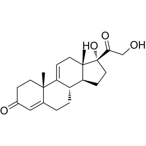
Anecortave desacetate

Based on 1 Customer Validation
Anecortave desacetate (AL-4940) is an angiogenesis inhibitor. Anecortave is indicated for the study of exudative (wet) age-related macular degeneration as well as retinal tumors.
For research use only. We do not sell to patients.
- Purity : 98.36%
- CAS No.: 10184-70-0
- Formula: C21H28O4
- Molecular Weight:344.44
-
Storage:Powder -20°C, 3 years , 4°C, 2 years ; In solvent -80°C, 6 months , -20°C, 1 month

To place orders, for customer services and technical support, please contact: MedChemExpress USA
Tel: 609-228-6898 E-mail: [email protected] [email protected]


-
 Biological Activity
-
Chemical Information
-
Solvent & Solubility
- Protocols
- Purity & Documentation
- References
-
 Help & FAQs




Biological Activity
Description
Cellular Effect
|
Cell Line

|
Type | Value | Description | References |
|---|---|---|---|---|
| C2C12 | EC50 |
6.29 x 10⁻⁸ M
Compound: VBP1
|
Inhibition of TNFalpha-induced NFkappaB activation in mouse C2C12 cells incubated for 24 hrs prior to TNFalpha induction measured after 24 hrs by luciferase reporter gene assay
Inhibition of TNFalpha-induced NFkappaB activation in mouse C2C12 cells incubated for 24 hrs prior to TNFalpha induction measured after 24 hrs by luciferase reporter gene assay
|
[PMID: 23498916] |
| C2C12 | EC50 |
6.29 x 10⁻² μM
Compound: VBP1
|
Inhibition of TNFalpha-induced NFkappaB activation in mouse C2C12 cells incubated for 24 hrs prior to TNFalpha induction measured after 24 hrs by luciferase reporter gene assay
Inhibition of TNFalpha-induced NFkappaB activation in mouse C2C12 cells incubated for 24 hrs prior to TNFalpha induction measured after 24 hrs by luciferase reporter gene assay
|
[PMID: 23498916] |
Clinical Trial
| NCT Number | Sponsor | Condition | Start Date |
Phase
|
|---|---|---|---|---|
| NCT01329991 | Plexxikon| | 2011-05 | PHASE1 |


Chemical Information
-
CAS No. 10184-70-0
-
Appearance Solid
-
Molecular Weight 344.44
-
Formula C21H28O4
-
Color White to off-white
-
SMILES
C[C@]12CCC(C=C1CC[C@@H]3C2=CC[C@]4([C@H]3CC[C@@]4(C(CO)=O)O)C)=O
-
Synonyms
AL-4940
-
Shipping
Room temperature in continental US; may vary elsewhere.
-
Storage
Powder -20°C 3 years 4°C 2 years In solvent -80°C 6 months -20°C 1 month


Solvent & Solubility
In Vitro:
DMSO : 50 mg/mL (145.16 mM; ultrasonic and warming and heat to 60°C; Hygroscopic DMSO has a significant impact on the solubility of product, please use newly opened DMSO)
Please refer to the solubility information to select the appropriate solvent. Once prepared, please aliquot and store the solution to prevent product inactivation from repeated freeze-thaw cycles.
Storage method and period of stock solution: -80°C, 6 months; -20°C, 1 month. When stored at -80°C, please use it within 6 months. When stored at -20°C, please use it within 1 month.
Please refer to the solubility information to select the appropriate solvent. Once prepared, please aliquot and store the solution to prevent product inactivation from repeated freeze-thaw cycles.
Storage method and period of stock solution: -80°C, 6 months; -20°C, 1 month. When stored at -80°C, please use it within 6 months. When stored at -20°C, please use it within 1 month.
Concentration (start) × Volume (start) = Concentration (final) × Volume (final)

Protocols
-
Human pluripotent stem cell endothelial-cell differentiation
Human pluripotent stem cell endothelial differentiation is based on stepwise developmental patterning: early activation of WNT/GSK3β inhibition promotes mesodermal or vascular progenitor entry, followed by endothelial specification using VEGF-related signaling, BMP4, FGF2, Notch modulation, or cAMP depending on the published protocol. Endothelial differentiation is read out by acquisition of CD31, CD34, VE-cadherin/CD144, KDR/VEGFR2, vWF, Tie2, NOS3, acetylated LDL uptake, tube/network formation, barrier function, and in vivo vessel-forming capacity where tested.
-
Vascular/Branching Fractal Analysis
Vascular/branching fractal analysis quantifies the geometric complexity of vessel trees or vascular networks from segmented 2D images, commonly by converting vessels into binary and/or skeletonized maps and estimating fractal dimension using box-counting or related approaches. Fractal dimension is interpreted as an image-derived readout of vascular branching complexity, space filling, or density, and has been applied to retinal photographs, fluorescein angiography, OCT angiography, capillary perfusion maps, and in vitro Matrigel angiogenesis networks. The assay readout is generated from vessel-positive pixels after image preprocessing, vessel segmentation, binarization, and optional skeletonization; reported outputs include fractal dimension, vessel density, branchpoint density, endpoint density, vessel length density, tortuosity, and generation-based branching metrics when VESGEN-style analysis is used. The biological interpretation is limited to quantitative vascular patterning and s
-
Endothelial Tube Formation Assay
Endothelial tube formation assay evaluates the ability of endothelial cells to attach, migrate, align, and organize into capillary-like networks when cultured on gelled basement membrane extract or Matrigel; the readout is the morphology and quantity of tube-like networks, which reflects an in vitro endothelial morphogenesis step related to angiogenesis. Basement membrane extract/Matrigel provides laminin-rich extracellular matrix cues that support endothelial differentiation into capillary-like structures, but it can contain biologically active growth factors, so growth-factor-reduced matrix is preferred when testing defined angiogenic stimulators or inhibitors.
-
Endothelial Cell Migration/Angiogenic Sprouting Assay
Endothelial cell migration and angiogenic sprouting assays are in vitro (and partially ex vivo-adapted) functional models that quantify the ability of endothelial cells to undergo coordinated migration, extracellular matrix invasion, and multicellular organization into capillary-like sprouts in response to pro-angiogenic stimuli such as VEGF, bFGF, or conditioned microenvironments. These assays are used to model early angiogenic events including tip-cell formation, directional migration, and lumen-like sprout extension, which collectively reflect angiogenic activation and vascular morphogenesis processes observed in vivo.


Purity & Documentation
-
Data Sheet (272 KB)
-
SDS (254 KB)
- English - EN (254 KB)
- Français - FR (254 KB)
- Deutsch - DE (254 KB)
- Norwegian - NO (254 KB)
- Español - ES (254 KB)
- Swedish - SV (254 KB)
- Italian - IT (254 KB)
- Korean - KR (254 KB)
- Portuguese - PT (254 KB)
-
Handling Instructions (2659 KB)
References
[1]. Kaiser PK, et al. Posterior juxtascleral depot administration of anecortave acetate. Surv Ophthalmol. 2007 Jan;52 Suppl 1:S62-9. [Content Brief]
[2]. Jockovich ME, et al. Anecortave acetate as single and adjuvant therapy in the treatment of retinal tumors of LH(BETA)T(AG) mice. Invest Ophthalmol Vis Sci. 2006 Apr;47(4):1264-8. [Content Brief]


Complete Stock Solution Preparation Table
Please refer to the solubility information to select the appropriate solvent. Once prepared, please aliquot and store the solution to prevent product inactivation from repeated freeze-thaw cycles.
Storage method and period of stock solution: -80°C, 6 months; -20°C, 1 month. When stored at -80°C, please use it within 6 months. When stored at -20°C, please use it within 1 month.
| Optional Solvent | Concentration Solvent Mass | 1 mg | 5 mg | 10 mg | 25 mg |
|---|---|---|---|---|---|
| DMSO | 1 mM | 2.9033 mL | 14.5163 mL | 29.0326 mL | 72.5816 mL |
| 5 mM | 0.5807 mL | 2.9033 mL | 5.8065 mL | 14.5163 mL | |
| 10 mM | 0.2903 mL | 1.4516 mL | 2.9033 mL | 7.2582 mL | |
| 15 mM | 0.1936 mL | 0.9678 mL | 1.9355 mL | 4.8388 mL | |
| 20 mM | 0.1452 mL | 0.7258 mL | 1.4516 mL | 3.6291 mL | |
| 25 mM | 0.1161 mL | 0.5807 mL | 1.1613 mL | 2.9033 mL | |
| 30 mM | 0.0968 mL | 0.4839 mL | 0.9678 mL | 2.4194 mL | |
| 40 mM | 0.0726 mL | 0.3629 mL | 0.7258 mL | 1.8145 mL | |
| 50 mM | 0.0581 mL | 0.2903 mL | 0.5807 mL | 1.4516 mL | |
| 60 mM | 0.0484 mL | 0.2419 mL | 0.4839 mL | 1.2097 mL | |
| 80 mM | 0.0363 mL | 0.1815 mL | 0.3629 mL | 0.9073 mL | |
| 100 mM | 0.0290 mL | 0.1452 mL | 0.2903 mL | 0.7258 mL |


Anecortave desacetate Related Classifications
HY-116098 Related Classifications
 Powered by Bioz